 Movie
Movie Controller


+ Open data

- Basic information
| Entry | 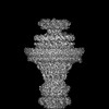 | |||||||||
|---|---|---|---|---|---|---|---|---|---|---|
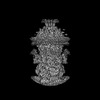
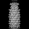
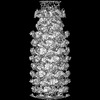
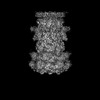
| Title | Portal and anchor DNA of phage 812 after tail contraction (C1) | |||||||||
 Map data Map data | Portal and anchor DNA of phage 812 after tail contraction (C1) | |||||||||
 Sample Sample |
| |||||||||
 Keywords Keywords | phage / neck / portal / anchor DNA / VIRUS | |||||||||
| Function / homology | Bacteriophage/Gene transfer agent portal protein / Phage portal protein / Portal protein Function and homology information Function and homology information | |||||||||
| Biological species |  Staphylococcus phage 812 (virus) Staphylococcus phage 812 (virus) | |||||||||
| Method | single particle reconstruction / cryo EM / Resolution: 3.94 Å | |||||||||
 Authors Authors | Cienikova Z / Siborova M / Fuzik T / Plevka P | |||||||||
| Funding support |  Czech Republic, 1 items Czech Republic, 1 items
| |||||||||
 Citation Citation | Journal: Commun Biol / Year: 2026 Title: Genome anchoring, retention, and release by neck proteins of Staphylococcus phage 812. Authors: Zuzana Cieniková / Jiří Nováček / Marta Šiborová / Barbora Popelářová / Tibor Füzik / Tibor Botka / Martin Benešík / Pavol Bárdy / Roman Pantůček / Pavel Plevka /   Abstract: The virion of Staphylococcus phage 812 is formed by a capsid and a contractile tail joined together by neck proteins. The neck proteins are crucial for virion assembly, DNA packaging, and the ...The virion of Staphylococcus phage 812 is formed by a capsid and a contractile tail joined together by neck proteins. The neck proteins are crucial for virion assembly, DNA packaging, and the regulation of genome release, but their functions are not completely understood. Here, we show that the neck of phage 812 consists of portal, adaptor, stopper, tail terminator, and two types of decoration proteins. A dodecameric DNA-binding site at the surface of the portal complex anchors the phage genome inside the capsid. The adaptor complex induces a local B-to-A form transition of the DNA in the neck channel that could slow or pause genome translocation during ejection. The central channel of a stopper complex that is not attached to the tail terminator complex is closed by gating loops. In contrast, in the phage 812 virion, the gating loops are in an open conformation, and the DNA extends into the tail. The structure of neck proteins is not affected by tail sheath contraction. Therefore, the expulsion of tail tape measure proteins triggers the genome release. | |||||||||
| History |
|
- Structure visualization
| Supplemental images |
|---|

- Downloads & links
-EMDB archive
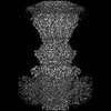
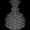
| Map data | emd_18395.map.gz | 24.2 MB | EMDB map data format | |
|---|---|---|---|---|
| Header (meta data) | emd-18395-v30.xmlemd-18395.xml | 20 KB 20 KB | Display Display | EMDB header |
| FSC (resolution estimation) | emd_18395_fsc.xml | 13.7 KB | Display | FSC data file |
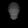
| Images |  emd_18395.png emd_18395.png | 76 KB | ||
| Masks | emd_18395_msk_1.map | 216 MB | Mask map | |
| Filedesc metadata | emd-18395.cif.gz | 5.9 KB | ||
| Others | emd_18395_half_map_1.map.gzemd_18395_half_map_2.map.gz | 170.8 MB 170.8 MB | ||
| Archive directory |  http://ftp.pdbj.org/pub/emdb/structures/EMD-18395ftp://ftp.pdbj.org/pub/emdb/structures/EMD-18395 http://ftp.pdbj.org/pub/emdb/structures/EMD-18395ftp://ftp.pdbj.org/pub/emdb/structures/EMD-18395 | HTTPS FTP |
-Related structure data
| Related structure data | 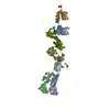 8q01C 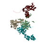 8q1iC 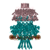 8q7dC  8qekC 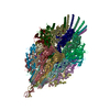 8qemC 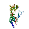 8qgrC  8qjeC  8qkhC  8r5gC  8r69C C: citing same article ( |
|---|---|
| Similar structure data |


-Links
| EMDB pages | EMDB (EBI/PDBe) / EMDataResource |
|---|
-Map
| File | Download / File: emd_18395.map.gz / Format: CCP4 / Size: 216 MB / Type: IMAGE STORED AS FLOATING POINT NUMBER (4 BYTES) | ||||||||||||||||||||||||||||||||||||
|---|---|---|---|---|---|---|---|---|---|---|---|---|---|---|---|---|---|---|---|---|---|---|---|---|---|---|---|---|---|---|---|---|---|---|---|---|---|
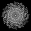
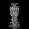
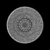
| Annotation | Portal and anchor DNA of phage 812 after tail contraction (C1) | ||||||||||||||||||||||||||||||||||||
| Projections & slices | Image control
Images are generated by Spider. | ||||||||||||||||||||||||||||||||||||
| Voxel size | X=Y=Z: 1.057 Å | ||||||||||||||||||||||||||||||||||||
| Density |
| ||||||||||||||||||||||||||||||||||||
| Symmetry | Space group: 1 | ||||||||||||||||||||||||||||||||||||
| Details | EMDB XML:
|
 Z (Sec.)
 Y (Row.)
 X (Col.)


-Supplemental data
-Mask #1
| File | emd_18395_msk_1.map | ||||||||||||
|---|---|---|---|---|---|---|---|---|---|---|---|---|---|
| Projections & Slices |
| ||||||||||||
| Density Histograms |


-Half map: Half-map 1
| File | emd_18395_half_map_1.map | ||||||||||||
|---|---|---|---|---|---|---|---|---|---|---|---|---|---|
| Annotation | Half-map 1 | ||||||||||||
| Projections & Slices |
| ||||||||||||
| Density Histograms |


-Half map: Half-map 2
| File | emd_18395_half_map_2.map | ||||||||||||
|---|---|---|---|---|---|---|---|---|---|---|---|---|---|
| Annotation | Half-map 2 | ||||||||||||
| Projections & Slices |
| ||||||||||||
| Density Histograms |


- Sample components
-Entire : Staphylococcus phage 812
| Entire | Name: Staphylococcus phage 812 (virus) |
|---|---|
| Components |
|
-Supramolecule #1: Staphylococcus phage 812
| Supramolecule | Name: Staphylococcus phage 812 / type: virus / ID: 1 / Parent: 0 / Macromolecule list: all Details: Purified phage virion was incubated in urea and LTA to induce tail contraction and genome ejection. NCBI-ID: 307898 / Sci species name: Staphylococcus phage 812 / Sci species strain: K1-420 / Virus type: VIRION / Virus isolate: SPECIES / Virus enveloped: No / Virus empty: Yes |
|---|---|
| Host (natural) | Organism:   Staphylococcus aureus (bacteria) / Strain: CCM 8428 Staphylococcus aureus (bacteria) / Strain: CCM 8428 |
| Virus shell | Shell ID: 1 / Diameter: 1100.0 Å |
-Macromolecule #1: Portal protein
| Macromolecule | Name: Portal protein / type: protein_or_peptide / ID: 1 / Details: Dodecamer / Enantiomer: LEVO |
|---|---|
| Source (natural) | Organism: Staphylococcus phage 812 (virus) / Strain: K1-420 |
| Sequence | String: MADLFKQFRL GKDYGNNSTI AQVPIDEGLQ ANIKKIEQDN KEYQDLTKSL YGQQQAYAEP FIEMMDTNPE FRDKRSYMKN EHNLHDVLKK FGNNPILNAI ILTRSNQVAM YCQPARYSEK GLGFEVRLRD LDAEPGRKEK EEMKRIEDFI VNTGKDKDVD RDSFQTFCKK ...String: MADLFKQFRL GKDYGNNSTI AQVPIDEGLQ ANIKKIEQDN KEYQDLTKSL YGQQQAYAEP FIEMMDTNPE FRDKRSYMKN EHNLHDVLKK FGNNPILNAI ILTRSNQVAM YCQPARYSEK GLGFEVRLRD LDAEPGRKEK EEMKRIEDFI VNTGKDKDVD RDSFQTFCKK IVRDTYIYDQ VNFEKVFNKN NKTKLEKFIA VDPSTIFYAT DKKGKIIKGG KRFVQVVDKR VVASFTSREL AMGIRNPRTE LSSSGYGLSE VEIAMKEFIA YNNTESFNDR FFSHGGTTRG ILQIRSDQQQ SQHALENFKR EWKSSLSGIN GSWQIPVVMA DDIKFVNMTP TANDMQFEKW LNYLINIISA LYGIDPAEIG FPNRGGATGS KGGSTLNEAD PGKKQQQSQN KGLQPLLRFI EDLVNRHIIS EYGDKYTFQF VGGDTKSATD KLNILKLETQ IFKTVNEARE EQGKKPIEGG DIILDASFLQ GTAQLQQDKQ YNDGKQKERL QMMMSLLEGD NDDSEEGQST DSSNDDKEIG TDAQIKGDDN VYRTQTSNKG QGRKGEKSSD FKH UniProtKB: Portal protein |
-Experimental details
-Structure determination
| Method | cryo EM |
|---|---|
 Processing Processing | single particle reconstruction |
| Aggregation state | particle |
-Sample preparation
| Buffer | pH: 8 Component:
| ||||||||||||
|---|---|---|---|---|---|---|---|---|---|---|---|---|---|
| Grid | Model: Quantifoil R2/1 / Material: COPPER / Mesh: 200 / Support film - Material: CARBON / Support film - topology: HOLEY / Pretreatment - Type: GLOW DISCHARGE / Pretreatment - Time: 30 sec. | ||||||||||||
| Vitrification | Cryogen name: ETHANE-PROPANE / Chamber humidity: 100 % / Instrument: FEI VITROBOT MARK IV |
- Electron microscopy
| Microscope | TFS KRIOS |
|---|---|
| Specialist optics | Energy filter - Slit width: 20 eV |
| Image recording | Film or detector model: GATAN K2 SUMMIT (4k x 4k) / Detector mode: SUPER-RESOLUTION / Digitization - Dimensions - Width: 4000 pixel / Digitization - Dimensions - Height: 4000 pixel / Number grids imaged: 1 / Number real images: 15371 / Average exposure time: 7.0 sec. / Average electron dose: 42.0 e/Å2 |
| Electron beam | Acceleration voltage: 300 kV / Electron source:  FIELD EMISSION GUN FIELD EMISSION GUN |
| Electron optics | C2 aperture diameter: 50.0 µm / Illumination mode: FLOOD BEAM / Imaging mode: BRIGHT FIELD / Cs: 2.7 mm / Nominal defocus max: 2.4 µm / Nominal defocus min: 1.1 µm / Nominal magnification: 130000 |
| Sample stage | Specimen holder model: FEI TITAN KRIOS AUTOGRID HOLDER / Cooling holder cryogen: NITROGEN |
| Experimental equipment |  Model: Titan Krios / Image courtesy: FEI Company |


