 Movie
Movie Controller

[English] 日本語
 Yorodumi
- EMDB-18122: A cryo-ET study of ciliary rootlet organization - purified rootle... -
+ Open data

- Basic information
| Entry |  | |||||||||
|---|---|---|---|---|---|---|---|---|---|---|
| Title | A cryo-ET study of ciliary rootlet organization - purified rootlet example 1 | |||||||||
 Map data Map data | purified rootlet, eman2 preprocessed cropped tomogram for segmentation. Object files are found on biostudies (S-BSST1164, https://www.ebi.ac.uk/biostudies/studies/S-BSST1164) | |||||||||
 Sample Sample |
| |||||||||
 Keywords Keywords | ciliary rootlet / rootlet / rootletin / centriole / cilia / STRUCTURAL PROTEIN | |||||||||
| Biological species |  | |||||||||
| Method | electron tomography / cryo EM | |||||||||
 Authors Authors | van Hoorn C / Carter AP | |||||||||
| Funding support |  United Kingdom, 2 items United Kingdom, 2 items
| |||||||||
 Citation Citation | Journal: Elife / Year: 2024 Title: A cryo-electron tomography study of ciliary rootlet organization. Authors: Chris van Hoorn / Andrew P Carter / Abstract: Ciliary rootlets are striated bundles of filaments that connect the base of cilia to internal cellular structures. Rootlets are critical for the sensory and motile functions of cilia. However, the ...Ciliary rootlets are striated bundles of filaments that connect the base of cilia to internal cellular structures. Rootlets are critical for the sensory and motile functions of cilia. However, the mechanisms underlying these functions remain unknown, in part due to a lack of structural information of rootlet organization. In this study, we obtain 3D reconstructions of membrane-associated and purified rootlets from mouse retina using cryo-electron tomography. We show that flexible protrusions on the rootlet surface, which emanate from the cross-striations, connect to intracellular membranes. In purified rootlets, the striations were classified into amorphous (A)-bands, associated with accumulations on the rootlet surface, and discrete (D)-bands corresponding to punctate lines of density that run through the rootlet. These striations connect a flexible network of longitudinal filaments. Subtomogram averaging suggests the filaments consist of two intertwined coiled coils. The rootlet's filamentous architecture, with frequent membrane-connecting cross-striations, lends itself well for anchoring large membranes in the cell. | |||||||||
| History |
|
- Structure visualization
| Supplemental images |
|---|

- Downloads & links
-EMDB archive
| Map data | emd_18122.map.gz | 54 MB |  EMDB map data format EMDB map data format | |
|---|---|---|---|---|
| Header (meta data) | emd-18122-v30.xmlemd-18122.xml | 8.4 KB 8.4 KB | Display Display | EMDB header |
| Images | 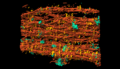 emd_18122.png emd_18122.png | 107 KB | ||
| Filedesc metadata | emd-18122.cif.gz | 4.3 KB | ||
| Archive directory |  http://ftp.pdbj.org/pub/emdb/structures/EMD-18122ftp://ftp.pdbj.org/pub/emdb/structures/EMD-18122 http://ftp.pdbj.org/pub/emdb/structures/EMD-18122ftp://ftp.pdbj.org/pub/emdb/structures/EMD-18122 | HTTPS FTP |
-Related structure data


-Links
| EMDB pages | EMDB (EBI/PDBe) / EMDataResource |
|---|
-Map
| File | Download / File: emd_18122.map.gz / Format: CCP4 / Size: 59.1 MB / Type: IMAGE STORED AS FLOATING POINT NUMBER (4 BYTES) | ||||||||||||||||||||||||||||||||
|---|---|---|---|---|---|---|---|---|---|---|---|---|---|---|---|---|---|---|---|---|---|---|---|---|---|---|---|---|---|---|---|---|---|
| Annotation | purified rootlet, eman2 preprocessed cropped tomogram for segmentation. Object files are found on biostudies (S-BSST1164, https://www.ebi.ac.uk/biostudies/studies/S-BSST1164) | ||||||||||||||||||||||||||||||||
| Projections & slices | Image control
Images are generated by Spider. generated in cubic-lattice coordinate | ||||||||||||||||||||||||||||||||
| Voxel size | X=Y=Z: 11.104 Å | ||||||||||||||||||||||||||||||||
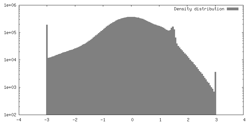
| Density |
| ||||||||||||||||||||||||||||||||
| Symmetry | Space group: 1 | ||||||||||||||||||||||||||||||||
| Details | EMDB XML:
|
 Z (Sec.)
 Y (Row.)
 X (Col.)


-Supplemental data
- Sample components
-Entire : Ciliary rootlet surrounded by cellular membranes
| Entire | Name: Ciliary rootlet surrounded by cellular membranes |
|---|---|
| Components |
|
-Supramolecule #1: Ciliary rootlet surrounded by cellular membranes
| Supramolecule | Name: Ciliary rootlet surrounded by cellular membranes / type: organelle_or_cellular_component / ID: 1 / Parent: 0 Details: The sample was purified according to https://doi.org/10.1016/j.cell.2012.10.038, maintaining cellular interactions with the rootlet. |
|---|---|
| Source (natural) | Organism: |
-Experimental details
-Structure determination
| Method | cryo EM |
|---|---|
 Processing Processing | electron tomography |
| Aggregation state | cell |
-Sample preparation
| Buffer | pH: 7.4 |
|---|---|
| Vitrification | Cryogen name: ETHANE / Chamber humidity: 100 % / Chamber temperature: 277 K / Instrument: FEI VITROBOT MARK III |
| Sectioning | Other: NO SECTIONING |
| Fiducial marker | Manufacturer: BBI / Diameter: 10 nm |
- Electron microscopy
| Microscope | FEI TITAN KRIOS |
|---|---|
| Image recording | Film or detector model: GATAN K3 (6k x 4k) / Average electron dose: 2.68 e/Å2 |
| Electron beam | Acceleration voltage: 300 kV / Electron source:  FIELD EMISSION GUN FIELD EMISSION GUN |
| Electron optics | Illumination mode: FLOOD BEAM / Imaging mode: BRIGHT FIELD / Nominal defocus max: 6.0 µm / Nominal defocus min: 1.0 µm |
| Experimental equipment |  Model: Titan Krios / Image courtesy: FEI Company |
-Image processing
| Details | For tomogram segmentation, tomograms were reconstructed as even/odd frame half tomograms using the above mentioned WARP pipeline and denoised using Noise2Map(Tegunov & Cramer, 2018). The tomograms were then deconvolved and isotropically reconstructed with denoising using IsoNet(Liu et al, 2021) with a cube size of 128 pixels. The tomograms were then preprocessed in EMAN2.2 for training of the TomoSeg CNN(Chen et al, 2017). |
|---|---|
| Final reconstruction | Number images used: 41 |
