 Movie
Movie Controller


+ Open data

- Basic information
| Entry | Database: EMDB / ID: EMD-1781 | |||||||||
|---|---|---|---|---|---|---|---|---|---|---|
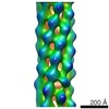
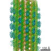
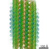
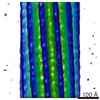
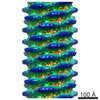
| Title | The nucleosome organization in 30nm fiber | |||||||||
 Map data Map data | sub-tomogram average | |||||||||
 Sample Sample |
| |||||||||
 Keywords Keywords | 30nm fiber | |||||||||
| Biological species |  | |||||||||
| Method | subtomogram averaging / cryo EM / Resolution: 43.1 Å | |||||||||
 Authors Authors | Scheffer MP / Eltsov M / Frangakis AS | |||||||||
 Citation Citation | Journal: Proc Natl Acad Sci U S A / Year: 2011 Title: Evidence for short-range helical order in the 30-nm chromatin fibers of erythrocyte nuclei. Authors: Margot P Scheffer / Mikhail Eltsov / Achilleas S Frangakis /  Abstract: Chromatin folding in eukaryotes fits the genome into the limited volume of the cell nucleus. Formation of higher-order chromatin structures attenuates DNA accessibility, thus contributing to the ...Chromatin folding in eukaryotes fits the genome into the limited volume of the cell nucleus. Formation of higher-order chromatin structures attenuates DNA accessibility, thus contributing to the control of essential genome functions such as transcription, DNA replication, and repair. The 30-nm fiber is thought to be the first hierarchical level of chromatin folding, but the nucleosome arrangement in the compact 30-nm fiber was previously unknown. We used cryoelectron tomography of vitreous sections to determine the structure of the compact, native 30-nm fiber of avian erythrocyte nuclei. The predominant geometry of the 30-nm fiber revealed by subtomogram averaging is a left-handed two-start helix with approximately 6.5 nucleosomes per 11 nm, in which the nucleosomes are juxtaposed face-to-face but are shifted off their superhelical axes with an axial translation of approximately 3.4 nm and an azimuthal rotation of approximately 54°. The nucleosomes produce a checkerboard pattern when observed in the direction perpendicular to the fiber axis but are not interdigitated. The nucleosome packing within the fibers shows larger center-to-center internucleosomal distances than previously anticipated, thus excluding the possibility of core-to-core interactions, explaining how transcription and regulation factors can access nucleosomes. | |||||||||
| History |
|
- Structure visualization
| Movie |
 Movie viewer Movie viewer |
|---|---|
| Structure viewer | EM map: SurfViewMolmilJmol/JSmol |
| Supplemental images |

- Downloads & links
-EMDB archive
| Map data | emd_1781.map.gz | 793.6 KB | EMDB map data format | |
|---|---|---|---|---|
| Header (meta data) | emd-1781-v30.xmlemd-1781.xml | 6.2 KB 6.2 KB | Display Display | EMDB header |
| Images | 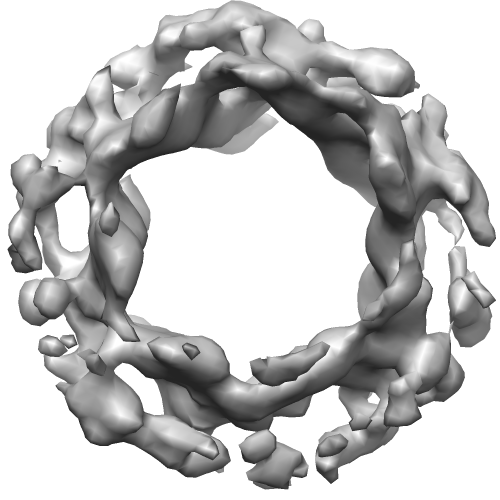 EMD-1781.png EMD-1781.png | 107.4 KB | ||
| Archive directory |  http://ftp.pdbj.org/pub/emdb/structures/EMD-1781ftp://ftp.pdbj.org/pub/emdb/structures/EMD-1781 http://ftp.pdbj.org/pub/emdb/structures/EMD-1781ftp://ftp.pdbj.org/pub/emdb/structures/EMD-1781 | HTTPS FTP |
-Validation report
| Summary document | emd_1781_validation.pdf.gz | 217.8 KB | Display | EMDB validaton report |
|---|---|---|---|---|
| Full document | emd_1781_full_validation.pdf.gz | 217 KB | Display | |
| Data in XML | emd_1781_validation.xml.gz | 4.7 KB | Display | |
| Arichive directory | https://ftp.pdbj.org/pub/emdb/validation_reports/EMD-1781ftp://ftp.pdbj.org/pub/emdb/validation_reports/EMD-1781 | HTTPS FTP |
-Related structure data

-Links
| EMDB pages | EMDB (EBI/PDBe) / EMDataResource |
|---|
-Map
| File | Download / File: emd_1781.map.gz / Format: CCP4 / Size: 1001 KB / Type: IMAGE STORED AS FLOATING POINT NUMBER (4 BYTES) | ||||||||||||||||||||||||||||||||||||||||||||||||||||||||||||||||||||
|---|---|---|---|---|---|---|---|---|---|---|---|---|---|---|---|---|---|---|---|---|---|---|---|---|---|---|---|---|---|---|---|---|---|---|---|---|---|---|---|---|---|---|---|---|---|---|---|---|---|---|---|---|---|---|---|---|---|---|---|---|---|---|---|---|---|---|---|---|---|
| Annotation | sub-tomogram average | ||||||||||||||||||||||||||||||||||||||||||||||||||||||||||||||||||||
| Projections & slices | Image control
Images are generated by Spider. | ||||||||||||||||||||||||||||||||||||||||||||||||||||||||||||||||||||
| Voxel size | X=Y=Z: 12 Å | ||||||||||||||||||||||||||||||||||||||||||||||||||||||||||||||||||||
| Density |
| ||||||||||||||||||||||||||||||||||||||||||||||||||||||||||||||||||||
| Symmetry | Space group: 1 | ||||||||||||||||||||||||||||||||||||||||||||||||||||||||||||||||||||
| Details | EMDB XML:
CCP4 map header:
| ||||||||||||||||||||||||||||||||||||||||||||||||||||||||||||||||||||
 Z (Sec.)
 Y (Row.)
 X (Col.)


-Supplemental data
- Sample components
-Entire : 30nm fiber
| Entire | Name: 30nm fiber |
|---|---|
| Components |
|
-Supramolecule #1000: 30nm fiber
| Supramolecule | Name: 30nm fiber / type: sample / ID: 1000 / Number unique components: 1 |
|---|
-Macromolecule #1: 30-nm fiber
| Macromolecule | Name: 30-nm fiber / type: protein_or_peptide / ID: 1 / Name.synonym: 30-nm fiber / Recombinant expression: No / Database: NCBI |
|---|---|
| Source (natural) | Organism: |
-Experimental details
-Structure determination
| Method | cryo EM |
|---|---|
 Processing Processing | subtomogram averaging |
-Sample preparation
| Vitrification | Cryogen name: NITROGEN / Instrument: OTHER |
|---|
- Electron microscopy
| Microscope | FEI POLARA 300 |
|---|---|
| Electron beam | Acceleration voltage: 300 kV / Electron source:  FIELD EMISSION GUN FIELD EMISSION GUN |
| Electron optics | Illumination mode: OTHER / Imaging mode: BRIGHT FIELD |
| Sample stage | Specimen holder: Eucentric / Specimen holder model: OTHER |
| Experimental equipment |  Model: Tecnai Polara / Image courtesy: FEI Company |
-Image processing
| Final reconstruction | Resolution.type: BY AUTHOR / Resolution: 43.1 Å / Resolution method: FSC 0.5 CUT-OFF |
|---|
