 Movie
Movie Controller

[English] 日本語
 Yorodumi
- EMDB-1780: High-resolution Cryo-EM structure of a programmed wheat germ ribosome -
+ Open data

- Basic information
| Entry | Database: EMDB / ID: EMD-1780 | |||||||||
|---|---|---|---|---|---|---|---|---|---|---|
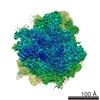
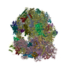
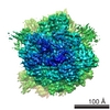
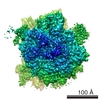
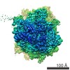
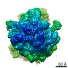
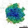
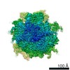
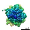
| Title | High-resolution Cryo-EM structure of a programmed wheat germ ribosome | |||||||||
 Map data Map data | Cryo-EM map of a programmed wheat germ ribosome containing a P-site tRNA | |||||||||
 Sample Sample |
| |||||||||
 Keywords Keywords | Eukaryotic Ribosome / Homology Modelling / RNA Expansion Segments / Novel Ribosomal Proteins | |||||||||
| Function / homology |  Function and homology information Function and homology informationtranslational elongation / protein kinase activator activity / ribonucleoprotein complex binding / cytosolic ribosome / maturation of LSU-rRNA from tricistronic rRNA transcript (SSU-rRNA, 5.8S rRNA, LSU-rRNA) / small-subunit processome / rRNA processing / ribosomal small subunit biogenesis / small ribosomal subunit / large ribosomal subunit rRNA binding ...translational elongation / protein kinase activator activity / ribonucleoprotein complex binding / cytosolic ribosome / maturation of LSU-rRNA from tricistronic rRNA transcript (SSU-rRNA, 5.8S rRNA, LSU-rRNA) / small-subunit processome / rRNA processing / ribosomal small subunit biogenesis / small ribosomal subunit / large ribosomal subunit rRNA binding / cytosolic small ribosomal subunit / cytosolic large ribosomal subunit / cytoplasmic translation / negative regulation of translation / structural constituent of ribosome / ribosome / translation / ribonucleoprotein complex / mRNA binding / signal transduction / RNA binding / zinc ion binding / plasma membrane / cytosol Similarity search - Function | |||||||||
| Biological species |  | |||||||||
| Method | single particle reconstruction / cryo EM / negative staining / Resolution: 5.5 Å | |||||||||
 Authors Authors | Armache JP / Jarasch A / Anger AM / Villa E / Becker T / Bhushan S / Jossinet F / Habeck M / Dindar G / Franckenberg S ...Armache JP / Jarasch A / Anger AM / Villa E / Becker T / Bhushan S / Jossinet F / Habeck M / Dindar G / Franckenberg S / Marquez V / Mielke T / Thomm M / Berninghausen O / Beatrix B / Soeding J / Westhof E / Wilson DN / Beckmann R | |||||||||
 Citation Citation | Journal: Proc Natl Acad Sci U S A / Year: 2010 Title: Localization of eukaryote-specific ribosomal proteins in a 5.5-Å cryo-EM map of the 80S eukaryotic ribosome. Authors: Jean-Paul Armache / Alexander Jarasch / Andreas M Anger / Elizabeth Villa / Thomas Becker / Shashi Bhushan / Fabrice Jossinet / Michael Habeck / Gülcin Dindar / Sibylle Franckenberg / Viter ...Authors: Jean-Paul Armache / Alexander Jarasch / Andreas M Anger / Elizabeth Villa / Thomas Becker / Shashi Bhushan / Fabrice Jossinet / Michael Habeck / Gülcin Dindar / Sibylle Franckenberg / Viter Marquez / Thorsten Mielke / Michael Thomm / Otto Berninghausen / Birgitta Beatrix / Johannes Söding / Eric Westhof / Daniel N Wilson / Roland Beckmann /  Abstract: Protein synthesis in all living organisms occurs on ribonucleoprotein particles, called ribosomes. Despite the universality of this process, eukaryotic ribosomes are significantly larger in size than ...Protein synthesis in all living organisms occurs on ribonucleoprotein particles, called ribosomes. Despite the universality of this process, eukaryotic ribosomes are significantly larger in size than their bacterial counterparts due in part to the presence of 80 r proteins rather than 54 in bacteria. Using cryoelectron microscopy reconstructions of a translating plant (Triticum aestivum) 80S ribosome at 5.5-Å resolution, together with a 6.1-Å map of a translating Saccharomyces cerevisiae 80S ribosome, we have localized and modeled 74/80 (92.5%) of the ribosomal proteins, encompassing 12 archaeal/eukaryote-specific small subunit proteins as well as the complete complement of the ribosomal proteins of the eukaryotic large subunit. Near-complete atomic models of the 80S ribosome provide insights into the structure, function, and evolution of the eukaryotic translational apparatus. | |||||||||
| History |
|
- Structure visualization
| Movie |
Movie viewer |
|---|---|
| Structure viewer | EM map: SurfViewMolmilJmol/JSmol |
| Supplemental images |

- Downloads & links
-EMDB archive
| Map data | emd_1780.map.gz | 24.8 MB | EMDB map data format | |
|---|---|---|---|---|
| Header (meta data) | emd-1780-v30.xmlemd-1780.xml | 10.5 KB 10.5 KB | Display Display | EMDB header |
| Images | 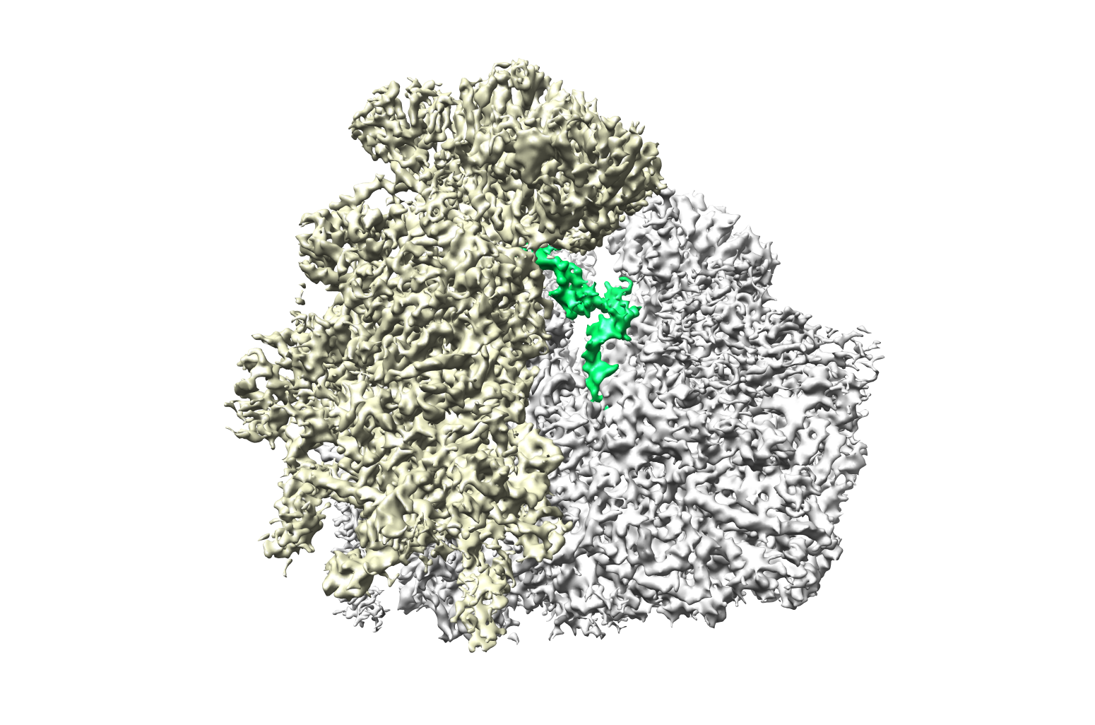 1780_wg_bf030_front.png 1780_wg_bf030_front.png | 958.4 KB | ||
| Archive directory |  http://ftp.pdbj.org/pub/emdb/structures/EMD-1780ftp://ftp.pdbj.org/pub/emdb/structures/EMD-1780 http://ftp.pdbj.org/pub/emdb/structures/EMD-1780ftp://ftp.pdbj.org/pub/emdb/structures/EMD-1780 | HTTPS FTP |
-Related structure data
| Related structure data |  4v7eM M: atomic model generated by this map |
|---|---|
| Similar structure data |

-Links
| EMDB pages | EMDB (EBI/PDBe) / EMDataResource |
|---|---|
| Related items in Molecule of the Month |


-Map
| File | Download / File: emd_1780.map.gz / Format: CCP4 / Size: 185.7 MB / Type: IMAGE STORED AS FLOATING POINT NUMBER (4 BYTES) | ||||||||||||||||||||||||||||||||||||||||||||||||||||||||||||||||||||
|---|---|---|---|---|---|---|---|---|---|---|---|---|---|---|---|---|---|---|---|---|---|---|---|---|---|---|---|---|---|---|---|---|---|---|---|---|---|---|---|---|---|---|---|---|---|---|---|---|---|---|---|---|---|---|---|---|---|---|---|---|---|---|---|---|---|---|---|---|---|
| Annotation | Cryo-EM map of a programmed wheat germ ribosome containing a P-site tRNA | ||||||||||||||||||||||||||||||||||||||||||||||||||||||||||||||||||||
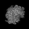
| Projections & slices | Image control
Images are generated by Spider. | ||||||||||||||||||||||||||||||||||||||||||||||||||||||||||||||||||||
| Voxel size | X=Y=Z: 1.2375 Å | ||||||||||||||||||||||||||||||||||||||||||||||||||||||||||||||||||||
| Density |
| ||||||||||||||||||||||||||||||||||||||||||||||||||||||||||||||||||||
| Symmetry | Space group: 1 | ||||||||||||||||||||||||||||||||||||||||||||||||||||||||||||||||||||
| Details | EMDB XML:
CCP4 map header:
| ||||||||||||||||||||||||||||||||||||||||||||||||||||||||||||||||||||
 Z (Sec.)
 X (Row.)
 Y (Col.)


-Supplemental data
- Sample components
-Entire : Programmed Wheat Germ 80S Ribosome
| Entire | Name: Programmed Wheat Germ 80S Ribosome |
|---|---|
| Components |
|
-Supramolecule #1000: Programmed Wheat Germ 80S Ribosome
| Supramolecule | Name: Programmed Wheat Germ 80S Ribosome / type: sample / ID: 1000 / Oligomeric state: One Ribosome / Number unique components: 1 |
|---|---|
| Molecular weight | Experimental: 4.2 MDa / Theoretical: 4.2 MDa / Method: Sedimentation |
-Supramolecule #1: Tritcum aestivum 80S ribosome
| Supramolecule | Name: Tritcum aestivum 80S ribosome / type: complex / ID: 1 / Name.synonym: Wheat Germ Ribosome / Recombinant expression: No / Ribosome-details: ribosome-eukaryote: ALL |
|---|---|
| Source (natural) | Organism: |
| Molecular weight | Experimental: 4.2 MDa / Theoretical: 4.2 MDa |
-Experimental details
-Structure determination
| Method | negative staining, cryo EM |
|---|---|
 Processing Processing | single particle reconstruction |
| Aggregation state | particle |
-Sample preparation
| Concentration | 0.02 mg/mL |
|---|---|
| Buffer | pH: 7.5 Details: 20 mM HEPES/KOH, pH 7.5, 100 mM KOAc, 10 mM Mg(OAc)2, 0.01 mg/ml cycloheximide, 1 mM DTT, 0.01 % Nikkol |
| Staining | Type: NEGATIVE / Details: Cryo-EM |
| Grid | Details: Quantifoil Grid with 2 nm carbon on top |
| Vitrification | Cryogen name: ETHANE / Chamber humidity: 100 % / Instrument: OTHER / Details: Vitrification instrument: Vitrobot Method: Blot for 10 seconds before plunging, use 2 layers of filter paper |
- Electron microscopy
| Microscope | FEI TECNAI F30 |
|---|---|
| Image recording | Category: FILM / Film or detector model: KODAK SO-163 FILM / Digitization - Scanner: PRIMESCAN / Digitization - Sampling interval: 4.76 µm / Number real images: 1374 / Average electron dose: 25 e/Å2 Details: Scanned at 5334 dpi on a Heidelberg Primescan Drum Scanner Od range: 1.2 / Bits/pixel: 16 |
| Electron beam | Acceleration voltage: 300 kV / Electron source:  FIELD EMISSION GUN FIELD EMISSION GUN |
| Electron optics | Calibrated magnification: 38900 / Illumination mode: FLOOD BEAM / Imaging mode: BRIGHT FIELD / Cs: 2.26 mm / Nominal defocus max: 4.5 µm / Nominal defocus min: 1.0 µm / Nominal magnification: 39000 |
| Sample stage | Specimen holder: FEI Polara Cartridge System / Specimen holder model: OTHER |
| Experimental equipment |  Model: Tecnai F30 / Image courtesy: FEI Company |
-Image processing
| Details | The reconstruction contains 2108230 particles in total |
|---|---|
| CTF correction | Details: Wiener Filter on 3D volumes (SPIDER) |
| Final reconstruction | Applied symmetry - Point group: C1 (asymmetric) / Algorithm: OTHER / Resolution.type: BY AUTHOR / Resolution: 5.5 Å / Resolution method: FSC 0.5 CUT-OFF / Software - Name: SPIDER |
