 Movie
Movie Controller


+ Open data

- Basic information
| Entry |  | |||||||||
|---|---|---|---|---|---|---|---|---|---|---|
| Title | Cryo electron tomography of human choriocarcinoma cells | |||||||||
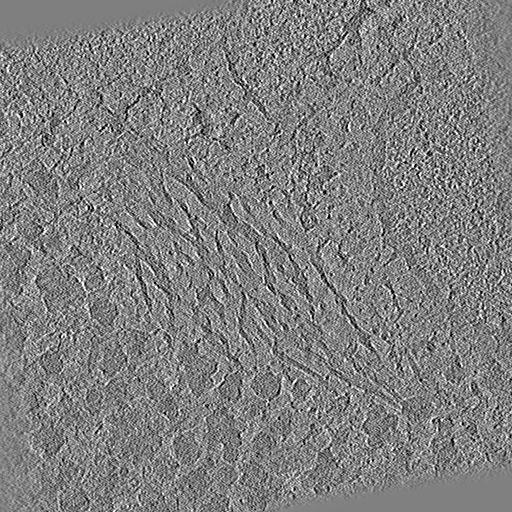
 Map data Map data | IMOD weighted back-projection reconstruction of tilt series from Position 09 using Ot2Rec. | |||||||||
 Sample Sample |
| |||||||||
 Keywords Keywords | human / choriocarcinoma / cell / trophoblast / CELL CYCLE | |||||||||
| Biological species |  Homo sapiens (human) Homo sapiens (human) | |||||||||
| Method | electron tomography / cryo EM | |||||||||
 Authors Authors | Tun WM / Yee NB-Y / Ho EML / Darrow MC / Basham M | |||||||||
| Funding support |  United Kingdom, 1 items United Kingdom, 1 items
| |||||||||
 Citation Citation | Journal: Biol Imaging / Year: 2023 Title: Ot2Rec: A semi-automatic, extensible, multi-software tomographic reconstruction workflow. Authors: Neville B-Y Yee / Elaine M L Ho / Win Tun / Jake L R Smith / Maud Dumoux / Michael Grange / Michele C Darrow / Mark Basham / Abstract: Electron cryo-tomography is an imaging technique for probing 3D structures with at the nanometer scale. This technique has been used extensively in the biomedical field to study the complex ...Electron cryo-tomography is an imaging technique for probing 3D structures with at the nanometer scale. This technique has been used extensively in the biomedical field to study the complex structures of proteins and other macromolecules. With the advancement in technology, microscopes are currently capable of producing images amounting to terabytes of data per day, posing great challenges for scientists as the speed of processing of the images cannot keep up with the ever-higher throughput of the microscopes. Therefore, automation is an essential and natural pathway on which image processing-from individual micrographs to full tomograms-is developing. In this paper, we present Ot2Rec, an open-source pipelining tool which aims to enable scientists to build their own processing workflows in a flexible and automatic manner. The basic building blocks of Ot2Rec are plugins which follow a unified application programming interface structure, making it simple for scientists to contribute to Ot2Rec by adding features which are not already available. In this paper, we also present three case studies of image processing using Ot2Rec, through which we demonstrate the speedup of using a semi-automatic workflow over a manual one, the possibility of writing and using custom (prototype) plugins, and the flexibility of Ot2Rec which enables the mix-and-match of plugins. We also demonstrate, in the Supplementary Material, a built-in reporting feature in Ot2Rec which aggregates the metadata from all process being run, and output them in the Jupyter Notebook and/or HTML formats for quick review of image processing quality. Ot2Rec can be found at https://github.com/rosalindfranklininstitute/ot2rec. | |||||||||
| History |
|
- Structure visualization
| Supplemental images |
|---|

- Downloads & links
-EMDB archive
| Map data | emd_17767.map.gz | 473.3 MB |  EMDB map data format EMDB map data format | |
|---|---|---|---|---|
| Header (meta data) | emd-17767-v30.xmlemd-17767.xml | 9.1 KB 9.1 KB | Display Display | EMDB header |
| Images |  emd_17767.png emd_17767.png | 268 KB | ||
| Filedesc metadata | emd-17767.cif.gz | 3.8 KB | ||
| Archive directory |  http://ftp.pdbj.org/pub/emdb/structures/EMD-17767ftp://ftp.pdbj.org/pub/emdb/structures/EMD-17767 http://ftp.pdbj.org/pub/emdb/structures/EMD-17767ftp://ftp.pdbj.org/pub/emdb/structures/EMD-17767 | HTTPS FTP |
-Validation report
| Summary document | emd_17767_validation.pdf.gz | 867.4 KB | Display | EMDB validaton report |
|---|---|---|---|---|
| Full document | emd_17767_full_validation.pdf.gz | 867 KB | Display | |
| Data in XML | emd_17767_validation.xml.gz | 8.5 KB | Display | |
| Data in CIF | emd_17767_validation.cif.gz | 9.7 KB | Display | |
| Arichive directory | https://ftp.pdbj.org/pub/emdb/validation_reports/EMD-17767ftp://ftp.pdbj.org/pub/emdb/validation_reports/EMD-17767 | HTTPS FTP |
-Links
| EMDB pages | EMDB (EBI/PDBe) / EMDataResource |
|---|
-Map
| File | Download / File: emd_17767.map.gz / Format: CCP4 / Size: 512 MB / Type: IMAGE STORED AS FLOATING POINT NUMBER (4 BYTES) | ||||||||||||||||||||||||||||||||
|---|---|---|---|---|---|---|---|---|---|---|---|---|---|---|---|---|---|---|---|---|---|---|---|---|---|---|---|---|---|---|---|---|---|
| Annotation | IMOD weighted back-projection reconstruction of tilt series from Position 09 using Ot2Rec. | ||||||||||||||||||||||||||||||||
| Projections & slices | Image control
Images are generated by Spider. | ||||||||||||||||||||||||||||||||
| Voxel size | X=Y=Z: 14.96 Å | ||||||||||||||||||||||||||||||||
| Density |
| ||||||||||||||||||||||||||||||||
| Symmetry | Space group: 1 | ||||||||||||||||||||||||||||||||
| Details | EMDB XML:
|
 Z (Sec.)
 Y (Row.)
 X (Col.)


-Supplemental data
- Sample components
-Entire : JEG-3 human choriocarcinoma cell line
| Entire | Name: JEG-3 human choriocarcinoma cell line |
|---|---|
| Components |
|
-Supramolecule #1: JEG-3 human choriocarcinoma cell line
| Supramolecule | Name: JEG-3 human choriocarcinoma cell line / type: cell / ID: 1 / Parent: 0 |
|---|---|
| Source (natural) | Organism: Homo sapiens (human) |
-Experimental details
-Structure determination
| Method | cryo EM |
|---|---|
 Processing Processing | electron tomography |
| Aggregation state | cell |
-Sample preparation
| Buffer | pH: 7.4 |
|---|---|
| Grid | Model: Quantifoil R2/2 / Mesh: 200 |
| Vitrification | Cryogen name: ETHANE / Chamber humidity: 80 % / Instrument: LEICA EM GP |
| Sectioning | Focused ion beam - Instrument: OTHER / Focused ion beam - Ion: OTHER / Focused ion beam - Voltage: 30 / Focused ion beam - Current: 0.3 / Focused ion beam - Duration: 1.0E-14 / Focused ion beam - Temperature: 105 K / Focused ion beam - Initial thickness: 1000 / Focused ion beam - Final thickness: 200 Focused ion beam - Details: Note: initial thickness and duration of FIB milling was not recorded.. The value given for _em_focused_ion_beam.instrument is ThermoFisher Scios. This is not in a list of ...Focused ion beam - Details: Note: initial thickness and duration of FIB milling was not recorded.. The value given for _em_focused_ion_beam.instrument is ThermoFisher Scios. This is not in a list of allowed values {'OTHER', 'DB235'} so OTHER is written into the XML file. |
- Electron microscopy
| Microscope | FEI TITAN KRIOS |
|---|---|
| Image recording | Film or detector model: TFS FALCON 4i (4k x 4k) / Average electron dose: 1.48 e/Å2 |
| Electron beam | Acceleration voltage: 300 kV / Electron source:  FIELD EMISSION GUN FIELD EMISSION GUN |
| Electron optics | Illumination mode: FLOOD BEAM / Imaging mode: BRIGHT FIELD / Nominal defocus max: 8.0 µm / Nominal defocus min: 4.0 µm / Nominal magnification: 64000 |
| Experimental equipment |  Model: Titan Krios / Image courtesy: FEI Company |
-Image processing
| Details | Images were motion-corrected with MotionCor2 1.4.0 using Ot2Rec. |
|---|---|
| Final reconstruction | Algorithm: BACK PROJECTION / Software - Name: IMOD (ver. 4.11.1) / Software - details: Weighted back-projection / Details: Bin factor 8 applied. / Number images used: 41 |
