 Movie
Movie Controller


+ Open data

- Basic information
| Entry |  | |||||||||
|---|---|---|---|---|---|---|---|---|---|---|
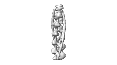
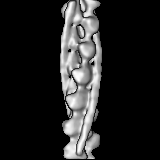
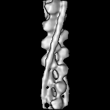
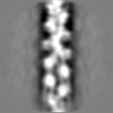
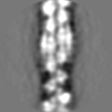
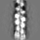
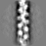
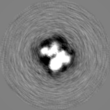
| Title | In situ tropomyosin decorated actin filament at focal adhesion | |||||||||
 Map data Map data | ||||||||||
 Sample Sample |
| |||||||||
 Keywords Keywords | focal adhesion / stress fiber / CELL ADHESION | |||||||||
| Biological species |  | |||||||||
| Method | subtomogram averaging / cryo EM / Resolution: 15.3 Å | |||||||||
 Authors Authors | Wen-Lu C / Ohad M | |||||||||
| Funding support | European Union, 1 items
| |||||||||
 Citation Citation | Journal: To Be Published Title: A mixed actin polarity network at focal adhesion Authors: Wen-Lu C / Ohad M | |||||||||
| History |
|
- Structure visualization
| Supplemental images |
|---|
- Downloads & links
-EMDB archive
| Map data | emd_17744.map.gz | 12 MB |  EMDB map data format EMDB map data format | |
|---|---|---|---|---|
| Header (meta data) | emd-17744-v30.xmlemd-17744.xml | 11 KB 11 KB | Display Display | EMDB header |
| Images |  emd_17744.png emd_17744.png | 16.3 KB | ||
| Filedesc metadata | emd-17744.cif.gz | 3.6 KB | ||
| Others | emd_17744_half_map_1.map.gzemd_17744_half_map_2.map.gz | 12 MB 12 MB | ||
| Archive directory |  http://ftp.pdbj.org/pub/emdb/structures/EMD-17744ftp://ftp.pdbj.org/pub/emdb/structures/EMD-17744 http://ftp.pdbj.org/pub/emdb/structures/EMD-17744ftp://ftp.pdbj.org/pub/emdb/structures/EMD-17744 | HTTPS FTP |
-Validation report
| Summary document | emd_17744_validation.pdf.gz | 625.1 KB | Display | EMDB validaton report |
|---|---|---|---|---|
| Full document | emd_17744_full_validation.pdf.gz | 624.7 KB | Display | |
| Data in XML | emd_17744_validation.xml.gz | 9.7 KB | Display | |
| Data in CIF | emd_17744_validation.cif.gz | 11.3 KB | Display | |
| Arichive directory | https://ftp.pdbj.org/pub/emdb/validation_reports/EMD-17744ftp://ftp.pdbj.org/pub/emdb/validation_reports/EMD-17744 | HTTPS FTP |
-Related structure data

-Links
| EMDB pages | EMDB (EBI/PDBe) / EMDataResource |
|---|
-Map
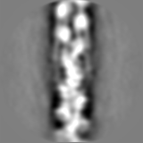
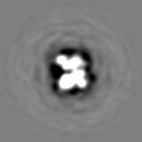
| File | Download / File: emd_17744.map.gz / Format: CCP4 / Size: 15.6 MB / Type: IMAGE STORED AS FLOATING POINT NUMBER (4 BYTES) | ||||||||||||||||||||||||||||||||||||
|---|---|---|---|---|---|---|---|---|---|---|---|---|---|---|---|---|---|---|---|---|---|---|---|---|---|---|---|---|---|---|---|---|---|---|---|---|---|
| Projections & slices | Image control
Images are generated by Spider. | ||||||||||||||||||||||||||||||||||||
| Voxel size | X=Y=Z: 2.20651 Å | ||||||||||||||||||||||||||||||||||||
| Density |
| ||||||||||||||||||||||||||||||||||||
| Symmetry | Space group: 1 | ||||||||||||||||||||||||||||||||||||
| Details | EMDB XML:
|
 Z (Sec.)
 Y (Row.)
 X (Col.)


-Supplemental data
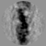
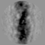
-Half map: #2
| File | emd_17744_half_map_1.map | ||||||||||||
|---|---|---|---|---|---|---|---|---|---|---|---|---|---|
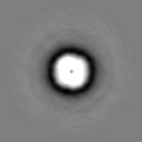
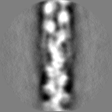
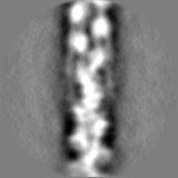
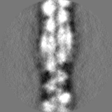
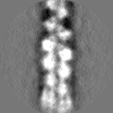
| Projections & Slices |
| ||||||||||||
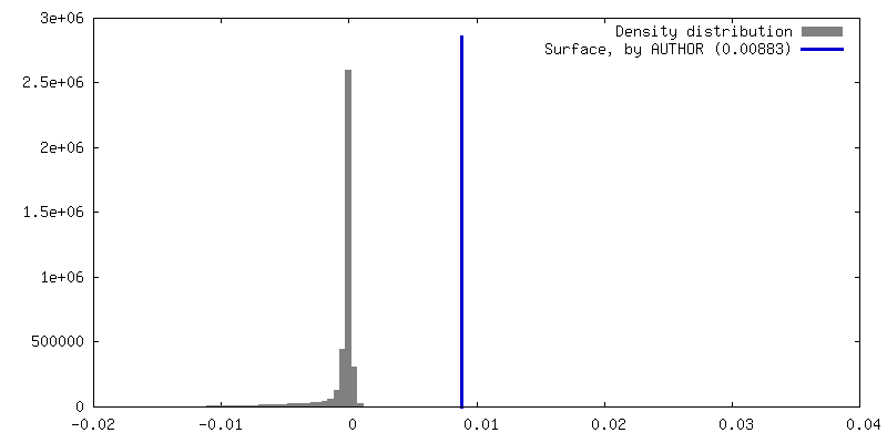
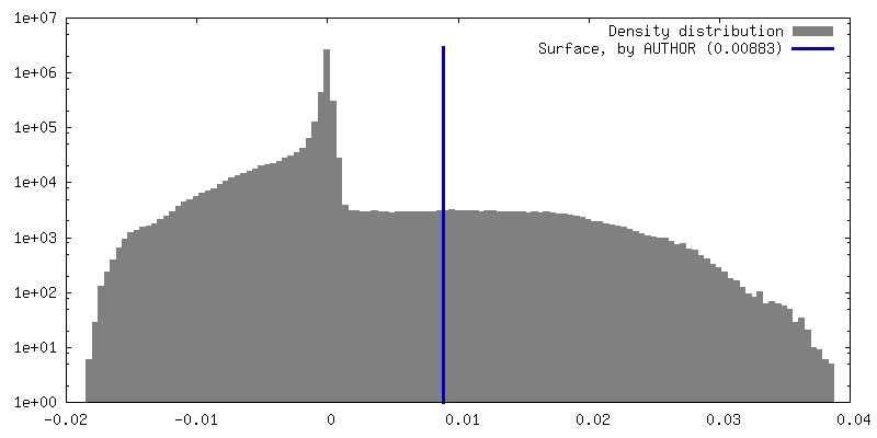
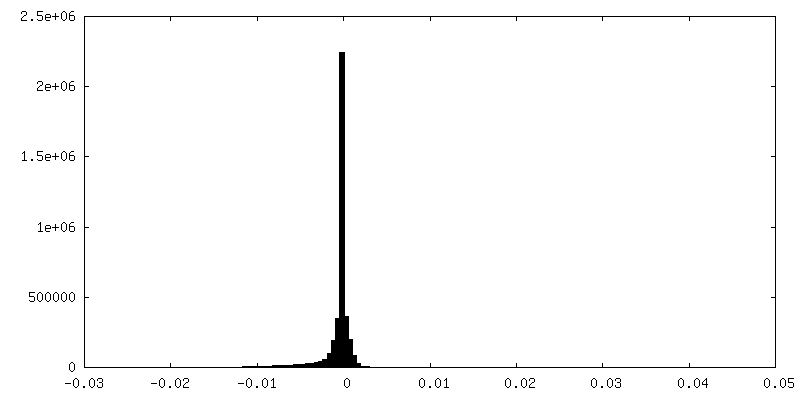
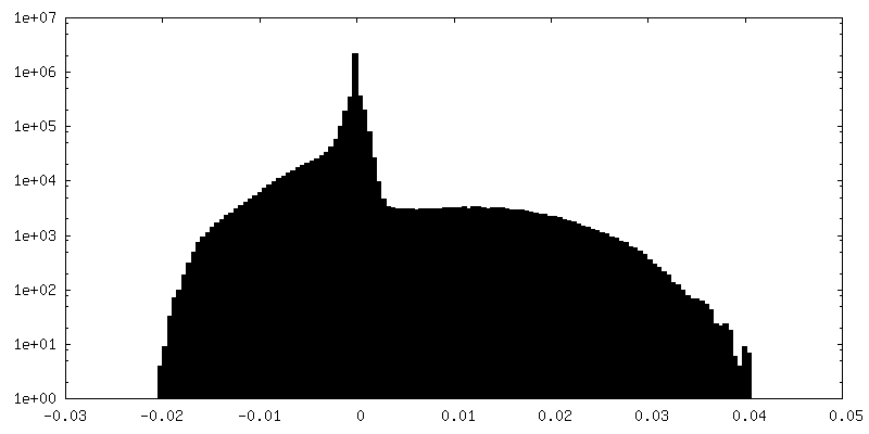
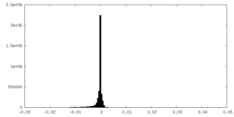
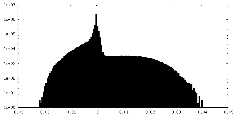
| Density Histograms |
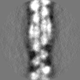

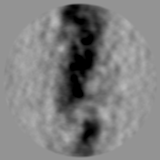
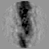
-Half map: #1
| File | emd_17744_half_map_2.map | ||||||||||||
|---|---|---|---|---|---|---|---|---|---|---|---|---|---|
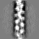
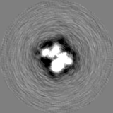
| Projections & Slices |
| ||||||||||||
| Density Histograms |

- Sample components
-Entire : Mouse embryonic fibroblast
| Entire | Name: Mouse embryonic fibroblast |
|---|---|
| Components |
|
-Supramolecule #1: Mouse embryonic fibroblast
| Supramolecule | Name: Mouse embryonic fibroblast / type: cell / ID: 1 / Parent: 0 |
|---|---|
| Source (natural) | Organism: |
-Experimental details
-Structure determination
| Method | cryo EM |
|---|---|
 Processing Processing | subtomogram averaging |
| Aggregation state | cell |
-Sample preparation
| Buffer | pH: 7.4 |
|---|---|
| Vitrification | Cryogen name: ETHANE |
- Electron microscopy
| Microscope | FEI TITAN KRIOS |
|---|---|
| Image recording | Film or detector model: GATAN K2 SUMMIT (4k x 4k) / Average electron dose: 2.9 e/Å2 |
| Electron beam | Acceleration voltage: 300 kV / Electron source:  FIELD EMISSION GUN FIELD EMISSION GUN |
| Electron optics | Illumination mode: FLOOD BEAM / Imaging mode: BRIGHT FIELD / Nominal defocus max: 5.0 µm / Nominal defocus min: 4.0 µm |
| Experimental equipment |  Model: Titan Krios / Image courtesy: FEI Company |
-Image processing
| Final reconstruction | Applied symmetry - Helical parameters - Δz: 28.0923 Å Applied symmetry - Helical parameters - Δ&Phi: -166.781 ° Applied symmetry - Helical parameters - Axial symmetry: C1 (asymmetric) Resolution.type: BY AUTHOR / Resolution: 15.3 Å / Resolution method: FSC 0.143 CUT-OFF / Number subtomograms used: 37330 |
|---|---|
| Extraction | Number tomograms: 189 / Number images used: 38900 |
| Final angle assignment | Type: ANGULAR RECONSTITUTION |
