 ムービー
ムービー コントローラー


+ データを開く

- 基本情報
基本情報
| 登録情報 | データベース: EMDB / ID: EMD-1750 | |||||||||
|---|---|---|---|---|---|---|---|---|---|---|
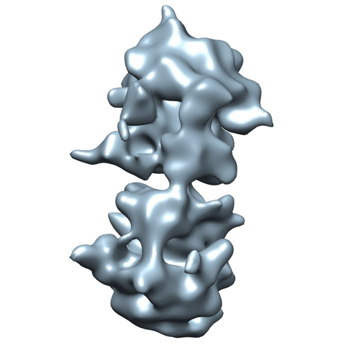
| タイトル | Structure of E. coli Hibernating Ribosomes in 'f-f' Organization | |||||||||
 マップデータ マップデータ | This is the density map of an E.coli 100S ribosome with 'f-f' organization obtained in vitro | |||||||||
 試料 試料 |
| |||||||||
 キーワード キーワード | Ribosome / dimers / hybernating / 100S / RMF / HPF | |||||||||
| 生物種 |  | |||||||||
| 手法 | サブトモグラム平均法 / クライオ電子顕微鏡法 / ネガティブ染色法 | |||||||||
 データ登録者 データ登録者 | Ortiz JO / Brandt F / Valerio M / Sennels L / Rappsilber J / Scheres SHW / Eibauer M / Hartl FU / Baumeister W | |||||||||
 引用 引用 | ジャーナル: J Cell Biol / 年: 2010 タイトル: Structure of hibernating ribosomes studied by cryoelectron tomography in vitro and in situ. 著者: Julio O Ortiz / Florian Brandt / Valério R F Matias / Lau Sennels / Juri Rappsilber / Sjors H W Scheres / Matthias Eibauer / F Ulrich Hartl / Wolfgang Baumeister /  要旨: Ribosomes arranged in pairs (100S) have been related with nutritional stress response and are believed to represent a "hibernation state." Several proteins have been identified that are associated ...Ribosomes arranged in pairs (100S) have been related with nutritional stress response and are believed to represent a "hibernation state." Several proteins have been identified that are associated with 100S ribosomes but their spatial organization has hitherto not been characterized. We have used cryoelectron tomography to reveal the three-dimensional configuration of 100S ribosomes isolated from starved Escherichia coli cells and we have described their mode of interaction. In situ studies with intact E. coli cells allowed us to demonstrate that 100S ribosomes do exist in vivo and represent an easily reversible state of quiescence; they readily vanish when the growth medium is replenished. | |||||||||
| 履歴 |
|
- 構造の表示
構造の表示
| ムービー |
 ムービービューア ムービービューア |
|---|---|
| 構造ビューア | EMマップ: SurfViewMolmilJmol/JSmol |
| 添付画像 |
- ダウンロードとリンク
-EMDBアーカイブ
| マップデータ | emd_1750.map.gz | 1.2 MB | EMDBマップデータ形式 | |
|---|---|---|---|---|
| ヘッダ (付随情報) | emd-1750-v30.xmlemd-1750.xml | 11 KB 11 KB | 表示 表示 | EMDBヘッダ |
| 画像 |  emd1750.jpg emd1750.jpg | 93.7 KB | ||
| アーカイブディレクトリ |  http://ftp.pdbj.org/pub/emdb/structures/EMD-1750ftp://ftp.pdbj.org/pub/emdb/structures/EMD-1750 http://ftp.pdbj.org/pub/emdb/structures/EMD-1750ftp://ftp.pdbj.org/pub/emdb/structures/EMD-1750 | HTTPS FTP |
-検証レポート
| 文書・要旨 | emd_1750_validation.pdf.gz | 194.1 KB | 表示 | EMDB検証レポート |
|---|---|---|---|---|
| 文書・詳細版 | emd_1750_full_validation.pdf.gz | 193.2 KB | 表示 | |
| XML形式データ | emd_1750_validation.xml.gz | 4.4 KB | 表示 | |
| アーカイブディレクトリ | https://ftp.pdbj.org/pub/emdb/validation_reports/EMD-1750ftp://ftp.pdbj.org/pub/emdb/validation_reports/EMD-1750 | HTTPS FTP |
-リンク
| EMDBのページ | EMDB (EBI/PDBe) / EMDataResource |
|---|---|
| 「今月の分子」の関連する項目 |

-マップ
| ファイル | ダウンロード / ファイル: emd_1750.map.gz / 形式: CCP4 / 大きさ: 12.6 MB / タイプ: IMAGE STORED AS FLOATING POINT NUMBER (4 BYTES) | ||||||||||||||||||||||||||||||||||||||||||||||||||||||||||||||||||||
|---|---|---|---|---|---|---|---|---|---|---|---|---|---|---|---|---|---|---|---|---|---|---|---|---|---|---|---|---|---|---|---|---|---|---|---|---|---|---|---|---|---|---|---|---|---|---|---|---|---|---|---|---|---|---|---|---|---|---|---|---|---|---|---|---|---|---|---|---|---|
| 注釈 | This is the density map of an E.coli 100S ribosome with 'f-f' organization obtained in vitro | ||||||||||||||||||||||||||||||||||||||||||||||||||||||||||||||||||||
| 投影像・断面図 | 画像のコントロール
画像は Spider により作成 | ||||||||||||||||||||||||||||||||||||||||||||||||||||||||||||||||||||
| ボクセルのサイズ | X=Y=Z: 5.6 Å | ||||||||||||||||||||||||||||||||||||||||||||||||||||||||||||||||||||
| 密度 |
| ||||||||||||||||||||||||||||||||||||||||||||||||||||||||||||||||||||
| 対称性 | 空間群: 1 | ||||||||||||||||||||||||||||||||||||||||||||||||||||||||||||||||||||
| 詳細 | EMDB XML:
CCP4マップ ヘッダ情報:
| ||||||||||||||||||||||||||||||||||||||||||||||||||||||||||||||||||||
 Z (Sec.)
 Y (Row.)
 X (Col.)


-添付データ
- 試料の構成要素
-全体 : 100S ribosomes in a 'f-f' 3D organization solved in vitro
| 全体 | 名称: 100S ribosomes in a 'f-f' 3D organization solved in vitro |
|---|---|
| 要素 |
|
-超分子 #1000: 100S ribosomes in a 'f-f' 3D organization solved in vitro
| 超分子 | 名称: 100S ribosomes in a 'f-f' 3D organization solved in vitro タイプ: sample / ID: 1000 詳細: Samples are crude fraction of sucrose gradient composed by a mix of ribosomes in different states of aggregation. 集合状態: Dimer / Number unique components: 2 |
|---|---|
| 分子量 | 理論値: 5.4 MDa |
-超分子 #1: 70S ribosome
| 超分子 | 名称: 70S ribosome / タイプ: complex / ID: 1 / 組換発現: No / データベース: NCBI / Ribosome-details: ribosome-prokaryote: ALL |
|---|---|
| 由来(天然) | 生物種: |
| 分子量 | 理論値: 2.7 MDa |
-実験情報
-構造解析
| 手法 | ネガティブ染色法, クライオ電子顕微鏡法 |
|---|---|
 解析 解析 | サブトモグラム平均法 |
| 試料の集合状態 | particle |
-試料調製
| 緩衝液 | pH: 7.6 詳細: 20 mM Tris-HCl, 15.2 mM (CH3COO)2Mg, 0.8 mM EDTA, 100 mM CH3COONH4, 3 mM DTT |
|---|---|
| 染色 | タイプ: NEGATIVE / 詳細: No staining |
| グリッド | 詳細: R2/2 Quantifoil cupper grids |
| 凍結 | 凍結剤: ETHANE / 装置: HOMEMADE PLUNGER 詳細: Vitrification instrument: Plunger. Vitrification carried out in air 手法: Blot for 2 seconds before plunging |
- 電子顕微鏡法
| 顕微鏡 | FEI/PHILIPS CM200FEG |
|---|---|
| アライメント法 | Legacy - 非点収差: Objective astigmatism was corrected using a quadrupole stigmator at 50,000 times magnification Legacy - Electron beam tilt params: -4 |
| 日付 | 2007年6月1日 |
| 撮影 | 実像数: 205 / 平均電子線量: 50 e/Å2 |
| 電子線 | 加速電圧: 200 kV / 電子線源:  FIELD EMISSION GUN FIELD EMISSION GUN |
| 電子光学系 | 倍率(補正後): 53960 / 照射モード: FLOOD BEAM / 撮影モード: BRIGHT FIELD / Cs: 2 mm / 最大 デフォーカス(公称値): 4.0 µm / 最小 デフォーカス(公称値): 3.0 µm / 倍率(公称値): 27500 |
| 試料ステージ | 試料ホルダー: Side entry liquid nitrogen / 試料ホルダーモデル: GATAN LIQUID NITROGEN / Tilt series - Axis1 - Min angle: -60 ° / Tilt series - Axis1 - Max angle: 60 ° |
-画像解析
| 詳細 | Average number of projections used in the 3D reconstructions: 35. |
|---|---|
| 最終 再構成 | アルゴリズム: OTHER / ソフトウェア - 名称: TOM ToolBox |
| CTF補正 | 詳細: Each projection |
-原子モデル構築 1
| 初期モデル | PDB ID:  2aw7 |
|---|---|
| ソフトウェア | 名称: Chimera |
| 詳細 | Protocol: Rigid Body |
| 精密化 | 空間: REAL / プロトコル: RIGID BODY FIT |
-原子モデル構築 2
| 初期モデル | PDB ID: 2awb |
|---|---|
| ソフトウェア | 名称: Chimera |
| 詳細 | Protocol: Rigid Body |
| 精密化 | 空間: REAL / プロトコル: RIGID BODY FIT |
