 Movie
Movie Controller

[English] 日本語
 Yorodumi
- EMDB-1750: Structure of E. coli Hibernating Ribosomes in 'f-f' Organization -
+ Open data

- Basic information
| Entry | Database: EMDB / ID: EMD-1750 | |||||||||
|---|---|---|---|---|---|---|---|---|---|---|
| Title | Structure of E. coli Hibernating Ribosomes in 'f-f' Organization | |||||||||
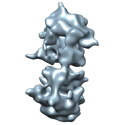
 Map data Map data | This is the density map of an E.coli 100S ribosome with 'f-f' organization obtained in vitro | |||||||||
 Sample Sample |
| |||||||||
 Keywords Keywords | Ribosome / dimers / hybernating / 100S / RMF / HPF | |||||||||
| Biological species |  | |||||||||
| Method | subtomogram averaging / cryo EM / negative staining | |||||||||
 Authors Authors | Ortiz JO / Brandt F / Valerio M / Sennels L / Rappsilber J / Scheres SHW / Eibauer M / Hartl FU / Baumeister W | |||||||||
 Citation Citation | Journal: J Cell Biol / Year: 2010 Title: Structure of hibernating ribosomes studied by cryoelectron tomography in vitro and in situ. Authors: Julio O Ortiz / Florian Brandt / Valério R F Matias / Lau Sennels / Juri Rappsilber / Sjors H W Scheres / Matthias Eibauer / F Ulrich Hartl / Wolfgang Baumeister /  Abstract: Ribosomes arranged in pairs (100S) have been related with nutritional stress response and are believed to represent a "hibernation state." Several proteins have been identified that are associated ...Ribosomes arranged in pairs (100S) have been related with nutritional stress response and are believed to represent a "hibernation state." Several proteins have been identified that are associated with 100S ribosomes but their spatial organization has hitherto not been characterized. We have used cryoelectron tomography to reveal the three-dimensional configuration of 100S ribosomes isolated from starved Escherichia coli cells and we have described their mode of interaction. In situ studies with intact E. coli cells allowed us to demonstrate that 100S ribosomes do exist in vivo and represent an easily reversible state of quiescence; they readily vanish when the growth medium is replenished. | |||||||||
| History |
|
- Structure visualization
| Movie |
 Movie viewer Movie viewer |
|---|---|
| Structure viewer | EM map: SurfViewMolmilJmol/JSmol |
| Supplemental images |
- Downloads & links
-EMDB archive
| Map data | emd_1750.map.gz | 1.2 MB | EMDB map data format | |
|---|---|---|---|---|
| Header (meta data) | emd-1750-v30.xmlemd-1750.xml | 11 KB 11 KB | Display Display | EMDB header |
| Images |  emd1750.jpg emd1750.jpg | 93.7 KB | ||
| Archive directory |  http://ftp.pdbj.org/pub/emdb/structures/EMD-1750ftp://ftp.pdbj.org/pub/emdb/structures/EMD-1750 http://ftp.pdbj.org/pub/emdb/structures/EMD-1750ftp://ftp.pdbj.org/pub/emdb/structures/EMD-1750 | HTTPS FTP |
-Links
| EMDB pages | EMDB (EBI/PDBe) / EMDataResource |
|---|---|
| Related items in Molecule of the Month |

-Map
| File | Download / File: emd_1750.map.gz / Format: CCP4 / Size: 12.6 MB / Type: IMAGE STORED AS FLOATING POINT NUMBER (4 BYTES) | ||||||||||||||||||||||||||||||||||||||||||||||||||||||||||||||||||||
|---|---|---|---|---|---|---|---|---|---|---|---|---|---|---|---|---|---|---|---|---|---|---|---|---|---|---|---|---|---|---|---|---|---|---|---|---|---|---|---|---|---|---|---|---|---|---|---|---|---|---|---|---|---|---|---|---|---|---|---|---|---|---|---|---|---|---|---|---|---|
| Annotation | This is the density map of an E.coli 100S ribosome with 'f-f' organization obtained in vitro | ||||||||||||||||||||||||||||||||||||||||||||||||||||||||||||||||||||
| Projections & slices | Image control
Images are generated by Spider. | ||||||||||||||||||||||||||||||||||||||||||||||||||||||||||||||||||||
| Voxel size | X=Y=Z: 5.6 Å | ||||||||||||||||||||||||||||||||||||||||||||||||||||||||||||||||||||
| Density |
| ||||||||||||||||||||||||||||||||||||||||||||||||||||||||||||||||||||
| Symmetry | Space group: 1 | ||||||||||||||||||||||||||||||||||||||||||||||||||||||||||||||||||||
| Details | EMDB XML:
CCP4 map header:
| ||||||||||||||||||||||||||||||||||||||||||||||||||||||||||||||||||||
 Z (Sec.)
 Y (Row.)
 X (Col.)


-Supplemental data
- Sample components
-Entire : 100S ribosomes in a 'f-f' 3D organization solved in vitro
| Entire | Name: 100S ribosomes in a 'f-f' 3D organization solved in vitro |
|---|---|
| Components |
|
-Supramolecule #1000: 100S ribosomes in a 'f-f' 3D organization solved in vitro
| Supramolecule | Name: 100S ribosomes in a 'f-f' 3D organization solved in vitro type: sample / ID: 1000 Details: Samples are crude fraction of sucrose gradient composed by a mix of ribosomes in different states of aggregation. Oligomeric state: Dimer / Number unique components: 2 |
|---|---|
| Molecular weight | Theoretical: 5.4 MDa |
-Supramolecule #1: 70S ribosome
| Supramolecule | Name: 70S ribosome / type: complex / ID: 1 / Recombinant expression: No / Database: NCBI / Ribosome-details: ribosome-prokaryote: ALL |
|---|---|
| Source (natural) | Organism: |
| Molecular weight | Theoretical: 2.7 MDa |
-Experimental details
-Structure determination
| Method | negative staining, cryo EM |
|---|---|
 Processing Processing | subtomogram averaging |
| Aggregation state | particle |
-Sample preparation
| Buffer | pH: 7.6 Details: 20 mM Tris-HCl, 15.2 mM (CH3COO)2Mg, 0.8 mM EDTA, 100 mM CH3COONH4, 3 mM DTT |
|---|---|
| Staining | Type: NEGATIVE / Details: No staining |
| Grid | Details: R2/2 Quantifoil cupper grids |
| Vitrification | Cryogen name: ETHANE / Instrument: HOMEMADE PLUNGER Details: Vitrification instrument: Plunger. Vitrification carried out in air Method: Blot for 2 seconds before plunging |
- Electron microscopy
| Microscope | FEI/PHILIPS CM200FEG |
|---|---|
| Alignment procedure | Legacy - Astigmatism: Objective astigmatism was corrected using a quadrupole stigmator at 50,000 times magnification Legacy - Electron beam tilt params: -4 |
| Date | Jun 1, 2007 |
| Image recording | Number real images: 205 / Average electron dose: 50 e/Å2 |
| Electron beam | Acceleration voltage: 200 kV / Electron source:  FIELD EMISSION GUN FIELD EMISSION GUN |
| Electron optics | Calibrated magnification: 53960 / Illumination mode: FLOOD BEAM / Imaging mode: BRIGHT FIELD / Cs: 2 mm / Nominal defocus max: 4.0 µm / Nominal defocus min: 3.0 µm / Nominal magnification: 27500 |
| Sample stage | Specimen holder: Side entry liquid nitrogen / Specimen holder model: GATAN LIQUID NITROGEN / Tilt series - Axis1 - Min angle: -60 ° / Tilt series - Axis1 - Max angle: 60 ° |
-Image processing
| Details | Average number of projections used in the 3D reconstructions: 35. |
|---|---|
| Final reconstruction | Algorithm: OTHER / Software - Name: TOM ToolBox |
| CTF correction | Details: Each projection |
-Atomic model buiding 1
| Initial model | PDB ID:  2aw7 |
|---|---|
| Software | Name: Chimera |
| Details | Protocol: Rigid Body |
| Refinement | Space: REAL / Protocol: RIGID BODY FIT |
-Atomic model buiding 2
| Initial model | PDB ID: 2awb |
|---|---|
| Software | Name: Chimera |
| Details | Protocol: Rigid Body |
| Refinement | Space: REAL / Protocol: RIGID BODY FIT |
