 Movie
Movie Controller

[English] 日本語
 Yorodumi
- EMDB-1731: The microtubule nucleating gamma-tubulin small complex assembles ... -
+ Open data

- Basic information
| Entry | Database: EMDB / ID: EMD-1731 | |||||||||
|---|---|---|---|---|---|---|---|---|---|---|
| Title | The microtubule nucleating gamma-tubulin small complex assembles into a structure with microtubule-like thirteen fold symmetry | |||||||||
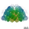
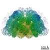
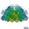
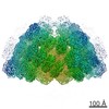
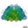
 Map data Map data | This is the map of the gamma-tubulin small complex - Spc110p filament. The filament axis is along the Z axis. | |||||||||
 Sample Sample |
| |||||||||
 Keywords Keywords | Microtubule / nucleation / tubulin / filament | |||||||||
| Function / homology |  Function and homology information Function and homology informationinner plaque of spindle pole body / microtubule nucleation by spindle pole body / outer plaque of spindle pole body / gamma-tubulin small complex / regulation of microtubule nucleation / microtubule nucleator activity / mitotic spindle pole body / gamma-tubulin complex / gamma-tubulin ring complex / meiotic spindle organization ...inner plaque of spindle pole body / microtubule nucleation by spindle pole body / outer plaque of spindle pole body / gamma-tubulin small complex / regulation of microtubule nucleation / microtubule nucleator activity / mitotic spindle pole body / gamma-tubulin complex / gamma-tubulin ring complex / meiotic spindle organization / microtubule nucleation / spindle pole body / positive regulation of cytoplasmic translation / gamma-tubulin binding / mitotic sister chromatid segregation / spindle assembly / cytoplasmic microtubule organization / mitotic spindle organization / meiotic cell cycle / structural constituent of cytoskeleton / spindle / spindle pole / mitotic cell cycle / microtubule / GTP binding / nucleus / cytoplasm Similarity search - Function | |||||||||
| Biological species |  | |||||||||
| Method | helical reconstruction / cryo EM / negative staining / Resolution: 8.0 Å | |||||||||
 Authors Authors | Kollman JM / Polka JK / Zelter A / Davis TN / Agard DA | |||||||||
 Citation Citation | Journal: Nature / Year: 2010 Title: Microtubule nucleating gamma-TuSC assembles structures with 13-fold microtubule-like symmetry. Authors: Justin M Kollman / Jessica K Polka / Alex Zelter / Trisha N Davis / David A Agard /  Abstract: Microtubules are nucleated in vivo by gamma-tubulin complexes. The 300-kDa gamma-tubulin small complex (gamma-TuSC), consisting of two molecules of gamma-tubulin and one copy each of the accessory ...Microtubules are nucleated in vivo by gamma-tubulin complexes. The 300-kDa gamma-tubulin small complex (gamma-TuSC), consisting of two molecules of gamma-tubulin and one copy each of the accessory proteins Spc97 and Spc98, is the conserved, essential core of the microtubule nucleating machinery. In metazoa multiple gamma-TuSCs assemble with other proteins into gamma-tubulin ring complexes (gamma-TuRCs). The structure of gamma-TuRC indicated that it functions as a microtubule template. Because each gamma-TuSC contains two molecules of gamma-tubulin, it was assumed that the gamma-TuRC-specific proteins are required to organize gamma-TuSCs to match 13-fold microtubule symmetry. Here we show that Saccharomyces cerevisiae gamma-TuSC forms rings even in the absence of other gamma-TuRC components. The yeast adaptor protein Spc110 stabilizes the rings into extended filaments and is required for oligomer formation under physiological buffer conditions. The 8-A cryo-electron microscopic reconstruction of the filament reveals 13 gamma-tubulins per turn, matching microtubule symmetry, with plus ends exposed for interaction with microtubules, implying that one turn of the filament constitutes a microtubule template. The domain structures of Spc97 and Spc98 suggest functions for conserved sequence motifs, with implications for the gamma-TuRC-specific proteins. The gamma-TuSC filaments nucleate microtubules at a low level, and the structure provides a strong hypothesis for how nucleation is regulated, converting this less active form to a potent nucleator. | |||||||||
| History |
|
- Structure visualization
| Movie |
Movie viewer |
|---|---|
| Structure viewer | EM map: SurfViewMolmilJmol/JSmol |
| Supplemental images |

- Downloads & links
-EMDB archive
| Map data | emd_1731.map.gz | 110.5 MB | EMDB map data format | |
|---|---|---|---|---|
| Header (meta data) | emd-1731-v30.xmlemd-1731.xml | 13.7 KB 13.7 KB | Display Display | EMDB header |
| Images | 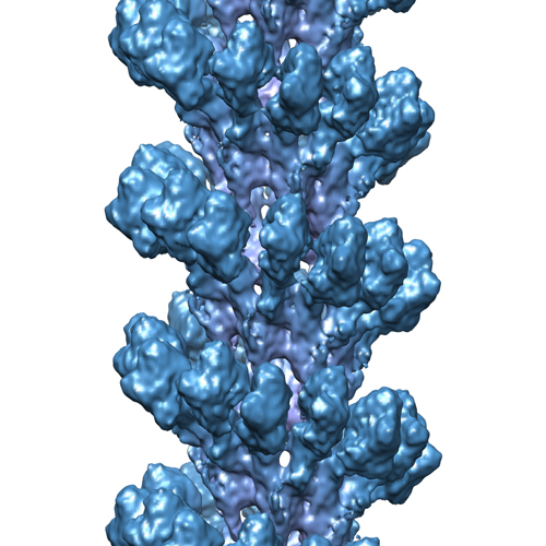 emd-1731.jpg emd-1731.jpg | 193.8 KB | ||
| Archive directory |  http://ftp.pdbj.org/pub/emdb/structures/EMD-1731ftp://ftp.pdbj.org/pub/emdb/structures/EMD-1731 http://ftp.pdbj.org/pub/emdb/structures/EMD-1731ftp://ftp.pdbj.org/pub/emdb/structures/EMD-1731 | HTTPS FTP |
-Related structure data
| Related structure data | 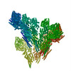 5fm1M M: atomic model generated by this map |
|---|---|
| Similar structure data |
-Links
| EMDB pages | EMDB (EBI/PDBe) / EMDataResource |
|---|---|
| Related items in Molecule of the Month |


-Map
| File | Download / File: emd_1731.map.gz / Format: CCP4 / Size: 122.1 MB / Type: IMAGE STORED AS FLOATING POINT NUMBER (4 BYTES) | ||||||||||||||||||||||||||||||||||||||||||||||||||||||||||||
|---|---|---|---|---|---|---|---|---|---|---|---|---|---|---|---|---|---|---|---|---|---|---|---|---|---|---|---|---|---|---|---|---|---|---|---|---|---|---|---|---|---|---|---|---|---|---|---|---|---|---|---|---|---|---|---|---|---|---|---|---|---|
| Annotation | This is the map of the gamma-tubulin small complex - Spc110p filament. The filament axis is along the Z axis. | ||||||||||||||||||||||||||||||||||||||||||||||||||||||||||||
| Projections & slices | Image control
Images are generated by Spider. | ||||||||||||||||||||||||||||||||||||||||||||||||||||||||||||
| Voxel size | X=Y=Z: 1.19 Å | ||||||||||||||||||||||||||||||||||||||||||||||||||||||||||||
| Density |
| ||||||||||||||||||||||||||||||||||||||||||||||||||||||||||||
| Symmetry | Space group: 1 | ||||||||||||||||||||||||||||||||||||||||||||||||||||||||||||
| Details | EMDB XML:
CCP4 map header:
| ||||||||||||||||||||||||||||||||||||||||||||||||||||||||||||
 Z (Sec.)
 Y (Row.)
 X (Col.)


-Supplemental data
- Sample components
-Entire : Gamma-tubulin small complex in complex with Spc110p(1-220)
| Entire | Name: Gamma-tubulin small complex in complex with Spc110p(1-220) |
|---|---|
| Components |
|
-Supramolecule #1000: Gamma-tubulin small complex in complex with Spc110p(1-220)
| Supramolecule | Name: Gamma-tubulin small complex in complex with Spc110p(1-220) type: sample / ID: 1000 Details: The stoichiometry of gamma-TuSC to Spc110p is not known. Number unique components: 4 |
|---|
-Macromolecule #1: Tub4p
| Macromolecule | Name: Tub4p / type: protein_or_peptide / ID: 1 / Name.synonym: gamma-tubulin / Number of copies: 2 / Oligomeric state: Helical filament / Recombinant expression: Yes |
|---|---|
| Source (natural) | Organism: |
| Molecular weight | Theoretical: 52.6 KDa |
| Recombinant expression | Organism: Spodoptera frugiperda (baculovirus infected) / Recombinant plasmid: pAZ37 |
| Sequence | InterPro: Gamma tubulin |
-Macromolecule #2: Spc97p
| Macromolecule | Name: Spc97p / type: protein_or_peptide / ID: 2 / Name.synonym: Spindle pole body component p97 / Number of copies: 1 / Oligomeric state: Helical filament / Recombinant expression: Yes |
|---|---|
| Source (natural) | Organism: |
| Molecular weight | Theoretical: 96.8 KDa |
| Recombinant expression | Organism: Spodoptera frugiperda (baculovirus infected) / Recombinant plasmid: pDV45 |
| Sequence | InterPro: INTERPRO: IPR015698 |
-Macromolecule #3: Spc98p
| Macromolecule | Name: Spc98p / type: protein_or_peptide / ID: 3 / Name.synonym: Spindle pole body component 98p / Number of copies: 1 / Oligomeric state: Helical filament / Recombinant expression: Yes |
|---|---|
| Source (natural) | Organism: |
| Molecular weight | Theoretical: 98.3 MDa |
| Recombinant expression | Organism: Spodoptera frugiperda (baculovirus infected) / Recombinant plasmid: pDV46 |
| Sequence | InterPro: INTERPRO: IPR015696 |
-Macromolecule #4: Spc110p
| Macromolecule | Name: Spc110p / type: protein_or_peptide / ID: 4 / Name.synonym: Spc110p Details: Construct includes the first 220 residues of Spc110p Number of copies: 1 / Oligomeric state: Helical filament / Recombinant expression: Yes |
|---|---|
| Source (natural) | Organism: |
| Molecular weight | Theoretical: 23 KDa |
| Recombinant expression | Organism: Spodoptera frugiperda (baculovirus infected) / Recombinant plasmid: pAZ34 |
-Experimental details
-Structure determination
| Method | negative staining, cryo EM |
|---|---|
 Processing Processing | helical reconstruction |
| Aggregation state | filament |
-Sample preparation
| Concentration | 2 mg/mL |
|---|---|
| Buffer | pH: 7.6 Details: 100 mM KCl, 1mM GTP, 1mM MgCl2, 2mM EGTA, 40 mM Hepes |
| Staining | Type: NEGATIVE Details: Sample was applied to C-FLAT holey carbon grids, blotted, and flash frozen in liquid ethane |
| Grid | Details: 200 mesh copper grid |
| Vitrification | Cryogen name: ETHANE / Chamber humidity: 100 % / Instrument: OTHER / Details: Vitrification instrument: Vitrobot / Method: Blotted 3s before plunging |
- Electron microscopy
| Microscope | FEI TECNAI F20 |
|---|---|
| Alignment procedure | Legacy - Astigmatism: Astigmatism corrected at 250,000 times magnification |
| Date | Sep 23, 2009 |
| Image recording | Category: CCD / Film or detector model: TVIPS TEMCAM-F816 (8k x 8k) / Average electron dose: 20 e/Å2 |
| Electron beam | Acceleration voltage: 200 kV / Electron source:  FIELD EMISSION GUN FIELD EMISSION GUN |
| Electron optics | Illumination mode: FLOOD BEAM / Imaging mode: BRIGHT FIELD / Cs: 2.2 mm / Nominal defocus max: 3.0 µm / Nominal defocus min: 0.8 µm / Nominal magnification: 60000 |
| Sample stage | Specimen holder: Oxford side-entry cryo stage / Specimen holder model: OTHER |
| Experimental equipment |  Model: Tecnai F20 / Image courtesy: FEI Company |
-Image processing
| Final reconstruction | Applied symmetry - Helical parameters - Δz: 22.2 Å Applied symmetry - Helical parameters - Δ&Phi: 54.3 ° Algorithm: OTHER / Resolution.type: BY AUTHOR / Resolution: 8.0 Å / Resolution method: FSC 0.5 CUT-OFF / Software - Name: Spider |
|---|---|
| CTF correction | Details: Whole micrograph |
| Final angle assignment | Details: SPIDER:theta 90 degrees, phi 90 degrees |
