[English] 日本語
 Yorodumi
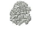
- EMDB-17216: CRYO-EM STRUCTURE OF LEISHMANIA MAJOR 80S RIBOSOME : PARENTAL STRAIN -
+ Open data
- Basic information
| Entry |  | |||||||||
|---|---|---|---|---|---|---|---|---|---|---|
| Title | CRYO-EM STRUCTURE OF LEISHMANIA MAJOR 80S RIBOSOME : PARENTAL STRAIN | |||||||||
 Map data Map data | ||||||||||
 Sample Sample |
| |||||||||
 Keywords Keywords | CRYO-EM LEISHMANIA MAJOR 80S RIBOSOME WILD TYPE / RIBOSOME | |||||||||
| Function / homology |  Function and homology information Function and homology informationciliary transition zone / nuclear lumen / ciliary plasm / negative regulation of translational frameshifting / endonucleolytic cleavage to generate mature 3'-end of SSU-rRNA from (SSU-rRNA, 5.8S rRNA, LSU-rRNA) / protein-RNA complex assembly / maturation of LSU-rRNA / endonucleolytic cleavage in ITS1 to separate SSU-rRNA from 5.8S rRNA and LSU-rRNA from tricistronic rRNA transcript (SSU-rRNA, 5.8S rRNA, LSU-rRNA) / translation regulator activity / rescue of stalled cytosolic ribosome ...ciliary transition zone / nuclear lumen / ciliary plasm / negative regulation of translational frameshifting / endonucleolytic cleavage to generate mature 3'-end of SSU-rRNA from (SSU-rRNA, 5.8S rRNA, LSU-rRNA) / protein-RNA complex assembly / maturation of LSU-rRNA / endonucleolytic cleavage in ITS1 to separate SSU-rRNA from 5.8S rRNA and LSU-rRNA from tricistronic rRNA transcript (SSU-rRNA, 5.8S rRNA, LSU-rRNA) / translation regulator activity / rescue of stalled cytosolic ribosome / protein kinase C binding / ribosomal large subunit biogenesis / maturation of LSU-rRNA from tricistronic rRNA transcript (SSU-rRNA, 5.8S rRNA, LSU-rRNA) / maturation of SSU-rRNA from tricistronic rRNA transcript (SSU-rRNA, 5.8S rRNA, LSU-rRNA) / maturation of SSU-rRNA / small-subunit processome / maintenance of translational fidelity / modification-dependent protein catabolic process / protein tag activity / rRNA processing / kinase activity / ribosome biogenesis / ribosome binding / ribosomal small subunit biogenesis / ribosomal small subunit assembly / 5S rRNA binding / ribosomal large subunit assembly / small ribosomal subunit / small ribosomal subunit rRNA binding / large ribosomal subunit rRNA binding / cytosolic small ribosomal subunit / cytosolic large ribosomal subunit / cytoplasmic translation / negative regulation of translation / rRNA binding / protein ubiquitination / structural constituent of ribosome / ribosome / translation / ribonucleoprotein complex / mRNA binding / ubiquitin protein ligase binding / nucleolus / RNA binding / zinc ion binding / nucleoplasm / nucleus / cytosol / cytoplasm Similarity search - Function | |||||||||
| Biological species |  Leishmania major strain Friedlin (eukaryote) Leishmania major strain Friedlin (eukaryote) | |||||||||
| Method | single particle reconstruction / cryo EM / Resolution: 2.4 Å | |||||||||
 Authors Authors | Rajan KS / Yonath A | |||||||||
| Funding support | European Union, 1 items
| |||||||||
 Citation Citation |  Journal: Cell Rep / Year: 2024 Journal: Cell Rep / Year: 2024Title: Structural and mechanistic insights into the function of Leishmania ribosome lacking a single pseudouridine modification. Authors: K Shanmugha Rajan / Saurav Aryal / Disha-Gajanan Hiregange / Anat Bashan / Hava Madmoni / Mika Olami / Tirza Doniger / Smadar Cohen-Chalamish / Pascal Pescher / Masato Taoka / Yuko Nobe / ...Authors: K Shanmugha Rajan / Saurav Aryal / Disha-Gajanan Hiregange / Anat Bashan / Hava Madmoni / Mika Olami / Tirza Doniger / Smadar Cohen-Chalamish / Pascal Pescher / Masato Taoka / Yuko Nobe / Aliza Fedorenko / Tanaya Bose / Ella Zimermann / Eric Prina / Noa Aharon-Hefetz / Yitzhak Pilpel / Toshiaki Isobe / Ron Unger / Gerald F Späth / Ada Yonath / Shulamit Michaeli /    Abstract: Leishmania is the causative agent of cutaneous and visceral diseases affecting millions of individuals worldwide. Pseudouridine (Ψ), the most abundant modification on rRNA, changes during the ...Leishmania is the causative agent of cutaneous and visceral diseases affecting millions of individuals worldwide. Pseudouridine (Ψ), the most abundant modification on rRNA, changes during the parasite life cycle. Alterations in the level of a specific Ψ in helix 69 (H69) affected ribosome function. To decipher the molecular mechanism of this phenotype, we determine the structure of ribosomes lacking the single Ψ and its parental strain at ∼2.4-3 Å resolution using cryo-EM. Our findings demonstrate the significance of a single Ψ on H69 to its structure and the importance for its interactions with helix 44 and specific tRNAs. Our study suggests that rRNA modification affects translation of mRNAs carrying codon bias due to selective accommodation of tRNAs by the ribosome. Based on the high-resolution structures, we propose a mechanism explaining how the ribosome selects specific tRNAs. | |||||||||
| History |
|
- Structure visualization
| Supplemental images |
|---|
- Downloads & links
-EMDB archive
| Map data |  emd_17216.map.gz emd_17216.map.gz | 376 MB |  EMDB map data format EMDB map data format | |
|---|---|---|---|---|
| Header (meta data) |  emd-17216-v30.xml emd-17216-v30.xml emd-17216.xml emd-17216.xml | 113.2 KB 113.2 KB | Display Display |  EMDB header EMDB header |
| Images |  emd_17216.png emd_17216.png | 79.6 KB | ||
| Filedesc metadata |  emd-17216.cif.gz emd-17216.cif.gz | 21.4 KB | ||
| Others |  emd_17216_additional_1.map.gz emd_17216_additional_1.map.gz emd_17216_half_map_1.map.gz emd_17216_half_map_1.map.gz emd_17216_half_map_2.map.gz emd_17216_half_map_2.map.gz | 336.9 MB 338.8 MB 338.8 MB | ||
| Archive directory |  http://ftp.pdbj.org/pub/emdb/structures/EMD-17216 http://ftp.pdbj.org/pub/emdb/structures/EMD-17216 ftp://ftp.pdbj.org/pub/emdb/structures/EMD-17216 ftp://ftp.pdbj.org/pub/emdb/structures/EMD-17216 | HTTPS FTP |
-Validation report
| Summary document |  emd_17216_validation.pdf.gz emd_17216_validation.pdf.gz | 965.3 KB | Display |  EMDB validaton report EMDB validaton report |
|---|---|---|---|---|
| Full document |  emd_17216_full_validation.pdf.gz emd_17216_full_validation.pdf.gz | 964.9 KB | Display | |
| Data in XML |  emd_17216_validation.xml.gz emd_17216_validation.xml.gz | 17.9 KB | Display | |
| Data in CIF |  emd_17216_validation.cif.gz emd_17216_validation.cif.gz | 21.5 KB | Display | |
| Arichive directory |  https://ftp.pdbj.org/pub/emdb/validation_reports/EMD-17216 https://ftp.pdbj.org/pub/emdb/validation_reports/EMD-17216 ftp://ftp.pdbj.org/pub/emdb/validation_reports/EMD-17216 ftp://ftp.pdbj.org/pub/emdb/validation_reports/EMD-17216 | HTTPS FTP |
-Related structure data
| Related structure data |  8ovjMC  8a98C  8rxhC  8rxxC C: citing same article ( M: atomic model generated by this map |
|---|---|
| Similar structure data | Similarity search - Function & homology  F&H Search F&H Search |
- Links
Links
| EMDB pages |  EMDB (EBI/PDBe) / EMDB (EBI/PDBe) /  EMDataResource EMDataResource |
|---|---|
| Related items in Molecule of the Month |
- Map
Map
| File |  Download / File: emd_17216.map.gz / Format: CCP4 / Size: 421.9 MB / Type: IMAGE STORED AS FLOATING POINT NUMBER (4 BYTES) Download / File: emd_17216.map.gz / Format: CCP4 / Size: 421.9 MB / Type: IMAGE STORED AS FLOATING POINT NUMBER (4 BYTES) | ||||||||||||||||||||||||||||||||||||
|---|---|---|---|---|---|---|---|---|---|---|---|---|---|---|---|---|---|---|---|---|---|---|---|---|---|---|---|---|---|---|---|---|---|---|---|---|---|
| Projections & slices | Image control
Images are generated by Spider. | ||||||||||||||||||||||||||||||||||||
| Voxel size | X=Y=Z: 0.85 Å | ||||||||||||||||||||||||||||||||||||
| Density |
| ||||||||||||||||||||||||||||||||||||
| Symmetry | Space group: 1 | ||||||||||||||||||||||||||||||||||||
| Details | EMDB XML:
|
-Supplemental data
-Additional map: #1
| File | emd_17216_additional_1.map | ||||||||||||
|---|---|---|---|---|---|---|---|---|---|---|---|---|---|
| Projections & Slices |
| ||||||||||||
| Density Histograms |
-Half map: #1
| File | emd_17216_half_map_1.map | ||||||||||||
|---|---|---|---|---|---|---|---|---|---|---|---|---|---|
| Projections & Slices |
| ||||||||||||
| Density Histograms |
-Half map: #2
| File | emd_17216_half_map_2.map | ||||||||||||
|---|---|---|---|---|---|---|---|---|---|---|---|---|---|
| Projections & Slices |
| ||||||||||||
| Density Histograms |
- Sample components
+Entire : 80S RIBOSOME FROM LEISHMANIA MAJOR
+Supramolecule #1: 80S RIBOSOME FROM LEISHMANIA MAJOR
+Macromolecule #1: LSUa_rRNA_chain_1
+Macromolecule #2: SR1_chain_3
+Macromolecule #3: SR2_chain_4
+Macromolecule #4: SR4_chain_5
+Macromolecule #5: SR6_chain_6
+Macromolecule #6: 5.8S_rRNA_chain_7
+Macromolecule #7: 5S_rRNA_chain_8
+Macromolecule #26: SSU_rRNA_chain_S1
+Macromolecule #27: E-site_tRNA_chain_S4
+Macromolecule #85: LSUb_rRNA_chain_2
+Macromolecule #8: Putative 60S ribosomal protein L2
+Macromolecule #9: Putative ribosomal protein L3
+Macromolecule #10: Putative ribosomal protein L1a
+Macromolecule #11: 60S ribosomal protein L11
+Macromolecule #12: Putative 60S ribosomal protein L9
+Macromolecule #13: Putative 60S ribosomal protein L6
+Macromolecule #14: 60S ribosomal protein L7a
+Macromolecule #15: Putative 60S ribosomal protein L13a
+Macromolecule #16: Putative 60S ribosomal protein L13
+Macromolecule #17: Putative 60S ribosomal protein L23
+Macromolecule #18: Putative 40S ribosomal protein L14
+Macromolecule #19: Putative 60S ribosomal protein L27A/L29
+Macromolecule #20: Ribosomal protein L15
+Macromolecule #21: Putative 60S ribosomal protein L10
+Macromolecule #22: Putative 60S ribosomal protein L5
+Macromolecule #23: 60S ribosomal protein L18
+Macromolecule #24: Putative 60S ribosomal protein L19
+Macromolecule #25: 60S ribosomal protein L18a
+Macromolecule #28: 40S ribosomal protein S3a
+Macromolecule #29: 40S ribosomal protein SA
+Macromolecule #30: Putative 40S ribosomal protein S3
+Macromolecule #31: Putative 40S ribosomal protein S9
+Macromolecule #32: 40S ribosomal protein S4
+Macromolecule #33: 40S ribosomal protein S2
+Macromolecule #34: 40S ribosomal protein S6
+Macromolecule #35: 40S ribosomal protein S5
+Macromolecule #36: 40S ribosomal protein S7
+Macromolecule #37: 40S ribosomal protein S8
+Macromolecule #38: Putative 40S ribosomal protein S16
+Macromolecule #39: Putative ribosomal protein S20
+Macromolecule #40: Putative 40S ribosomal protein S10
+Macromolecule #41: 40S ribosomal protein S14
+Macromolecule #42: Putative 40S ribosomal protein S23
+Macromolecule #43: 40S ribosomal protein S12
+Macromolecule #44: Putative 40S ribosomal protein S18
+Macromolecule #45: Putative ribosomal protein S29
+Macromolecule #46: Putative 40S ribosomal protein S13
+Macromolecule #47: Putative 40S ribosomal protein S11
+Macromolecule #48: Putative 40S ribosomal protein S17
+Macromolecule #49: Putative 40S ribosomal protein S15
+Macromolecule #50: 40S ribosomal protein S19-like protein
+Macromolecule #51: Putative 40S ribosomal protein S21
+Macromolecule #52: 40S ribosomal protein S24
+Macromolecule #53: Putative 60S ribosomal protein L21
+Macromolecule #54: 40S ribosomal protein S25
+Macromolecule #55: Putative 40S ribosomal protein S27-1
+Macromolecule #56: 40S ribosomal protein S26
+Macromolecule #57: Putative 40S ribosomal protein S33
+Macromolecule #58: 40S ribosomal protein S30
+Macromolecule #59: Guanine nucleotide-binding protein subunit beta-like protein
+Macromolecule #60: Putative RNA binding protein
+Macromolecule #61: Putative 40S ribosomal protein S15A
+Macromolecule #62: Putative 60S ribosomal protein L17
+Macromolecule #63: Putative 60S ribosomal protein L22
+Macromolecule #64: Putative 60S ribosomal protein L23a
+Macromolecule #65: Putative 60S ribosomal protein L26
+Macromolecule #66: Putative ribosomal protein L24
+Macromolecule #67: 60S ribosomal protein L27
+Macromolecule #68: Putative 60S ribosomal protein L28
+Macromolecule #69: Putative 60S ribosomal protein L35
+Macromolecule #70: 60S ribosomal protein L29
+Macromolecule #71: Putative 60S ribosomal protein L7
+Macromolecule #72: 60S ribosomal protein L30
+Macromolecule #73: Putative 60S ribosomal subunit protein L31
+Macromolecule #74: 60S ribosomal protein L32
+Macromolecule #75: Putative ribosomal protein l35a
+Macromolecule #76: Putative 60S ribosomal protein L34
+Macromolecule #77: Putative 60S Ribosomal protein L36
+Macromolecule #78: Ribosomal protein L37
+Macromolecule #79: Putative ribosomal protein L38
+Macromolecule #80: Putative 60S ribosomal protein L39
+Macromolecule #81: Ubiquitin-60S ribosomal protein L40
+Macromolecule #82: 60S ribosomal protein L41
+Macromolecule #83: 60S ribosomal protein L37a
+Macromolecule #84: Putative 60S ribosomal protein L44
+Macromolecule #86: MAGNESIUM ION
+Macromolecule #87: POTASSIUM ION
+Macromolecule #88: SODIUM ION
+Macromolecule #89: ZINC ION
+Macromolecule #90: water
-Experimental details
-Structure determination
| Method | cryo EM |
|---|---|
 Processing Processing | single particle reconstruction |
| Aggregation state | particle |
- Sample preparation
| Buffer | pH: 7.6 |
|---|---|
| Vitrification | Cryogen name: ETHANE |
- Electron microscopy
| Microscope | FEI TITAN KRIOS |
|---|---|
| Image recording | Film or detector model: GATAN K3 BIOQUANTUM (6k x 4k) / Average electron dose: 0.83 e/Å2 |
| Electron beam | Acceleration voltage: 300 kV / Electron source:  FIELD EMISSION GUN FIELD EMISSION GUN |
| Electron optics | Illumination mode: FLOOD BEAM / Imaging mode: BRIGHT FIELD / Nominal defocus max: 1.3 µm / Nominal defocus min: 0.7000000000000001 µm |
| Experimental equipment |  Model: Titan Krios / Image courtesy: FEI Company |
 Movie
Movie Controller


 Z (Sec.)
 X (Row.)
 Y (Col.)


