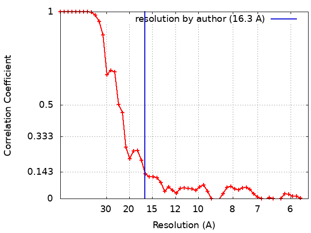
Journal: Sci Rep / Year: 2023 Title: Determining the structure of the bacterial voltage-gated sodium channel NaChBac embedded in liposomes by cryo electron tomography and subtomogram averaging. Authors: Shih-Ying Scott Chang / Patricia M Dijkman / Simon A Wiessing / Misha Kudryashev / Abstract: Voltage-gated sodium channels shape action potentials that propagate signals along cells. When the membrane potential reaches a certain threshold, the channels open and allow sodium ions to flow ...Voltage-gated sodium channels shape action potentials that propagate signals along cells. When the membrane potential reaches a certain threshold, the channels open and allow sodium ions to flow through the membrane depolarizing it, followed by the deactivation of the channels. Opening and closing of the channels is important for cellular signalling and regulates various physiological processes in muscles, heart and brain. Mechanistic insights into the voltage-gated channels are difficult to achieve as the proteins are typically extracted from membranes for structural analysis which results in the loss of the transmembrane potential that regulates their activity. Here, we report the structural analysis of a bacterial voltage-gated sodium channel, NaChBac, reconstituted in liposomes under an electrochemical gradient by cryo electron tomography and subtomogram averaging. We show that the small channel, most of the residues of which are embedded in the membrane, can be localized using a genetically fused GFP. GFP can aid the initial alignment to an average resulting in a correct structure, but does not help for the final refinement. At a moderate resolution of ˜16 Å the structure of NaChBac in an unrestricted membrane bilayer is 10% wider than the structure of the purified protein previously solved in nanodiscs, suggesting the potential movement of the peripheral voltage-sensing domains. Our study explores the limits of structural analysis of membrane proteins in membranes.
In the structure databanks used in Yorodumi, some data are registered as the other names, "COVID-19 virus" and "2019-nCoV". Here are the details of the virus and the list of structure data.
Jan 31, 2019. EMDB accession codes are about to change! (news from PDBe EMDB page)
EMDB accession codes are about to change! (news from PDBe EMDB page)
The allocation of 4 digits for EMDB accession codes will soon come to an end. Whilst these codes will remain in use, new EMDB accession codes will include an additional digit and will expand incrementally as the available range of codes is exhausted. The current 4-digit format prefixed with “EMD-” (i.e. EMD-XXXX) will advance to a 5-digit format (i.e. EMD-XXXXX), and so on. It is currently estimated that the 4-digit codes will be depleted around Spring 2019, at which point the 5-digit format will come into force.
The EM Navigator/Yorodumi systems omit the EMD- prefix.
Related info.:Q: What is EMD? / ID/Accession-code notation in Yorodumi/EM Navigator
Yorodumi is a browser for structure data from EMDB, PDB, SASBDB, etc.
This page is also the successor to EM Navigator detail page, and also detail information page/front-end page for Omokage search.
The word "yorodu" (or yorozu) is an old Japanese word meaning "ten thousand". "mi" (miru) is to see.
Related info.:EMDB / PDB / SASBDB / Comparison of 3 databanks / Yorodumi Search / Aug 31, 2016. New EM Navigator & Yorodumi / Yorodumi Papers / Jmol/JSmol / Function and homology information / Changes in new EM Navigator and Yorodumi
 Movie
Movie Controller

 Yorodumi
 Open data
 Basic information
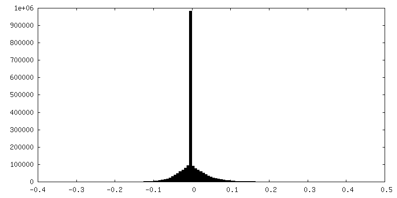
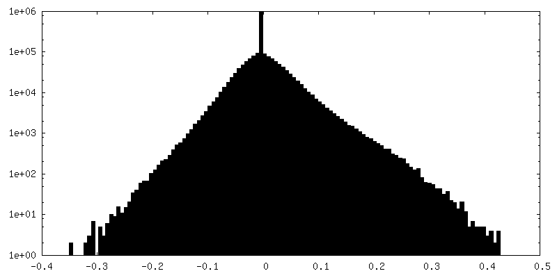
 Map data
 Sample
 Keywords
 Halalkalibacterium halodurans C-125 (bacteria)
 Authors
 Germany, 1 items
  Citation
 Structure visualization
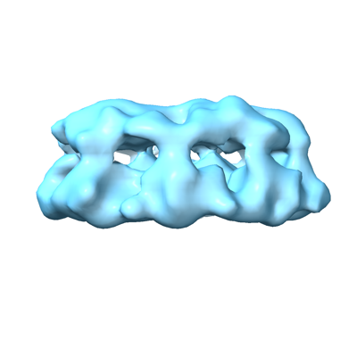
 Downloads & links
 EMDB map data format
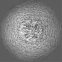
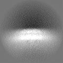
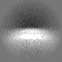
 emd_17163.png
 http://ftp.pdbj.org/pub/emdb/structures/EMD-17163
 Z (Sec.)
 Y (Row.)
 X (Col.)


 Sample components
 Processing
 Electron microscopy
 FIELD EMISSION GUN