 Movie
Movie Controller

[English] 日本語
 Yorodumi
- EMDB-17127: Cryo-EM structure of human tRNA ligase RTCB in complex with human... -
+ Open data

- Basic information
| Entry |  | |||||||||
|---|---|---|---|---|---|---|---|---|---|---|
| Title | Cryo-EM structure of human tRNA ligase RTCB in complex with human PYROXD1. | |||||||||
 Map data Map data | Unsharpened map | |||||||||
 Sample Sample |
| |||||||||
 Keywords Keywords | tRNA ligase / PYROXD1 / RTCB / Flavoprotein / Ligase | |||||||||
| Function / homology |  Function and homology information Function and homology informationtRNA exon ligation / tRNA-splicing ligase complex / 3'-phosphate/5'-hydroxy nucleic acid ligase / RNA ligase (GTP) activity / RNA splicing, via endonucleolytic cleavage and ligation / mRNA splicing, via endonucleolytic cleavage and ligation / NADPH dehydrogenase / molecular sensor activity / tRNA splicing, via endonucleolytic cleavage and ligation / NADPH dehydrogenase activity ...tRNA exon ligation / tRNA-splicing ligase complex / 3'-phosphate/5'-hydroxy nucleic acid ligase / RNA ligase (GTP) activity / RNA splicing, via endonucleolytic cleavage and ligation / mRNA splicing, via endonucleolytic cleavage and ligation / NADPH dehydrogenase / molecular sensor activity / tRNA splicing, via endonucleolytic cleavage and ligation / NADPH dehydrogenase activity / NAD(P)H oxidase H2O2-forming activity / vinculin binding / tRNA processing in the nucleus / IRE1-mediated unfolded protein response / NADH dehydrogenase activity / catalytic complex / placenta development / sarcomere / nuclear envelope / in utero embryonic development / cellular response to oxidative stress / endoplasmic reticulum membrane / GTP binding / RNA binding / nucleoplasm / metal ion binding / nucleus / cytosol / cytoplasm Similarity search - Function | |||||||||
| Biological species |  Homo sapiens (human) Homo sapiens (human) | |||||||||
| Method | single particle reconstruction / cryo EM / Resolution: 3.3 Å | |||||||||
 Authors Authors | Loeff L / Kroupova A / Asanovic I / Boneberg F / Pfleiderer MM / Ferdigg A / Ackle F / Martinez J / Jinek M | |||||||||
| Funding support |  Switzerland, 1 items Switzerland, 1 items
| |||||||||
 Citation Citation | Journal: Nat Struct Mol Biol / Year: 2025 Title: Mechanistic basis for PYROXD1-mediated protection of the human tRNA ligase complex against oxidative inactivation. Authors: Luuk Loeff / Alena Kroupova / Igor Asanović / Franziska M Boneberg / Moritz M Pfleiderer / Luca Riermeier / Alexander Leitner / Andrè Ferdigg / Fabian Ackle / Javier Martinez / Martin Jinek /   Abstract: The metazoan tRNA ligase complex (tRNA-LC) has essential roles in tRNA biogenesis and unfolded protein response. Its catalytic subunit RTCB contains a conserved active-site cysteine that is ...The metazoan tRNA ligase complex (tRNA-LC) has essential roles in tRNA biogenesis and unfolded protein response. Its catalytic subunit RTCB contains a conserved active-site cysteine that is susceptible to metal ion-induced oxidative inactivation. The flavin-containing oxidoreductase PYROXD1 preserves the activity of human tRNA-LC in a NAD(P)H-dependent manner, but its protective mechanism remains elusive. Here, we report a cryogenic electron microscopic structure of the human RTCB-PYROXD1 complex, revealing that PYROXD1 directly interacts with the catalytic center of RTCB through its carboxy-terminal tail. NAD(P)H binding and FAD reduction allosterically control PYROXD1 activity and RTCB recruitment, while reoxidation of PYROXD1 enables timed release of RTCB. PYROXD1 interaction is mutually exclusive with Archease-mediated RTCB guanylylation, and guanylylated RTCB is intrinsically protected from oxidative inactivation. Together, these findings provide a mechanistic framework for the protective function of PYROXD1 that maintains the activity of the tRNA-LC under aerobic conditions. #1: Journal: BioRxiv / Year: 2023Title: Mechanistic basis for oxidative stress protection of the human tRNA ligase complex by the oxidoreductase PYROXD1 Authors: Loeff L / Kroupova A / Asanovic I / Boneberg F / Pfleiderer MM / Ferdigg A / Ackle F / Martinez J / Jinek M | |||||||||
| History |
|
- Structure visualization
| Supplemental images |
|---|

- Downloads & links
-EMDB archive
| Map data | emd_17127.map.gz | 45.1 MB | EMDB map data format | |
|---|---|---|---|---|
| Header (meta data) | emd-17127-v30.xmlemd-17127.xml | 27.7 KB 27.7 KB | Display Display | EMDB header |
| FSC (resolution estimation) | emd_17127_fsc.xml | 9.5 KB | Display | FSC data file |
| Images |  emd_17127.png emd_17127.png | 511.1 KB | ||
| Masks | emd_17127_msk_1.map | 91.1 MB | Mask map | |
| Filedesc metadata | emd-17127.cif.gz | 7.6 KB | ||
| Others | emd_17127_additional_1.map.gzemd_17127_half_map_1.map.gzemd_17127_half_map_2.map.gz | 85.7 MB 84.5 MB 84.5 MB | ||
| Archive directory |  http://ftp.pdbj.org/pub/emdb/structures/EMD-17127ftp://ftp.pdbj.org/pub/emdb/structures/EMD-17127 http://ftp.pdbj.org/pub/emdb/structures/EMD-17127ftp://ftp.pdbj.org/pub/emdb/structures/EMD-17127 | HTTPS FTP |
-Related structure data
| Related structure data | 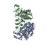 8orjMC M: atomic model generated by this map C: citing same article ( |
|---|---|
| Similar structure data |
-Links
| EMDB pages | EMDB (EBI/PDBe) / EMDataResource |
|---|---|
| Related items in Molecule of the Month |


-Map
| File | Download / File: emd_17127.map.gz / Format: CCP4 / Size: 91.1 MB / Type: IMAGE STORED AS FLOATING POINT NUMBER (4 BYTES) | ||||||||||||||||||||||||||||||||||||
|---|---|---|---|---|---|---|---|---|---|---|---|---|---|---|---|---|---|---|---|---|---|---|---|---|---|---|---|---|---|---|---|---|---|---|---|---|---|
| Annotation | Unsharpened map | ||||||||||||||||||||||||||||||||||||
| Projections & slices | Image control
Images are generated by Spider. | ||||||||||||||||||||||||||||||||||||
| Voxel size | X=Y=Z: 0.65 Å | ||||||||||||||||||||||||||||||||||||
| Density |
| ||||||||||||||||||||||||||||||||||||
| Symmetry | Space group: 1 | ||||||||||||||||||||||||||||||||||||
| Details | EMDB XML:
|
 Z (Sec.)
 Y (Row.)
 X (Col.)


-Supplemental data
-Mask #1
| File | emd_17127_msk_1.map | ||||||||||||
|---|---|---|---|---|---|---|---|---|---|---|---|---|---|
| Projections & Slices |
| ||||||||||||
| Density Histograms |


-Additional map: Unsharpened map
| File | emd_17127_additional_1.map | ||||||||||||
|---|---|---|---|---|---|---|---|---|---|---|---|---|---|
| Annotation | Unsharpened map | ||||||||||||
| Projections & Slices |
| ||||||||||||
| Density Histograms |


-Half map: Unsharpened map
| File | emd_17127_half_map_1.map | ||||||||||||
|---|---|---|---|---|---|---|---|---|---|---|---|---|---|
| Annotation | Unsharpened map | ||||||||||||
| Projections & Slices |
| ||||||||||||
| Density Histograms |


-Half map: Unsharpened map
| File | emd_17127_half_map_2.map | ||||||||||||
|---|---|---|---|---|---|---|---|---|---|---|---|---|---|
| Annotation | Unsharpened map | ||||||||||||
| Projections & Slices |
| ||||||||||||
| Density Histograms |


- Sample components
-Entire : Cryo-EM structure of human RTCB in complex with human PYROXD1.
| Entire | Name: Cryo-EM structure of human RTCB in complex with human PYROXD1. |
|---|---|
| Components |
|
-Supramolecule #1: Cryo-EM structure of human RTCB in complex with human PYROXD1.
| Supramolecule | Name: Cryo-EM structure of human RTCB in complex with human PYROXD1. type: complex / ID: 1 / Parent: 0 / Macromolecule list: #1-#2 / Details: In vitro reconstituted protein complex |
|---|---|
| Source (natural) | Organism: Homo sapiens (human) |
-Macromolecule #1: RNA-splicing ligase RtcB homolog
| Macromolecule | Name: RNA-splicing ligase RtcB homolog / type: protein_or_peptide / ID: 1 / Number of copies: 1 / Enantiomer: LEVO / EC number: 3'-phosphate/5'-hydroxy nucleic acid ligase |
|---|---|
| Source (natural) | Organism: Homo sapiens (human) |
| Molecular weight | Theoretical: 55.556445 KDa |
| Recombinant expression | Organism:   Spodoptera frugiperda (fall armyworm) Spodoptera frugiperda (fall armyworm) |
| Sequence | String: SNAMSRSYND ELQFLEKINK NCWRIKKGFV PNMQVEGVFY VNDALEKLMF EELRNACRGG GVGGFLPAMK QIGNVAALPG IVHRSIGLP DVHSGYGFAI GNMAAFDMND PEAVVSPGGV GFDINCGVRL LRTNLDESDV QPVKEQLAQA MFDHIPVGVG S KGVIPMNA ...String: SNAMSRSYND ELQFLEKINK NCWRIKKGFV PNMQVEGVFY VNDALEKLMF EELRNACRGG GVGGFLPAMK QIGNVAALPG IVHRSIGLP DVHSGYGFAI GNMAAFDMND PEAVVSPGGV GFDINCGVRL LRTNLDESDV QPVKEQLAQA MFDHIPVGVG S KGVIPMNA KDLEEALEMG VDWSLREGYA WAEDKEHCEE YGRMLQADPN KVSARAKKRG LPQLGTLGAG NHYAEIQVVD EI FNEYAAK KMGIDHKGQV CVMIHSGSRG LGHQVATDAL VAMEKAMKRD KIIVNDRQLA CARIASPEGQ DYLKGMAAAG NYA WVNRSS MTFLTRQAFA KVFNTTPDDL DLHVIYDVSH NIAKVEQHVV DGKERTLLVH RKGSTRAFPP HHPLIAVDYQ LTGQ PVLIG GTMGTCSYVL TGTEQGMTET FGTTCHGAGR ALSRAKSRRN LDFQDVLDKL ADMGIAIRVA SPKLVMEEAP ESYKN VTDV VNTCHDAGIS KKAIKLRPIA VIKG UniProtKB: RNA-splicing ligase RTCB |
-Macromolecule #2: Pyridine nucleotide-disulfide oxidoreductase domain-containing pr...
| Macromolecule | Name: Pyridine nucleotide-disulfide oxidoreductase domain-containing protein 1 type: protein_or_peptide / ID: 2 / Number of copies: 1 / Enantiomer: LEVO EC number: Oxidoreductases; Acting on a sulfur group of donors; With NAD+ or NADP+ as acceptor |
|---|---|
| Source (natural) | Organism: Homo sapiens (human) |
| Molecular weight | Theoretical: 56.016859 KDa |
| Recombinant expression | Organism:  |
| Sequence | String: GPMEAARPPP TAGKFVVVGG GIAGVTCAEQ LATHFPSEDI LLVTASPVIK AVTNFKQISK ILEEFDVEEQ SSTMLGKRFP NIKVIESGV KQLKSEEHCI VTEDGNQHVY KKLCLCAGAK PKLICEGNPY VLGIRDTDSA QEFQKQLTKA KRIMIIGNGG I ALELVYEI ...String: GPMEAARPPP TAGKFVVVGG GIAGVTCAEQ LATHFPSEDI LLVTASPVIK AVTNFKQISK ILEEFDVEEQ SSTMLGKRFP NIKVIESGV KQLKSEEHCI VTEDGNQHVY KKLCLCAGAK PKLICEGNPY VLGIRDTDSA QEFQKQLTKA KRIMIIGNGG I ALELVYEI EGCEVIWAIK DKAIGNTFFD AGAAEFLTSK LIAEKSEAKI AHKRTRYTTE GRKKEARSKS KADNVGSALG PD WHEGLNL KGTKEFSHKI HLETMCEVKK IYLQDEFRIL KKKSFTFPRD HKSVTADTEM WPVYVELTNE KIYGCDFIVS ATG VTPNVE PFLHGNSFDL GEDGGLKVDD HMHTSLPDIY AAGDICTTSW QLSPVWQQMR LWTQARQMGW YAAKCMAAAS SGDS IDMDF SFELFAHVTK FFNYKVVLLG KYNAQGLGSD HELMLRCTKG REYIKVVMQN GRMMGAVLIG ETDLEETFEN LILNQ MNLS SYGEDLLDPN IDIEDYFD UniProtKB: tRNA ligase complex-associated NAD(P)H dehydrogenase PYROXD1 |
-Macromolecule #3: MAGNESIUM ION
| Macromolecule | Name: MAGNESIUM ION / type: ligand / ID: 3 / Number of copies: 2 / Formula: MG |
|---|---|
| Molecular weight | Theoretical: 24.305 Da |
-Macromolecule #4: NICOTINAMIDE-ADENINE-DINUCLEOTIDE
| Macromolecule | Name: NICOTINAMIDE-ADENINE-DINUCLEOTIDE / type: ligand / ID: 4 / Number of copies: 1 / Formula: NAD |
|---|---|
| Molecular weight | Theoretical: 663.425 Da |
| Chemical component information |  ChemComp-NAD: |
-Macromolecule #5: DIHYDROFLAVINE-ADENINE DINUCLEOTIDE
| Macromolecule | Name: DIHYDROFLAVINE-ADENINE DINUCLEOTIDE / type: ligand / ID: 5 / Number of copies: 1 / Formula: FDA |
|---|---|
| Molecular weight | Theoretical: 787.566 Da |
| Chemical component information |  ChemComp-FDA: |
-Experimental details
-Structure determination
| Method | cryo EM |
|---|---|
 Processing Processing | single particle reconstruction |
| Aggregation state | particle |
-Sample preparation
| Concentration | 0.6 mg/mL | ||||||||||||||||||
|---|---|---|---|---|---|---|---|---|---|---|---|---|---|---|---|---|---|---|---|
| Buffer | pH: 8 Component:
Details: Dataset 1: 20 mM HEPES pH 8.0, 150 mM NaCl, 0.5 mM TCEP, 5 mM MgCl2 and 0.5 mM NADH, 0.01% Octyl-beta-Glucoside Dataset 2: 20 mM HEPES pH 8.0, 150 mM NaCl, 0.5 mM TCEP, 5 mM MgCl2 and 0.5 mM NADH | ||||||||||||||||||
| Vitrification | Cryogen name: ETHANE / Chamber humidity: 80 % / Chamber temperature: 277.15 K / Instrument: FEI VITROBOT MARK IV | ||||||||||||||||||
| Details | The complex was purified over size exclusion chromatography, prior to grid freezing. Protein concentration per dataset: Dataset 1: 1.5 mg/ ml Dataset 2: 0.6 mg/ ml |
- Electron microscopy
| Microscope | FEI TITAN KRIOS |
|---|---|
| Image recording | #0 - Image recording ID: 1 / #0 - Film or detector model: GATAN K3 (6k x 4k) / #0 - Number grids imaged: 1 / #0 - Number real images: 5114 / #0 - Average electron dose: 64.592 e/Å2 / #1 - Image recording ID: 2 / #1 - Film or detector model: GATAN K3 (6k x 4k) / #1 - Number grids imaged: 1 / #1 - Number real images: 9302 / #1 - Average electron dose: 66.459 e/Å2 |
| Electron beam | Acceleration voltage: 300 kV / Electron source:  FIELD EMISSION GUN FIELD EMISSION GUN |
| Electron optics | C2 aperture diameter: 50.0 µm / Illumination mode: FLOOD BEAM / Imaging mode: BRIGHT FIELD / Cs: 2.7 mm / Nominal defocus max: 2.4 µm / Nominal defocus min: 1.0 µm / Nominal magnification: 130000 |
| Sample stage | Specimen holder model: FEI TITAN KRIOS AUTOGRID HOLDER / Cooling holder cryogen: NITROGEN |
| Experimental equipment |  Model: Titan Krios / Image courtesy: FEI Company |

