 Movie
Movie Controller

[English] 日本語
 Yorodumi
- EMDB-16732: Cryo-EM structure of the Photosystem I - LHCI supercomplex from C... -
+ Open data

- Basic information
| Entry |  | |||||||||
|---|---|---|---|---|---|---|---|---|---|---|
| Title | Cryo-EM structure of the Photosystem I - LHCI supercomplex from Coelastrella sp. | |||||||||
 Map data Map data | ||||||||||
 Sample Sample |
| |||||||||
 Keywords Keywords | Green alga / PSI / Coelastrella / membrane protein / Cryo-EM / PHOTOSYNTHESIS | |||||||||
| Biological species |  Coelastrella (plant) Coelastrella (plant) | |||||||||
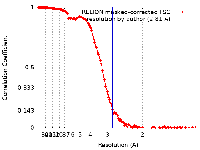
| Method | single particle reconstruction / cryo EM / Resolution: 2.81 Å | |||||||||
 Authors Authors | Fadeeva M / Klaiman D / Nelson N | |||||||||
| Funding support |  Israel, 1 items Israel, 1 items
| |||||||||
 Citation Citation | Journal: To Be Published Title: Cryo-EM structure of the Photosystem I - LHCI supercomplex from Coelastrella sp. Authors: Fadeeva M / Klaiman D / Nelson N | |||||||||
| History |
|
- Structure visualization
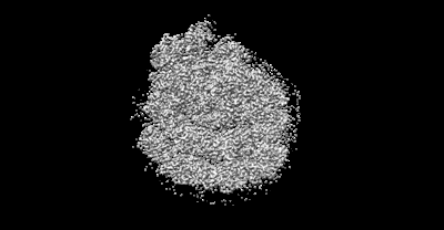
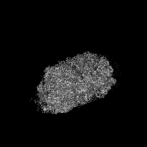
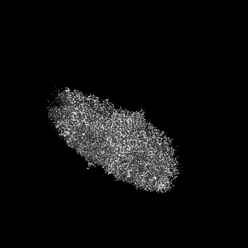
| Supplemental images |
|---|
- Downloads & links
-EMDB archive
| Map data | emd_16732.map.gz | 444.1 MB |  EMDB map data format EMDB map data format | |
|---|---|---|---|---|
| Header (meta data) | emd-16732-v30.xmlemd-16732.xml | 42.8 KB 42.8 KB | Display Display | EMDB header |
| FSC (resolution estimation) | emd_16732_fsc.xml | 17.8 KB | Display | FSC data file |
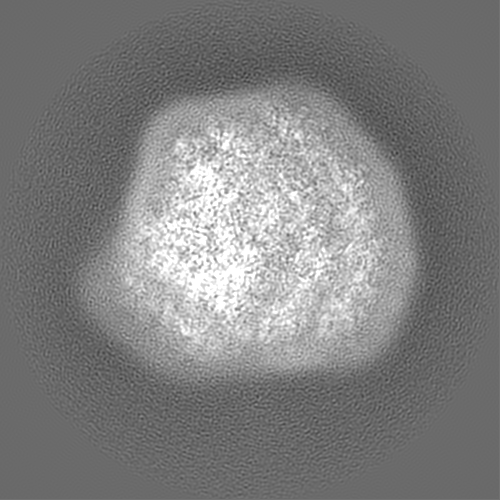
| Images |  emd_16732.png emd_16732.png | 45.9 KB | ||
| Filedesc metadata | emd-16732.cif.gz | 10.3 KB | ||
| Others | emd_16732_half_map_1.map.gzemd_16732_half_map_2.map.gz | 382.2 MB 382.2 MB | ||
| Archive directory |  http://ftp.pdbj.org/pub/emdb/structures/EMD-16732ftp://ftp.pdbj.org/pub/emdb/structures/EMD-16732 http://ftp.pdbj.org/pub/emdb/structures/EMD-16732ftp://ftp.pdbj.org/pub/emdb/structures/EMD-16732 | HTTPS FTP |
-Related structure data
-Links
| EMDB pages | EMDB (EBI/PDBe) / EMDataResource |
|---|
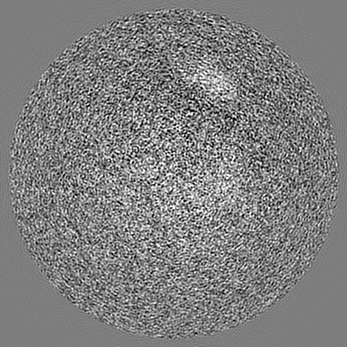
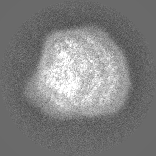
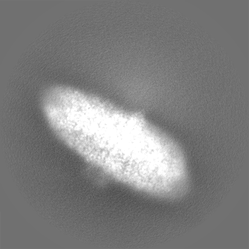
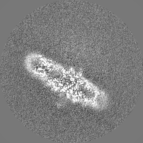
-Map
| File | Download / File: emd_16732.map.gz / Format: CCP4 / Size: 476.8 MB / Type: IMAGE STORED AS FLOATING POINT NUMBER (4 BYTES) | ||||||||||||||||||||||||||||||||||||
|---|---|---|---|---|---|---|---|---|---|---|---|---|---|---|---|---|---|---|---|---|---|---|---|---|---|---|---|---|---|---|---|---|---|---|---|---|---|
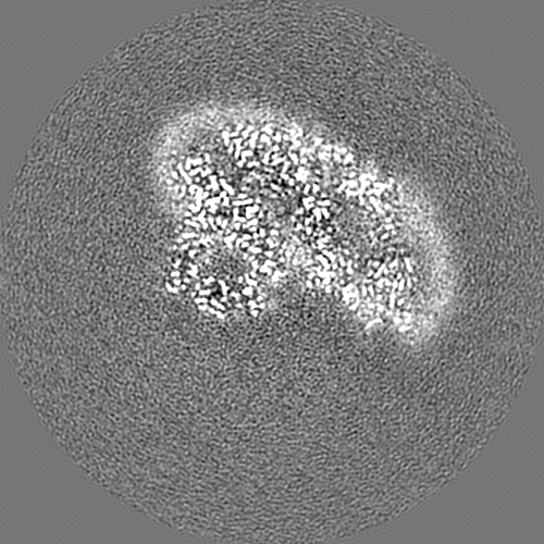
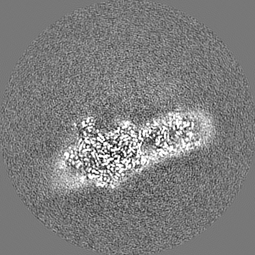
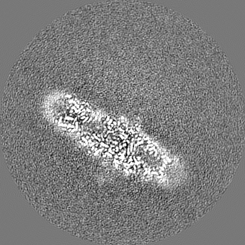
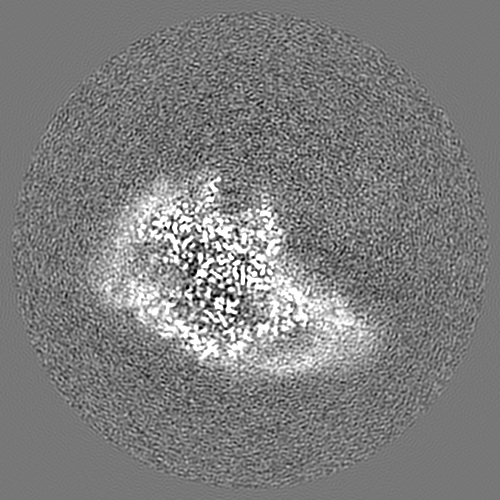
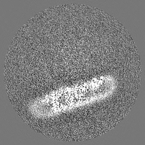
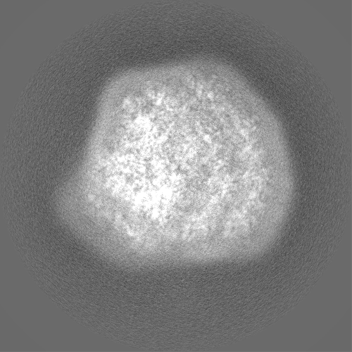
| Projections & slices | Image control
Images are generated by Spider. | ||||||||||||||||||||||||||||||||||||
| Voxel size | X=Y=Z: 0.651 Å | ||||||||||||||||||||||||||||||||||||
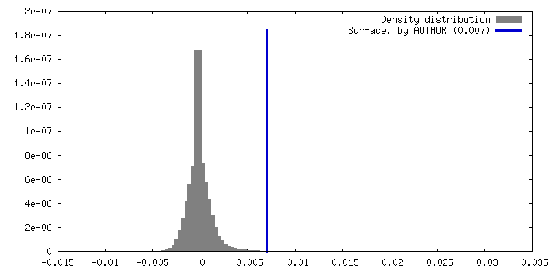
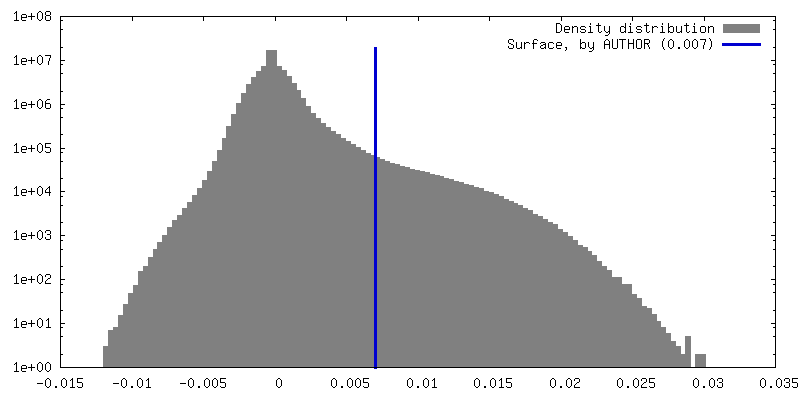
| Density |
| ||||||||||||||||||||||||||||||||||||
| Symmetry | Space group: 1 | ||||||||||||||||||||||||||||||||||||
| Details | EMDB XML:
|
 Z (Sec.)
 Y (Row.)
 X (Col.)


-Supplemental data
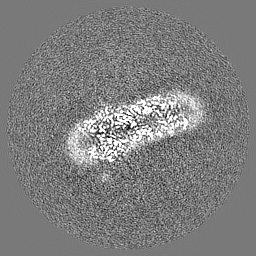
-Half map: #2
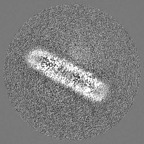
| File | emd_16732_half_map_1.map | ||||||||||||
|---|---|---|---|---|---|---|---|---|---|---|---|---|---|
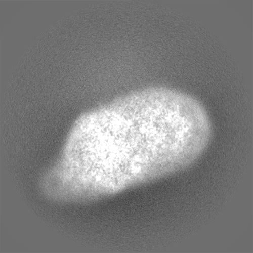
| Projections & Slices |
| ||||||||||||
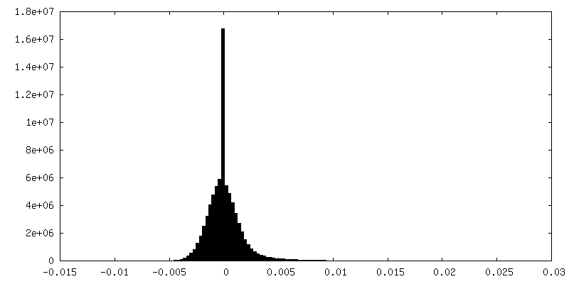
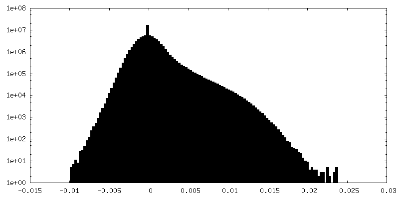
| Density Histograms |


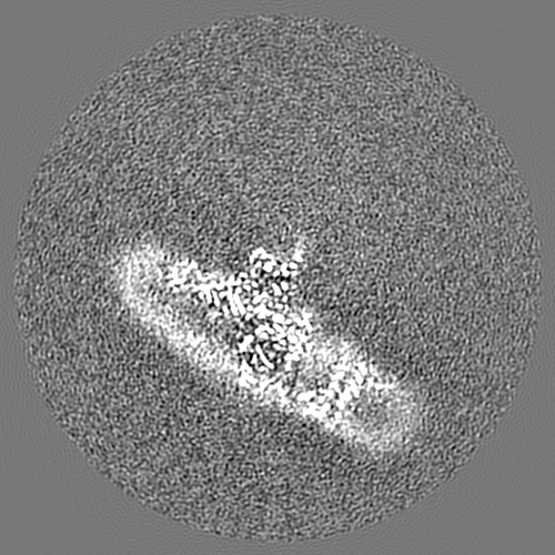
-Half map: #1
| File | emd_16732_half_map_2.map | ||||||||||||
|---|---|---|---|---|---|---|---|---|---|---|---|---|---|
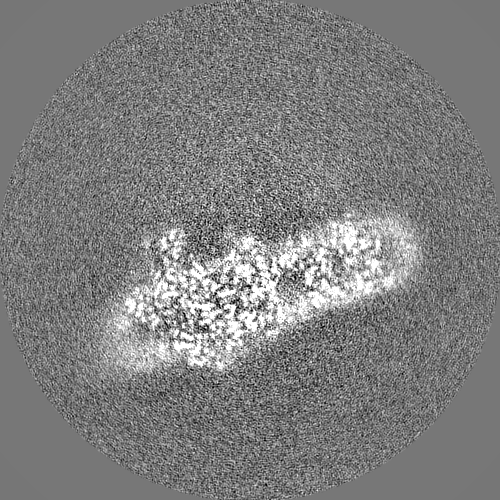
| Projections & Slices |
| ||||||||||||
| Density Histograms |


- Sample components
+Entire : Coelastrella sp. Photosystem I
+Supramolecule #1: Coelastrella sp. Photosystem I
+Macromolecule #1: Photosystem I P700 chlorophyll a apoprotein A1 PsaA
+Macromolecule #2: Photosystem I P700 chlorophyll a apoprotein A2 PsaB
+Macromolecule #3: Photosystem I subunit VII PsaC
+Macromolecule #4: Photosystem I reaction center subunit II PsaD
+Macromolecule #5: Photosystem I reaction center subunit IV PsaE
+Macromolecule #6: Photosystem I reaction center subunit III PsaF
+Macromolecule #7: Photosystem I reaction center subunit V PsaG
+Macromolecule #8: Photosystem I reaction center subunit VIII PsaI
+Macromolecule #9: Photosystem I subunit IX PsaJ
+Macromolecule #10: Photosystem I reaction center subunit X psaK
+Macromolecule #11: Photosystem I reaction centre subunit XI PsaL
+Macromolecule #12: Light-harvesting protein of photosystem I Lhca1
+Macromolecule #13: Light-harvesting protein of photosystem I Lhca3
+Macromolecule #14: Light-harvesting protein of photosystem I Lhca7
+Macromolecule #15: Light-harvesting protein of photosystem I Lhca8
+Macromolecule #16: Light-harvesting protein of photosystem I Lhca4
+Macromolecule #17: Light-harvesting protein of photosystem I Lhca5
+Macromolecule #18: Light-harvesting protein of photosystem I Lhca6
+Macromolecule #19: Light-harvesting protein of photosystem I Lhca9
+Macromolecule #20: Light-harvesting protein of photosystem I Lhca2 partial
+Macromolecule #21: CHLOROPHYLL A ISOMER

+Macromolecule #22: CHLOROPHYLL A

+Macromolecule #23: PHYLLOQUINONE

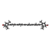
+Macromolecule #24: BETA-CAROTENE

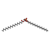
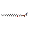
+Macromolecule #25: 1,2-DIPALMITOYL-PHOSPHATIDYL-GLYCEROLE

+Macromolecule #26: (2R)-2-hydroxy-3-(phosphonooxy)propyl (9E)-octadec-9-enoate
+Macromolecule #27: DIACYL GLYCEROL
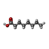
+Macromolecule #28: OCTANOIC ACID (CAPRYLIC ACID)
+Macromolecule #29: IRON/SULFUR CLUSTER

+Macromolecule #30: CALCIUM ION
+Macromolecule #31: DIGALACTOSYL DIACYL GLYCEROL (DGDG)

+Macromolecule #32: (3R)-beta,beta-caroten-3-ol

+Macromolecule #33: DODECYL-BETA-D-MALTOSIDE

+Macromolecule #34: 1,2-DISTEAROYL-MONOGALACTOSYL-DIGLYCERIDE

+Macromolecule #35: Phosphatidylinositol
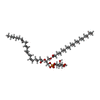
+Macromolecule #36: (1~{S})-3,5,5-trimethyl-4-[(1~{E},3~{E},5~{E},7~{E},9~{E},11~{E},...
+Macromolecule #37: LAURIC ACID

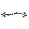
+Macromolecule #38: (3R,3'R,6S)-4,5-DIDEHYDRO-5,6-DIHYDRO-BETA,BETA-CAROTENE-3,3'-DIOL
+Macromolecule #39: CHLOROPHYLL B

+Macromolecule #40: 1,2-DI-O-ACYL-3-O-[6-DEOXY-6-SULFO-ALPHA-D-GLUCOPYRANOSYL]-SN-GLYCEROL

+Macromolecule #41: 1,2-DIACYL-GLYCEROL-3-SN-PHOSPHATE
+Macromolecule #42: SPHINGOSINE

+Macromolecule #43: (2S)-3-{[(R)-(2-aminoethoxy)(hydroxy)phosphoryl]oxy}-2-hydroxypro...
+Macromolecule #44: water

-Experimental details
-Structure determination
| Method | cryo EM |
|---|---|
 Processing Processing | single particle reconstruction |
| Aggregation state | particle |
-Sample preparation
| Concentration | 2 mg/mL |
|---|---|
| Buffer | pH: 8 |
| Grid | Model: Quantifoil R1.2/1.3 / Material: COPPER / Mesh: 300 / Support film - Material: CARBON / Support film - topology: HOLEY / Support film - Film thickness: 12 / Pretreatment - Type: PLASMA CLEANING / Pretreatment - Time: 40 sec. / Pretreatment - Atmosphere: AIR / Pretreatment - Pressure: 1.0 kPa / Details: Harrick Plasma cleaner PDC-32G-2 |
| Vitrification | Cryogen name: ETHANE / Chamber humidity: 90 % / Chamber temperature: 295 K / Instrument: LEICA EM GP / Details: blot for 3 seconds before plunging. |
| Details | Chlorophyll concentration was provided |
- Electron microscopy
| Microscope | FEI TITAN KRIOS |
|---|---|
| Image recording | Film or detector model: GATAN K3 BIOQUANTUM (6k x 4k) / Number real images: 31707 / Average exposure time: 1.38 sec. / Average electron dose: 50.0 e/Å2 |
| Electron beam | Acceleration voltage: 300 kV / Electron source:  FIELD EMISSION GUN FIELD EMISSION GUN |
| Electron optics | C2 aperture diameter: 70.0 µm / Illumination mode: FLOOD BEAM / Imaging mode: BRIGHT FIELD / Cs: 2.7 mm / Nominal defocus max: 1.75 µm / Nominal defocus min: 0.15 µm / Nominal magnification: 130000 |
| Sample stage | Cooling holder cryogen: NITROGEN |
| Experimental equipment |  Model: Titan Krios / Image courtesy: FEI Company |
+Image processing
-Atomic model buiding 1
| Refinement | Space: REAL / Protocol: RIGID BODY FIT / Overall B value: 50 |
|---|---|
| Output model | 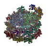 PDB-8cmo: |
