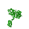
Journal: Nat Commun / Year: 2023 Title: Acetylation regulates the oligomerization state and activity of RNase J, the Helicobacter pylori major ribonuclease. Authors: Alejandro Tejada-Arranz / Aleksei Lulla / Maxime Bouilloux-Lafont / Evelyne Turlin / Xue-Yuan Pei / Thibaut Douché / Mariette Matondo / Allison H Williams / Bertrand Raynal / Ben F Luisi / Hilde De Reuse / Abstract: In the gastric pathogen Helicobacter pylori, post-transcriptional regulation relies strongly on the activity of the essential ribonuclease RNase J. Here, we elucidated the crystal and cryo-EM ...In the gastric pathogen Helicobacter pylori, post-transcriptional regulation relies strongly on the activity of the essential ribonuclease RNase J. Here, we elucidated the crystal and cryo-EM structures of RNase J and determined that it assembles into dimers and tetramers in vitro. We found that RNase J extracted from H. pylori is acetylated on multiple lysine residues. Alanine substitution of several of these residues impacts on H. pylori morphology, and thus on RNase J function in vivo. Mutations of Lysine 649 modulates RNase J oligomerization in vitro, which in turn influences ribonuclease activity in vitro. Our structural analyses of RNase J reveal loops that gate access to the active site and rationalizes how acetylation state of K649 can influence activity. We propose acetylation as a regulatory level controlling the activity of RNase J and its potential cooperation with other enzymes of RNA metabolism in H. pylori.
In the structure databanks used in Yorodumi, some data are registered as the other names, "COVID-19 virus" and "2019-nCoV". Here are the details of the virus and the list of structure data.
Jan 31, 2019. EMDB accession codes are about to change! (news from PDBe EMDB page)
EMDB accession codes are about to change! (news from PDBe EMDB page)
The allocation of 4 digits for EMDB accession codes will soon come to an end. Whilst these codes will remain in use, new EMDB accession codes will include an additional digit and will expand incrementally as the available range of codes is exhausted. The current 4-digit format prefixed with “EMD-” (i.e. EMD-XXXX) will advance to a 5-digit format (i.e. EMD-XXXXX), and so on. It is currently estimated that the 4-digit codes will be depleted around Spring 2019, at which point the 5-digit format will come into force.
The EM Navigator/Yorodumi systems omit the EMD- prefix.
Related info.:Q: What is EMD? / ID/Accession-code notation in Yorodumi/EM Navigator
Yorodumi is a browser for structure data from EMDB, PDB, SASBDB, etc.
This page is also the successor to EM Navigator detail page, and also detail information page/front-end page for Omokage search.
The word "yorodu" (or yorozu) is an old Japanese word meaning "ten thousand". "mi" (miru) is to see.
Related info.:EMDB / PDB / SASBDB / Comparison of 3 databanks / Yorodumi Search / Aug 31, 2016. New EM Navigator & Yorodumi / Yorodumi Papers / Jmol/JSmol / Function and homology information / Changes in new EM Navigator and Yorodumi
 Movie
Movie Controller


 Open data
 Basic information
 Map data
 Sample
 Keywords
 Function and homology information
 Helicobacter pylori (bacteria) /
  Authors
 United Kingdom, 1 items
  Citation


 Structure visualization
 Downloads & links
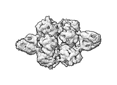 emd_16647.png
 http://ftp.pdbj.org/pub/emdb/structures/EMD-16647
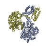


 Z (Sec.)
 Y (Row.)
 X (Col.)


 Sample components
 Processing
 Electron microscopy
 FIELD EMISSION GUN