 Movie
Movie Controller

[English] 日本語
 Yorodumi
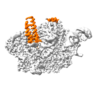
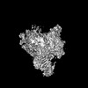
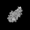
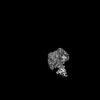
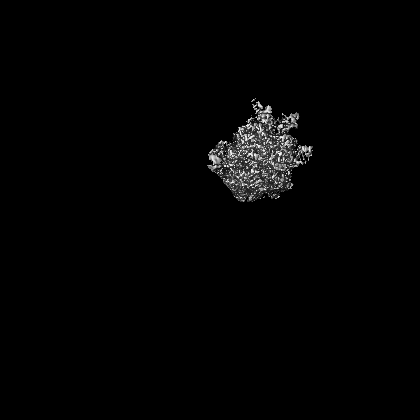
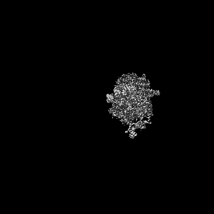
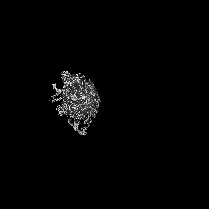
- EMDB-16542: Local refinement map of Otu2 N-terminal domain bound to yeast 40S... -
+ Open data

- Basic information
| Entry |  | ||||||||||||||||||
|---|---|---|---|---|---|---|---|---|---|---|---|---|---|---|---|---|---|---|---|
| Title | Local refinement map of Otu2 N-terminal domain bound to yeast 40S ribosome | ||||||||||||||||||
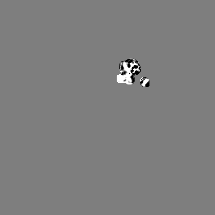
 Map data Map data | Local refinement map of Otu2 N-terminal domain bound to yeast 40S ribosome | ||||||||||||||||||
 Sample Sample |
| ||||||||||||||||||
| Biological species |  | ||||||||||||||||||
| Method | single particle reconstruction / cryo EM / Resolution: 2.9 Å | ||||||||||||||||||
 Authors Authors | Ikeuchi K / Buschauer R / Cheng J / Berninghausen O / Becker T / Beckmann R | ||||||||||||||||||
| Funding support |  Germany, Germany,  Japan, 5 items Japan, 5 items
| ||||||||||||||||||
 Citation Citation | Journal: Nat Commun / Year: 2023 Title: Molecular basis for recognition and deubiquitination of 40S ribosomes by Otu2. Authors: Ken Ikeuchi / Nives Ivic / Robert Buschauer / Jingdong Cheng / Thomas Fröhlich / Yoshitaka Matsuo / Otto Berninghausen / Toshifumi Inada / Thomas Becker / Roland Beckmann /   Abstract: In actively translating 80S ribosomes the ribosomal protein eS7 of the 40S subunit is monoubiquitinated by the E3 ligase Not4 and deubiquitinated by Otu2 upon ribosomal subunit recycling. Despite its ...In actively translating 80S ribosomes the ribosomal protein eS7 of the 40S subunit is monoubiquitinated by the E3 ligase Not4 and deubiquitinated by Otu2 upon ribosomal subunit recycling. Despite its importance for translation efficiency the exact role and structural basis for this translational reset is poorly understood. Here, structural analysis by cryo-electron microscopy of native and reconstituted Otu2-bound ribosomal complexes reveals that Otu2 engages 40S subunits mainly between ribosome recycling and initiation stages. Otu2 binds to several sites on the intersubunit surface of the 40S that are not occupied by any other 40S-binding factors. This binding mode explains the discrimination against 80S ribosomes via the largely helical N-terminal domain of Otu2 as well as the specificity for mono-ubiquitinated eS7 on 40S. Collectively, this study reveals mechanistic insights into the Otu2-driven deubiquitination steps for translational reset during ribosome recycling/(re)initiation. | ||||||||||||||||||
| History |
|
- Structure visualization
| Supplemental images |
|---|
- Downloads & links
-EMDB archive
| Map data | emd_16542.map.gz | 3.3 MB |  EMDB map data format EMDB map data format | |
|---|---|---|---|---|
| Header (meta data) | emd-16542-v30.xmlemd-16542.xml | 15.4 KB 15.4 KB | Display Display | EMDB header |
| FSC (resolution estimation) | emd_16542_fsc.xml | 13.9 KB | Display | FSC data file |
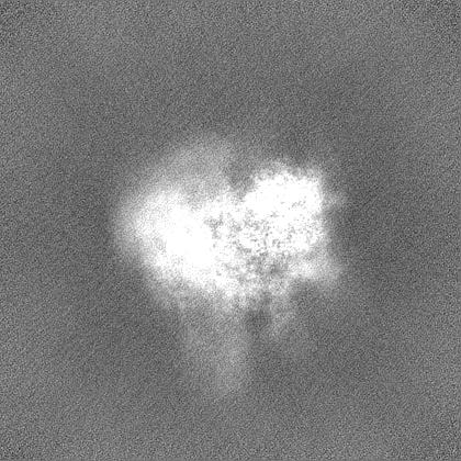
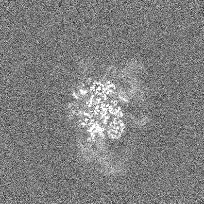
| Images |  emd_16542.png emd_16542.png | 94.1 KB | ||
| Others | emd_16542_half_map_1.map.gzemd_16542_half_map_2.map.gz | 262 MB 262 MB | ||
| Archive directory |  http://ftp.pdbj.org/pub/emdb/structures/EMD-16542ftp://ftp.pdbj.org/pub/emdb/structures/EMD-16542 http://ftp.pdbj.org/pub/emdb/structures/EMD-16542ftp://ftp.pdbj.org/pub/emdb/structures/EMD-16542 | HTTPS FTP |
-Validation report
| Summary document | emd_16542_validation.pdf.gz | 850.2 KB | Display | EMDB validaton report |
|---|---|---|---|---|
| Full document | emd_16542_full_validation.pdf.gz | 849.8 KB | Display | |
| Data in XML | emd_16542_validation.xml.gz | 23.2 KB | Display | |
| Data in CIF | emd_16542_validation.cif.gz | 30.3 KB | Display | |
| Arichive directory | https://ftp.pdbj.org/pub/emdb/validation_reports/EMD-16542ftp://ftp.pdbj.org/pub/emdb/validation_reports/EMD-16542 | HTTPS FTP |
-Related structure data


-Links
| EMDB pages | EMDB (EBI/PDBe) / EMDataResource |
|---|
-Map
| File | Download / File: emd_16542.map.gz / Format: CCP4 / Size: 282.6 MB / Type: IMAGE STORED AS FLOATING POINT NUMBER (4 BYTES) | ||||||||||||||||||||||||||||||||||||
|---|---|---|---|---|---|---|---|---|---|---|---|---|---|---|---|---|---|---|---|---|---|---|---|---|---|---|---|---|---|---|---|---|---|---|---|---|---|
| Annotation | Local refinement map of Otu2 N-terminal domain bound to yeast 40S ribosome | ||||||||||||||||||||||||||||||||||||
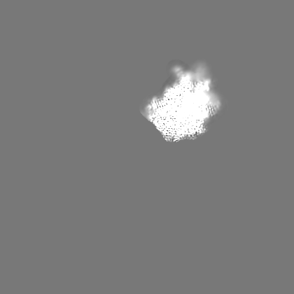
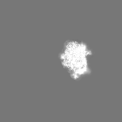
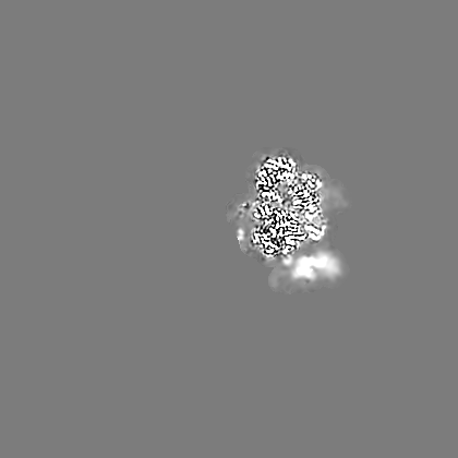
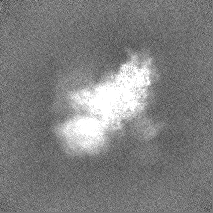
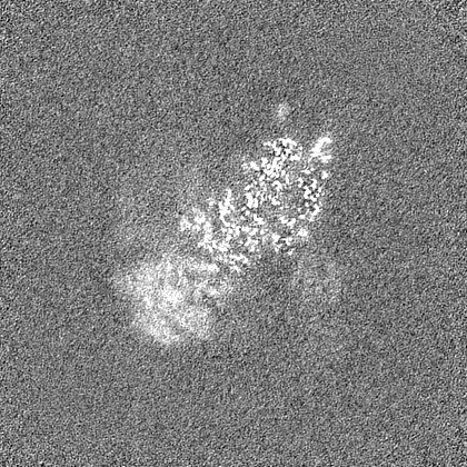
| Projections & slices | Image control
Images are generated by Spider. | ||||||||||||||||||||||||||||||||||||
| Voxel size | X=Y=Z: 1.045 Å | ||||||||||||||||||||||||||||||||||||
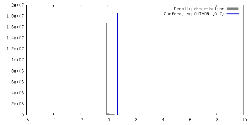
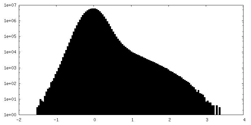
| Density |
| ||||||||||||||||||||||||||||||||||||
| Symmetry | Space group: 1 | ||||||||||||||||||||||||||||||||||||
| Details | EMDB XML:
|
 Z (Sec.)
 Y (Row.)
 X (Col.)


-Supplemental data
-Half map: Half-map B
| File | emd_16542_half_map_1.map | ||||||||||||
|---|---|---|---|---|---|---|---|---|---|---|---|---|---|
| Annotation | Half-map B | ||||||||||||
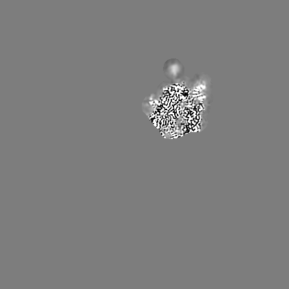
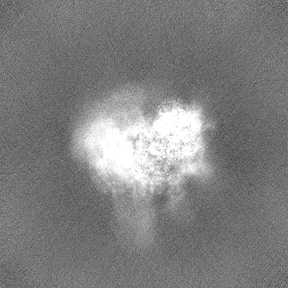
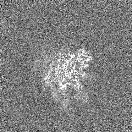
| Projections & Slices |
| ||||||||||||
| Density Histograms |

-Half map: Half-map A
| File | emd_16542_half_map_2.map | ||||||||||||
|---|---|---|---|---|---|---|---|---|---|---|---|---|---|
| Annotation | Half-map A | ||||||||||||
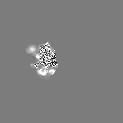
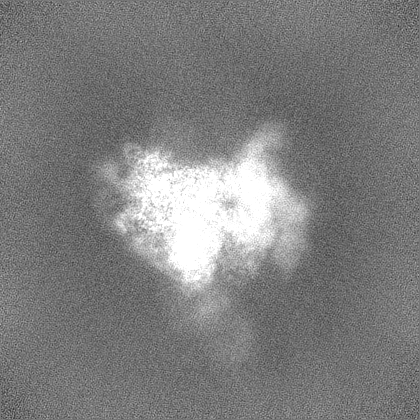
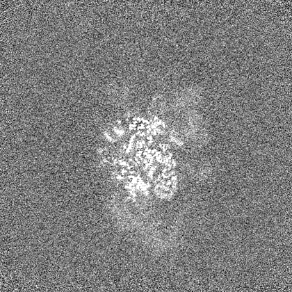
| Projections & Slices |
| ||||||||||||
| Density Histograms |


- Sample components
-Entire : Local refinement map of Otu2 N-terminal domain bound to yeast 40S...
| Entire | Name: Local refinement map of Otu2 N-terminal domain bound to yeast 40S ribosome |
|---|---|
| Components |
|
-Supramolecule #1: Local refinement map of Otu2 N-terminal domain bound to yeast 40S...
| Supramolecule | Name: Local refinement map of Otu2 N-terminal domain bound to yeast 40S ribosome type: complex / ID: 1 / Chimera: Yes / Parent: 0 / Macromolecule list: all |
|---|---|
| Source (natural) | Organism: |
-Macromolecule #1: OTU domain-containing protein 2 (Otu2) C178S
| Macromolecule | Name: OTU domain-containing protein 2 (Otu2) C178S / type: protein_or_peptide / ID: 1 / Enantiomer: LEVO |
|---|---|
| Source (natural) | Organism: |
| Recombinant expression | Organism: |
| Sequence | String: MTGMESGENL ENMEDILARH RKENKDLQNK ITGMKKQATK SKRKEVNSKC LDLQDKLKTK QENEIRDWKI ANNEVFDAEQ EDEVTPEKL LEQLSISRDE KEQQNVPVQQ QQQGQTKKRR NRQKERLAKR DAAIAKMKEE AALEASKQPD LKKMEQESID Q LCELKKLK ...String: MTGMESGENL ENMEDILARH RKENKDLQNK ITGMKKQATK SKRKEVNSKC LDLQDKLKTK QENEIRDWKI ANNEVFDAEQ EDEVTPEKL LEQLSISRDE KEQQNVPVQQ QQQGQTKKRR NRQKERLAKR DAAIAKMKEE AALEASKQPD LKKMEQESID Q LCELKKLK QFDIQPDGHS LFASILDQLK LRHDPKKLDQ DMDVMKLRWL SCNYVQEHRD DFIPYLFDEE TMKMKDIDEY TK EMEHTAQ WGGEIEILAL SHVFDCPISI LMSGRPIQVY NECGKNPELK LVYYKHSYAL GEHYNSLHDS |
-Experimental details
-Structure determination
| Method | cryo EM |
|---|---|
 Processing Processing | single particle reconstruction |
| Aggregation state | particle |
-Sample preparation
| Buffer | pH: 7.5 |
|---|---|
| Vitrification | Cryogen name: ETHANE |
- Electron microscopy
| Microscope | FEI TITAN KRIOS |
|---|---|
| Image recording | Film or detector model: GATAN K2 SUMMIT (4k x 4k) / Detector mode: COUNTING / Average electron dose: 46.4 e/Å2 |
| Electron beam | Acceleration voltage: 300 kV / Electron source:  FIELD EMISSION GUN FIELD EMISSION GUN |
| Electron optics | Illumination mode: FLOOD BEAM / Imaging mode: BRIGHT FIELD / Nominal defocus max: 3.0 µm / Nominal defocus min: 0.5 µm |
| Experimental equipment |  Model: Titan Krios / Image courtesy: FEI Company |
-Image processing
| Final reconstruction | Resolution.type: BY AUTHOR / Resolution: 2.9 Å / Resolution method: FSC 0.143 CUT-OFF / Number images used: 36086 |
|---|---|
| Initial angle assignment | Type: OTHER / Details: Relion |
| Final angle assignment | Type: OTHER / Details: Relion |
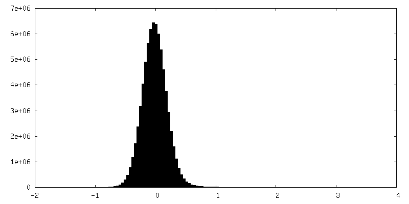
| FSC plot (resolution estimation) | 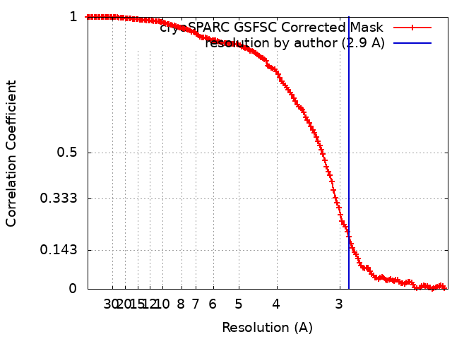 |
