 Movie
Movie Controller

[English] 日本語
 Yorodumi
- EMDB-16472: In situ cryo-electron tomogram of cytoplasmic lattice filaments f... -
+ Open data

- Basic information
| Entry |  | ||||||||||||
|---|---|---|---|---|---|---|---|---|---|---|---|---|---|
| Title | In situ cryo-electron tomogram of cytoplasmic lattice filaments from a mouse oocyte | ||||||||||||
 Map data Map data | In situ cryo-electron tomogram of cytoplasmic lattice filaments from a mouse oocyte (unprocessed tomogram) | ||||||||||||
 Sample Sample |
| ||||||||||||
 Keywords Keywords | mammalian oocytes / cytoplasmic lattices / protein storage / filaments / CYTOSOLIC PROTEIN | ||||||||||||
| Biological species |  | ||||||||||||
| Method | electron tomography / cryo EM | ||||||||||||
 Authors Authors | Bauerlein FJB / Jentoft IMA / Petrovic A / Fernandez-Busnadiego R / Schuh M | ||||||||||||
| Funding support |  Germany, 3 items Germany, 3 items
| ||||||||||||
 Citation Citation | Journal: Cell / Year: 2023 Title: Mammalian oocytes store proteins for the early embryo on cytoplasmic lattices. Authors: Ida M A Jentoft / Felix J B Bäuerlein / Luisa M Welp / Benjamin H Cooper / Arsen Petrovic / Chun So / Sarah Mae Penir / Antonio Z Politi / Yehor Horokhovskyi / Iina Takala / Heike Eckel / ...Authors: Ida M A Jentoft / Felix J B Bäuerlein / Luisa M Welp / Benjamin H Cooper / Arsen Petrovic / Chun So / Sarah Mae Penir / Antonio Z Politi / Yehor Horokhovskyi / Iina Takala / Heike Eckel / Rüdiger Moltrecht / Peter Lénárt / Tommaso Cavazza / Juliane Liepe / Nils Brose / Henning Urlaub / Rubén Fernández-Busnadiego / Melina Schuh / Abstract: Mammalian oocytes are filled with poorly understood structures called cytoplasmic lattices. First discovered in the 1960s and speculated to correspond to mammalian yolk, ribosomal arrays, or ...Mammalian oocytes are filled with poorly understood structures called cytoplasmic lattices. First discovered in the 1960s and speculated to correspond to mammalian yolk, ribosomal arrays, or intermediate filaments, their function has remained enigmatic to date. Here, we show that cytoplasmic lattices are sites where oocytes store essential proteins for early embryonic development. Using super-resolution light microscopy and cryoelectron tomography, we show that cytoplasmic lattices are composed of filaments with a high surface area, which contain PADI6 and subcortical maternal complex proteins. The lattices associate with many proteins critical for embryonic development, including proteins that control epigenetic reprogramming of the preimplantation embryo. Loss of cytoplasmic lattices by knocking out PADI6 or the subcortical maternal complex prevents the accumulation of these proteins and results in early embryonic arrest. Our work suggests that cytoplasmic lattices enrich maternally provided proteins to prevent their premature degradation and cellular activity, thereby enabling early mammalian development. | ||||||||||||
| History |
|
- Structure visualization
| Supplemental images |
|---|
- Downloads & links
-EMDB archive
| Map data | emd_16472.map.gz | 236.3 MB |  EMDB map data format EMDB map data format | |
|---|---|---|---|---|
| Header (meta data) | emd-16472-v30.xmlemd-16472.xml | 13.7 KB 13.7 KB | Display Display | EMDB header |
| Images |  emd_16472.png emd_16472.png | 267.2 KB | ||
| Filedesc metadata | emd-16472.cif.gz | 4.9 KB | ||
| Others | emd_16472_additional_1.map.gz | 241.5 MB | ||
| Archive directory |  http://ftp.pdbj.org/pub/emdb/structures/EMD-16472ftp://ftp.pdbj.org/pub/emdb/structures/EMD-16472 http://ftp.pdbj.org/pub/emdb/structures/EMD-16472ftp://ftp.pdbj.org/pub/emdb/structures/EMD-16472 | HTTPS FTP |
-Related structure data
-Links
| EMDB pages | EMDB (EBI/PDBe) / EMDataResource |
|---|
-Map
| File | Download / File: emd_16472.map.gz / Format: CCP4 / Size: 450 MB / Type: IMAGE STORED AS SIGNED INTEGER (2 BYTES) | ||||||||||||||||||||||||||||||||
|---|---|---|---|---|---|---|---|---|---|---|---|---|---|---|---|---|---|---|---|---|---|---|---|---|---|---|---|---|---|---|---|---|---|
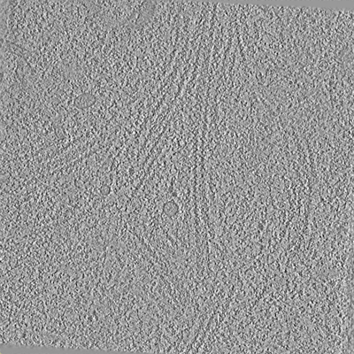
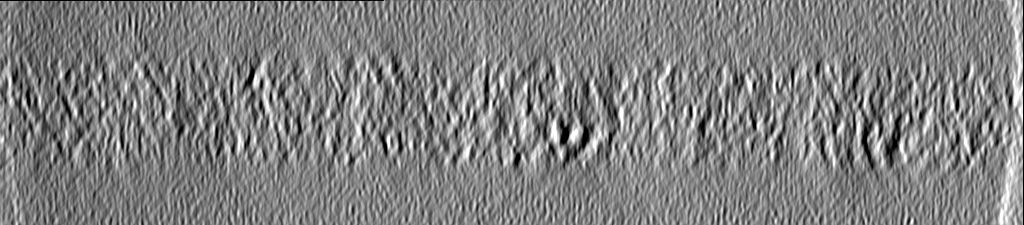
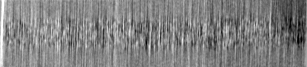
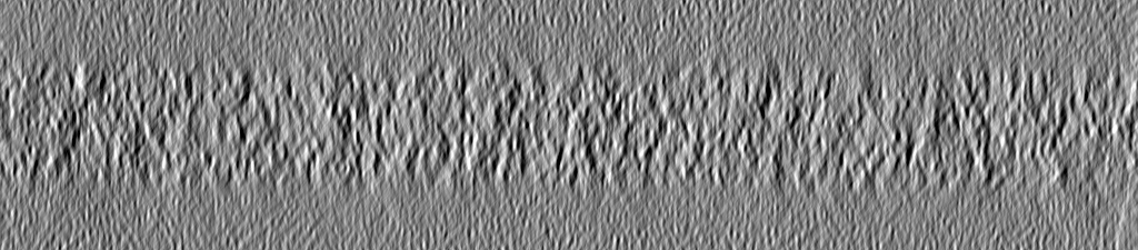
| Annotation | In situ cryo-electron tomogram of cytoplasmic lattice filaments from a mouse oocyte (unprocessed tomogram) | ||||||||||||||||||||||||||||||||
| Projections & slices | Image control
Images are generated by Spider. generated in cubic-lattice coordinate | ||||||||||||||||||||||||||||||||
| Voxel size | X=Y=Z: 14.61 Å | ||||||||||||||||||||||||||||||||
| Density |
| ||||||||||||||||||||||||||||||||
| Symmetry | Space group: 1 | ||||||||||||||||||||||||||||||||
| Details | EMDB XML:
|
 Z (Sec.)
 Y (Row.)
 X (Col.)


-Supplemental data
-Additional map: In situ cryo-electron tomogram of cytoplasmic lattice filaments...
| File | emd_16472_additional_1.map | ||||||||||||
|---|---|---|---|---|---|---|---|---|---|---|---|---|---|
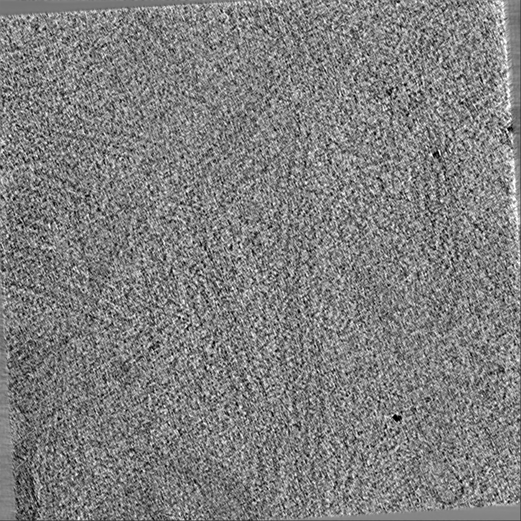
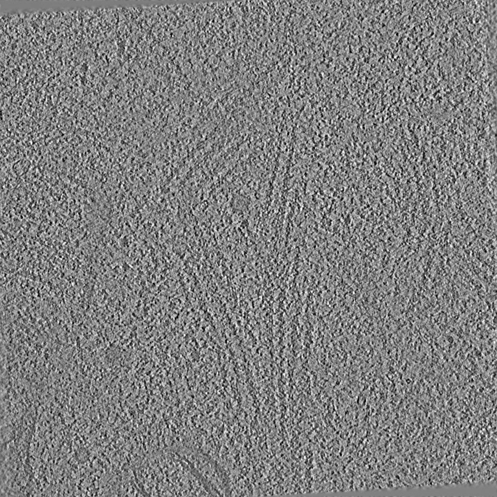
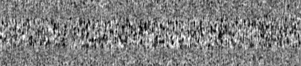
| Annotation | In situ cryo-electron tomogram of cytoplasmic lattice filaments from a mouse oocyte (deconvoluted tomogram) | ||||||||||||
| Projections & Slices |
| ||||||||||||
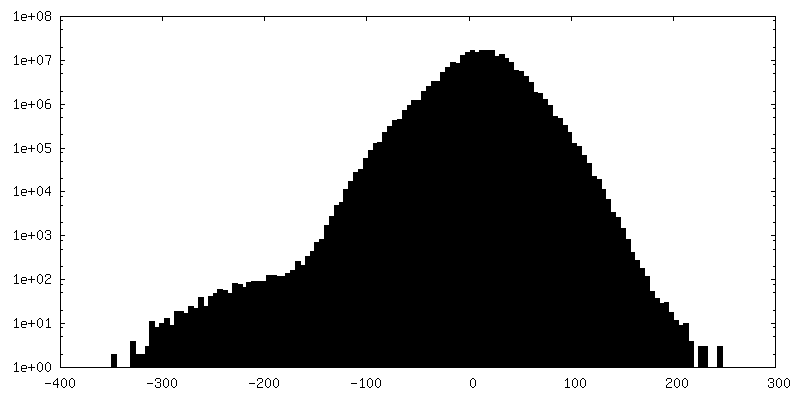
| Density Histograms |

- Sample components
-Entire : mouse oocyte
| Entire | Name: mouse oocyte |
|---|---|
| Components |
|
-Supramolecule #1: mouse oocyte
| Supramolecule | Name: mouse oocyte / type: cell / ID: 1 / Parent: 0 |
|---|---|
| Source (natural) | Organism: |
-Experimental details
-Structure determination
| Method | cryo EM |
|---|---|
 Processing Processing | electron tomography |
| Aggregation state | cell |
-Sample preparation
| Buffer | pH: 7 |
|---|---|
| Grid | Model: Quantifoil R2/1 / Material: MOLYBDENUM / Mesh: 200 / Support film - Material: CARBON / Support film - topology: HOLEY / Pretreatment - Type: PLASMA CLEANING / Pretreatment - Time: 30 sec. / Pretreatment - Atmosphere: AIR |
| Vitrification | Cryogen name: ETHANE-PROPANE / Instrument: HOMEMADE PLUNGER |
| Details | FIB milled lamella of mouse oocytes |
| Cryo protectant | 7.5%/7.5% DMSO/EG |
| Sectioning | Focused ion beam - Instrument: OTHER / Focused ion beam - Ion: OTHER / Focused ion beam - Voltage: 30 / Focused ion beam - Current: 0.05 / Focused ion beam - Duration: 3600 / Focused ion beam - Temperature: 83 K / Focused ion beam - Initial thickness: 80000 / Focused ion beam - Final thickness: 150 Focused ion beam - Details: We develop a 'deep-FIBing' approach, by which the cells were first milled perpendicular to the EM grid to reveal a clean surface from which lamellae were subsequently ...Focused ion beam - Details: We develop a 'deep-FIBing' approach, by which the cells were first milled perpendicular to the EM grid to reveal a clean surface from which lamellae were subsequently prepared (see publication).. The value given for _em_focused_ion_beam.instrument is Aquilos II. This is not in a list of allowed values {'OTHER', 'DB235'} so OTHER is written into the XML file. |
- Electron microscopy
| Microscope | TFS KRIOS |
|---|---|
| Image recording | Film or detector model: FEI FALCON IV (4k x 4k) / Digitization - Dimensions - Width: 4096 pixel / Digitization - Dimensions - Height: 4096 pixel / Average electron dose: 3.34 e/Å2 |
| Electron beam | Acceleration voltage: 300 kV / Electron source:  FIELD EMISSION GUN FIELD EMISSION GUN |
| Electron optics | C2 aperture diameter: 50.0 µm / Illumination mode: FLOOD BEAM / Imaging mode: BRIGHT FIELD / Cs: 2.7 mm / Nominal defocus max: 5.0 µm / Nominal defocus min: 3.0 µm / Nominal magnification: 33000 |
| Sample stage | Specimen holder model: FEI TITAN KRIOS AUTOGRID HOLDER / Cooling holder cryogen: NITROGEN |
| Experimental equipment |  Model: Titan Krios / Image courtesy: FEI Company |
-Image processing
| Final reconstruction | Algorithm: BACK PROJECTION / Software - Name: IMOD (ver. 4.11.13) / Number images used: 30 |
|---|
