Movie
Movie Controller

[English] 日本語
 Yorodumi
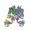
- EMDB-1645: Structure of human erythrocyte anion exchanger 1 revealed by elec... -
+ Open data

- Basic information
| Entry | Database: EMDB / ID: EMD-1645 | |||||||||
|---|---|---|---|---|---|---|---|---|---|---|
| Title | Structure of human erythrocyte anion exchanger 1 revealed by electron crystallography. | |||||||||
 Map data Map data | 3D map of AE1 solved by electron crystallography. | |||||||||
 Sample Sample |
| |||||||||
 Keywords Keywords | AE1 / anion exchanger 1 / band 3 / SLC4A1 | |||||||||
| Biological species |  Homo sapiens (human) Homo sapiens (human) | |||||||||
| Method | electron crystallography / cryo EM / Resolution: 7.5 Å | |||||||||
 Authors Authors | Yamaguchi T / Ikeda Y / Abe Y / Kuma H / Kang D / Hamasaki N / Hirai T | |||||||||
 Citation Citation | Journal: J Mol Biol / Year: 2010 Title: Structure of the membrane domain of human erythrocyte anion exchanger 1 revealed by electron crystallography. Authors: Tomohiro Yamaguchi / Yohei Ikeda / Yoshito Abe / Hiroyuki Kuma / Dongchon Kang / Naotaka Hamasaki / Teruhisa Hirai /  Abstract: The membrane domain of human erythrocyte anion exchanger 1 (AE1) works as a Cl(-)/HCO(3)(-) antiporter. This exchange is a key step for CO(2)/O(2) circulation in the blood. In spite of their ...The membrane domain of human erythrocyte anion exchanger 1 (AE1) works as a Cl(-)/HCO(3)(-) antiporter. This exchange is a key step for CO(2)/O(2) circulation in the blood. In spite of their importance, structural information about AE1 and the AE (anion exchanger) family are still very limited. We used electron microscopy to solve the three-dimensional structure of the AE1 membrane domain, fixed in an outward-open conformation by cross-linking, at 7.5-A resolution. A dimer of AE1 membrane domains packed in two-dimensional array showed a projection map similar to that of the prokaryotic homolog of the ClC chloride channel, a Cl(-)/H(+) antiporter. In a three-dimensional map, there are V-shaped densities near the center of the dimer and slightly narrower V-shaped clusters at a greater distance from the center of the dimer. These appear to be inserted into the membrane from opposite sides. The structural motifs, two homologous pairs of helices in internal repeats of the ClC transporter (helices B+C and J+K), are well fitted to those AE1 densities after simple domain movement. | |||||||||
| History |
|
- Structure visualization
| Movie |
 Movie viewer Movie viewer |
|---|---|
| Structure viewer | EM map: SurfViewMolmilJmol/JSmol |
| Supplemental images |

- Downloads & links
-EMDB archive
| Map data | emd_1645.map.gz | 13.2 MB | EMDB map data format | |
|---|---|---|---|---|
| Header (meta data) | emd-1645-v30.xmlemd-1645.xml | 11.1 KB 11.1 KB | Display Display | EMDB header |
| Images |  EMD-1645.png EMD-1645.png | 377.9 KB | ||
| Archive directory |  http://ftp.pdbj.org/pub/emdb/structures/EMD-1645ftp://ftp.pdbj.org/pub/emdb/structures/EMD-1645 http://ftp.pdbj.org/pub/emdb/structures/EMD-1645ftp://ftp.pdbj.org/pub/emdb/structures/EMD-1645 | HTTPS FTP |
-Links
| EMDB pages | EMDB (EBI/PDBe) / EMDataResource |
|---|
-Map
| File | Download / File: emd_1645.map.gz / Format: CCP4 / Size: 13.9 MB / Type: IMAGE STORED AS FLOATING POINT NUMBER (4 BYTES) | ||||||||||||||||||||||||||||||||||||||||||||||||||||||||||||||||||||
|---|---|---|---|---|---|---|---|---|---|---|---|---|---|---|---|---|---|---|---|---|---|---|---|---|---|---|---|---|---|---|---|---|---|---|---|---|---|---|---|---|---|---|---|---|---|---|---|---|---|---|---|---|---|---|---|---|---|---|---|---|---|---|---|---|---|---|---|---|---|
| Annotation | 3D map of AE1 solved by electron crystallography. | ||||||||||||||||||||||||||||||||||||||||||||||||||||||||||||||||||||
| Projections & slices | Image control
Images are generated by Spider. generated in cubic-lattice coordinate | ||||||||||||||||||||||||||||||||||||||||||||||||||||||||||||||||||||
| Voxel size | X: 1.21562 Å / Y: 1.26389 Å / Z: 1.23457 Å | ||||||||||||||||||||||||||||||||||||||||||||||||||||||||||||||||||||
| Density |
| ||||||||||||||||||||||||||||||||||||||||||||||||||||||||||||||||||||
| Symmetry | Space group: 1 | ||||||||||||||||||||||||||||||||||||||||||||||||||||||||||||||||||||
| Details | EMDB XML:
CCP4 map header:
| ||||||||||||||||||||||||||||||||||||||||||||||||||||||||||||||||||||
 Z (Sec.)
 Y (Row.)
 X (Col.)


-Supplemental data
- Sample components
-Entire : Membrane domain of anion exchanger 1
| Entire | Name: Membrane domain of anion exchanger 1 |
|---|---|
| Components |
|
-Supramolecule #1000: Membrane domain of anion exchanger 1
| Supramolecule | Name: Membrane domain of anion exchanger 1 / type: sample / ID: 1000 Details: The sample is cross-linked by H2DIDS. N-terminal cytoplasmic domain is cleaved off by trypsin digestion. Glycosylation is removed by glycosidase. Oligomeric state: Dimer / Number unique components: 1 |
|---|---|
| Molecular weight | Experimental: 55 KDa / Theoretical: 55 KDa / Method: SDS-PAGE |
-Macromolecule #1: Anion Exchanger 1
| Macromolecule | Name: Anion Exchanger 1 / type: protein_or_peptide / ID: 1 / Name.synonym: Band 3 / Number of copies: 2 / Oligomeric state: Dimer / Recombinant expression: No / Database: NCBI |
|---|---|
| Source (natural) | Organism: Homo sapiens (human) / synonym: Human / Tissue: Blood / Cell: Erythrocyte / Location in cell: Membrane |
| Molecular weight | Experimental: 55 KDa / Theoretical: 55 KDa |
-Experimental details
-Structure determination
| Method | cryo EM |
|---|---|
 Processing Processing | electron crystallography |
| Aggregation state | 2D array |
-Sample preparation
| Concentration | 2.5 mg/mL |
|---|---|
| Grid | Details: Mo grid |
| Vitrification | Cryogen name: NITROGEN / Chamber temperature: 77 K / Instrument: HOMEMADE PLUNGER Details: Vitrification instrument: Manual. Excess buffer was removed by pipetting and blotting with filter paper, and then the grid was manually plunged into liquid nitrogen. These operations were ...Details: Vitrification instrument: Manual. Excess buffer was removed by pipetting and blotting with filter paper, and then the grid was manually plunged into liquid nitrogen. These operations were carried out at 4 degrees celsius. Method: Carbon sandwich method |
- Electron microscopy
| Microscope | JEOL 3000SFF |
|---|---|
| Temperature | Average: 4 K |
| Image recording | Category: FILM / Film or detector model: KODAK SO-163 FILM / Digitization - Scanner: ZEISS SCAI / Digitization - Sampling interval: 7 µm / Number real images: 31 / Bits/pixel: 12 |
| Electron beam | Acceleration voltage: 300 kV / Electron source:  FIELD EMISSION GUN FIELD EMISSION GUN |
| Electron optics | Illumination mode: FLOOD BEAM / Imaging mode: BRIGHT FIELD / Cs: 1.6 mm / Nominal defocus max: 2.5 µm / Nominal defocus min: 1.0 µm / Nominal magnification: 40000 |
| Sample stage | Specimen holder: Top entry / Specimen holder model: JEOL / Tilt angle min: -60 / Tilt angle max: 60 / Tilt series - Axis1 - Min angle: -60 ° / Tilt series - Axis1 - Max angle: 60 ° |
-Image processing
| Final reconstruction | Resolution.type: BY AUTHOR / Resolution: 7.5 Å / Resolution method: OTHER / Software - Name: MRC |
|---|---|
| Crystal parameters | Unit cell - A: 194.5 Å / Unit cell - B: 182.0 Å / Unit cell - C: 200 Å / Unit cell - γ: 90 ° / Unit cell - α: 90 ° / Unit cell - β: 90 ° / Plane group: P 2 21 21 |
| CTF correction | Details: Each film |
-Atomic model buiding 1
| Initial model | PDB ID: |
|---|---|
| Software | Name: CNS |
| Details | Protocol: Rigid Body |
| Refinement | Space: RECIPROCAL / Protocol: RIGID BODY FIT / Target criteria: Free R |