 ムービー
ムービー コントローラー


+ データを開く

- 基本情報
基本情報
| 登録情報 | データベース: EMDB / ID: EMD-1643 | |||||||||
|---|---|---|---|---|---|---|---|---|---|---|
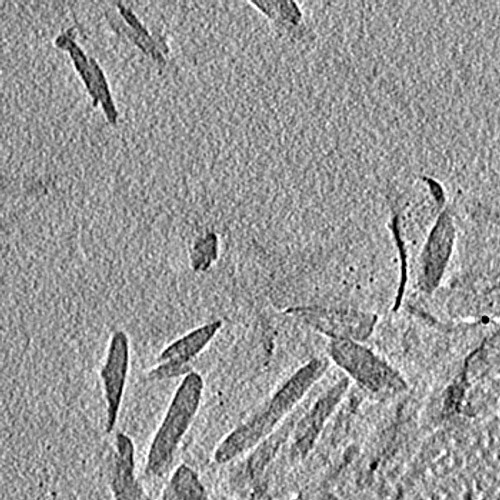
| タイトル | Structure of chlorosomes from the green filamentous bacterium Chloroflexus aurantiacus | |||||||||
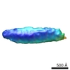
 マップデータ マップデータ | This is a tomogram of a field of chlorosomes from Chloroflexus aurantiacus. | |||||||||
 試料 試料 |
| |||||||||
 キーワード キーワード | Chlorosome / light-harvesting complex / Chloroflexus | |||||||||
| 生物種 |   Chloroflexus aurantiacus (バクテリア) Chloroflexus aurantiacus (バクテリア) | |||||||||
| 手法 | 電子線トモグラフィー法 / クライオ電子顕微鏡法 | |||||||||
 データ登録者 データ登録者 | Psencik J / Collins AM / Liljeroos L / Torkkeli M / Laurinmaki P / Ansink HM / Ikonen TP / Serimaa RE / Blankenship RE / Tuma R / Butcher SJ | |||||||||
 引用 引用 | ジャーナル: J Bacteriol / 年: 2009 タイトル: Structure of chlorosomes from the green filamentous bacterium Chloroflexus aurantiacus. 著者: Jakub Psencík / Aaron M Collins / Lassi Liljeroos / Mika Torkkeli / Pasi Laurinmäki / Hermanus M Ansink / Teemu P Ikonen / Ritva E Serimaa / Robert E Blankenship / Roman Tuma / Sarah J Butcher /  要旨: The green filamentous bacterium Chloroflexus aurantiacus employs chlorosomes as photosynthetic antennae. Chlorosomes contain bacteriochlorophyll aggregates and are attached to the inner side of a ...The green filamentous bacterium Chloroflexus aurantiacus employs chlorosomes as photosynthetic antennae. Chlorosomes contain bacteriochlorophyll aggregates and are attached to the inner side of a plasma membrane via a protein baseplate. The structure of chlorosomes from C. aurantiacus was investigated by using a combination of cryo-electron microscopy and X-ray diffraction and compared with that of Chlorobi species. Cryo-electron tomography revealed thin chlorosomes for which a distinct crystalline baseplate lattice was visualized in high-resolution projections. The baseplate is present only on one side of the chlorosome, and the lattice dimensions suggest that a dimer of the CsmA protein is the building block. The bacteriochlorophyll aggregates inside the chlorosome are arranged in lamellae, but the spacing is much greater than that in Chlorobi species. A comparison of chlorosomes from different species suggested that the lamellar spacing is proportional to the chain length of the esterifying alcohols. C. aurantiacus chlorosomes accumulate larger quantities of carotenoids under high-light conditions, presumably to provide photoprotection. The wider lamellae allow accommodation of the additional carotenoids and lead to increased disorder within the lamellae. | |||||||||
| 履歴 |
|
- 構造の表示
構造の表示
| ムービー |
 ムービービューア ムービービューア |
|---|---|
| 構造ビューア | EMマップ: SurfViewMolmilJmol/JSmol |
| 添付画像 |
- ダウンロードとリンク
-EMDBアーカイブ
| マップデータ | emd_1643.map.gz | 7.8 MB | EMDBマップデータ形式 | |
|---|---|---|---|---|
| ヘッダ (付随情報) | emd-1643-v30.xmlemd-1643.xml | 9.7 KB 9.7 KB | 表示 表示 | EMDBヘッダ |
| 画像 |  1643.jpg 1643.jpg | 89.4 KB | ||
| アーカイブディレクトリ |  http://ftp.pdbj.org/pub/emdb/structures/EMD-1643ftp://ftp.pdbj.org/pub/emdb/structures/EMD-1643 http://ftp.pdbj.org/pub/emdb/structures/EMD-1643ftp://ftp.pdbj.org/pub/emdb/structures/EMD-1643 | HTTPS FTP |
-検証レポート
| 文書・要旨 | emd_1643_validation.pdf.gz | 147.4 KB | 表示 | EMDB検証レポート |
|---|---|---|---|---|
| 文書・詳細版 | emd_1643_full_validation.pdf.gz | 146.5 KB | 表示 | |
| XML形式データ | emd_1643_validation.xml.gz | 3.1 KB | 表示 | |
| アーカイブディレクトリ | https://ftp.pdbj.org/pub/emdb/validation_reports/EMD-1643ftp://ftp.pdbj.org/pub/emdb/validation_reports/EMD-1643 | HTTPS FTP |
-関連構造データ
-リンク
| EMDBのページ | EMDB (EBI/PDBe) / EMDataResource |
|---|
-マップ
| ファイル | ダウンロード / ファイル: emd_1643.map.gz / 形式: CCP4 / 大きさ: 12.8 MB / タイプ: IMAGE STORED AS SIGNED INTEGER (2 BYTES) | ||||||||||||||||||||||||||||||||||||||||||||||||||||||||||||||||||||
|---|---|---|---|---|---|---|---|---|---|---|---|---|---|---|---|---|---|---|---|---|---|---|---|---|---|---|---|---|---|---|---|---|---|---|---|---|---|---|---|---|---|---|---|---|---|---|---|---|---|---|---|---|---|---|---|---|---|---|---|---|---|---|---|---|---|---|---|---|---|
| 注釈 | This is a tomogram of a field of chlorosomes from Chloroflexus aurantiacus. | ||||||||||||||||||||||||||||||||||||||||||||||||||||||||||||||||||||
| ボクセルのサイズ | X=Y=Z: 23 Å | ||||||||||||||||||||||||||||||||||||||||||||||||||||||||||||||||||||
| 密度 |
| ||||||||||||||||||||||||||||||||||||||||||||||||||||||||||||||||||||
| 対称性 | 空間群: 1 | ||||||||||||||||||||||||||||||||||||||||||||||||||||||||||||||||||||
| 詳細 | EMDB XML:
CCP4マップ ヘッダ情報:
| ||||||||||||||||||||||||||||||||||||||||||||||||||||||||||||||||||||
-添付データ
- 試料の構成要素
-全体 : Chlorosomes of Chloroflexus aurantiacus
| 全体 | 名称: Chlorosomes of Chloroflexus aurantiacus |
|---|---|
| 要素 |
|
-超分子 #1000: Chlorosomes of Chloroflexus aurantiacus
| 超分子 | 名称: Chlorosomes of Chloroflexus aurantiacus / タイプ: sample / ID: 1000 / 集合状態: Monomeric / Number unique components: 1 |
|---|
-超分子 #1: Chlorosome
| 超分子 | 名称: Chlorosome / タイプ: organelle_or_cellular_component / ID: 1 / Name.synonym: Chlorosome / 集合状態: Monomeric / 組換発現: No / データベース: NCBI |
|---|---|
| 由来(天然) | 生物種: Chloroflexus aurantiacus (バクテリア) / 細胞: Chloroflexus aurantiacus |
-実験情報
-構造解析
| 手法 | クライオ電子顕微鏡法 |
|---|---|
 解析 解析 | 電子線トモグラフィー法 |
-試料調製
| 緩衝液 | pH: 8 / 詳細: Sample was dialyzed against water |
|---|---|
| グリッド | 詳細: Quantifoil R2/2 holey carbon film, copper grid 400 mesh |
| 凍結 | 凍結剤: ETHANE / 装置: HOMEMADE PLUNGER / 詳細: Vitrification instrument: Homemade plunger / 手法: Blot for 1.5 seconds before plunging |
- 電子顕微鏡法
| 顕微鏡 | FEI TECNAI F20 |
|---|---|
| 温度 | 最低: 93 K / 最高: 103 K / 平均: 93 K |
| アライメント法 | Legacy - 非点収差: Objective lens astigmatism was corrected at 80,000 times magnification |
| 詳細 | Low-dose illumination |
| 日付 | 2008年2月15日 |
| 撮影 | カテゴリ: CCD フィルム・検出器のモデル: GATAN ULTRASCAN 4000 (4k x 4k) デジタル化 - サンプリング間隔: 1.50 µm / 実像数: 65 / 平均電子線量: 160 e/Å2 / ビット/ピクセル: 8 |
| 電子線 | 加速電圧: 200 kV / 電子線源:  FIELD EMISSION GUN FIELD EMISSION GUN |
| 電子光学系 | 倍率(補正後): 25840 / 照射モード: FLOOD BEAM / 撮影モード: BRIGHT FIELD / Cs: 2.0 mm / 倍率(公称値): 25840 |
| 試料ステージ | 試料ホルダー: Eucentric / 試料ホルダーモデル: GATAN LIQUID NITROGEN / Tilt series - Axis1 - Min angle: -64 ° / Tilt series - Axis1 - Max angle: 64 ° / Tilt series - Axis1 - Angle increment: 2 ° |
| 実験機器 |  モデル: Tecnai F20 / 画像提供: FEI Company |
-画像解析
| 詳細 | Particles were selected manually |
|---|---|
| 最終 再構成 | アルゴリズム: OTHER / ソフトウェア - 名称: IMOD / 使用した粒子像数: 65 |
