 Movie
Movie Controller

[English] 日本語
 Yorodumi
- EMDB-1643: Structure of chlorosomes from the green filamentous bacterium Chl... -
+ Open data

- Basic information
| Entry | Database: EMDB / ID: EMD-1643 | |||||||||
|---|---|---|---|---|---|---|---|---|---|---|
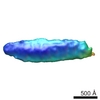
| Title | Structure of chlorosomes from the green filamentous bacterium Chloroflexus aurantiacus | |||||||||
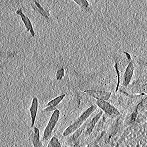
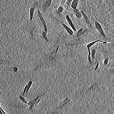
 Map data Map data | This is a tomogram of a field of chlorosomes from Chloroflexus aurantiacus. | |||||||||
 Sample Sample |
| |||||||||
 Keywords Keywords | Chlorosome / light-harvesting complex / Chloroflexus | |||||||||
| Biological species |   Chloroflexus aurantiacus (bacteria) Chloroflexus aurantiacus (bacteria) | |||||||||
| Method | electron tomography / cryo EM | |||||||||
 Authors Authors | Psencik J / Collins AM / Liljeroos L / Torkkeli M / Laurinmaki P / Ansink HM / Ikonen TP / Serimaa RE / Blankenship RE / Tuma R / Butcher SJ | |||||||||
 Citation Citation | Journal: J Bacteriol / Year: 2009 Title: Structure of chlorosomes from the green filamentous bacterium Chloroflexus aurantiacus. Authors: Jakub Psencík / Aaron M Collins / Lassi Liljeroos / Mika Torkkeli / Pasi Laurinmäki / Hermanus M Ansink / Teemu P Ikonen / Ritva E Serimaa / Robert E Blankenship / Roman Tuma / Sarah J Butcher /  Abstract: The green filamentous bacterium Chloroflexus aurantiacus employs chlorosomes as photosynthetic antennae. Chlorosomes contain bacteriochlorophyll aggregates and are attached to the inner side of a ...The green filamentous bacterium Chloroflexus aurantiacus employs chlorosomes as photosynthetic antennae. Chlorosomes contain bacteriochlorophyll aggregates and are attached to the inner side of a plasma membrane via a protein baseplate. The structure of chlorosomes from C. aurantiacus was investigated by using a combination of cryo-electron microscopy and X-ray diffraction and compared with that of Chlorobi species. Cryo-electron tomography revealed thin chlorosomes for which a distinct crystalline baseplate lattice was visualized in high-resolution projections. The baseplate is present only on one side of the chlorosome, and the lattice dimensions suggest that a dimer of the CsmA protein is the building block. The bacteriochlorophyll aggregates inside the chlorosome are arranged in lamellae, but the spacing is much greater than that in Chlorobi species. A comparison of chlorosomes from different species suggested that the lamellar spacing is proportional to the chain length of the esterifying alcohols. C. aurantiacus chlorosomes accumulate larger quantities of carotenoids under high-light conditions, presumably to provide photoprotection. The wider lamellae allow accommodation of the additional carotenoids and lead to increased disorder within the lamellae. | |||||||||
| History |
|
- Structure visualization
| Movie |
 Movie viewer Movie viewer |
|---|---|
| Structure viewer | EM map: SurfViewMolmilJmol/JSmol |
| Supplemental images |
- Downloads & links
-EMDB archive
| Map data | emd_1643.map.gz | 7.8 MB | EMDB map data format | |
|---|---|---|---|---|
| Header (meta data) | emd-1643-v30.xmlemd-1643.xml | 9.7 KB 9.7 KB | Display Display | EMDB header |
| Images |  1643.jpg 1643.jpg | 89.4 KB | ||
| Archive directory |  http://ftp.pdbj.org/pub/emdb/structures/EMD-1643ftp://ftp.pdbj.org/pub/emdb/structures/EMD-1643 http://ftp.pdbj.org/pub/emdb/structures/EMD-1643ftp://ftp.pdbj.org/pub/emdb/structures/EMD-1643 | HTTPS FTP |
-Validation report
| Summary document | emd_1643_validation.pdf.gz | 147.4 KB | Display | EMDB validaton report |
|---|---|---|---|---|
| Full document | emd_1643_full_validation.pdf.gz | 146.5 KB | Display | |
| Data in XML | emd_1643_validation.xml.gz | 3.1 KB | Display | |
| Arichive directory | https://ftp.pdbj.org/pub/emdb/validation_reports/EMD-1643ftp://ftp.pdbj.org/pub/emdb/validation_reports/EMD-1643 | HTTPS FTP |
-Related structure data
-Links
| EMDB pages | EMDB (EBI/PDBe) / EMDataResource |
|---|
-Map
| File | Download / File: emd_1643.map.gz / Format: CCP4 / Size: 12.8 MB / Type: IMAGE STORED AS SIGNED INTEGER (2 BYTES) | ||||||||||||||||||||||||||||||||||||||||||||||||||||||||||||||||||||
|---|---|---|---|---|---|---|---|---|---|---|---|---|---|---|---|---|---|---|---|---|---|---|---|---|---|---|---|---|---|---|---|---|---|---|---|---|---|---|---|---|---|---|---|---|---|---|---|---|---|---|---|---|---|---|---|---|---|---|---|---|---|---|---|---|---|---|---|---|---|
| Annotation | This is a tomogram of a field of chlorosomes from Chloroflexus aurantiacus. | ||||||||||||||||||||||||||||||||||||||||||||||||||||||||||||||||||||
| Projections & slices | Image control
Images are generated by Spider. generated in cubic-lattice coordinate | ||||||||||||||||||||||||||||||||||||||||||||||||||||||||||||||||||||
| Voxel size | X=Y=Z: 23 Å | ||||||||||||||||||||||||||||||||||||||||||||||||||||||||||||||||||||
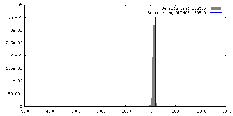
| Density |
| ||||||||||||||||||||||||||||||||||||||||||||||||||||||||||||||||||||
| Symmetry | Space group: 1 | ||||||||||||||||||||||||||||||||||||||||||||||||||||||||||||||||||||
| Details | EMDB XML:
CCP4 map header:
| ||||||||||||||||||||||||||||||||||||||||||||||||||||||||||||||||||||
 Z (Sec.)
 Y (Row.)
 X (Col.)


-Supplemental data
- Sample components
-Entire : Chlorosomes of Chloroflexus aurantiacus
| Entire | Name: Chlorosomes of Chloroflexus aurantiacus |
|---|---|
| Components |
|
-Supramolecule #1000: Chlorosomes of Chloroflexus aurantiacus
| Supramolecule | Name: Chlorosomes of Chloroflexus aurantiacus / type: sample / ID: 1000 / Oligomeric state: Monomeric / Number unique components: 1 |
|---|
-Supramolecule #1: Chlorosome
| Supramolecule | Name: Chlorosome / type: organelle_or_cellular_component / ID: 1 / Name.synonym: Chlorosome / Oligomeric state: Monomeric / Recombinant expression: No / Database: NCBI |
|---|---|
| Source (natural) | Organism: Chloroflexus aurantiacus (bacteria) / Cell: Chloroflexus aurantiacus |
-Experimental details
-Structure determination
| Method | cryo EM |
|---|---|
 Processing Processing | electron tomography |
-Sample preparation
| Buffer | pH: 8 / Details: Sample was dialyzed against water |
|---|---|
| Grid | Details: Quantifoil R2/2 holey carbon film, copper grid 400 mesh |
| Vitrification | Cryogen name: ETHANE / Instrument: HOMEMADE PLUNGER / Details: Vitrification instrument: Homemade plunger / Method: Blot for 1.5 seconds before plunging |
- Electron microscopy
| Microscope | FEI TECNAI F20 |
|---|---|
| Temperature | Min: 93 K / Max: 103 K / Average: 93 K |
| Alignment procedure | Legacy - Astigmatism: Objective lens astigmatism was corrected at 80,000 times magnification |
| Details | Low-dose illumination |
| Date | Feb 15, 2008 |
| Image recording | Category: CCD / Film or detector model: GATAN ULTRASCAN 4000 (4k x 4k) / Digitization - Sampling interval: 1.50 µm / Number real images: 65 / Average electron dose: 160 e/Å2 / Bits/pixel: 8 |
| Electron beam | Acceleration voltage: 200 kV / Electron source:  FIELD EMISSION GUN FIELD EMISSION GUN |
| Electron optics | Calibrated magnification: 25840 / Illumination mode: FLOOD BEAM / Imaging mode: BRIGHT FIELD / Cs: 2.0 mm / Nominal magnification: 25840 |
| Sample stage | Specimen holder: Eucentric / Specimen holder model: GATAN LIQUID NITROGEN / Tilt series - Axis1 - Min angle: -64 ° / Tilt series - Axis1 - Max angle: 64 ° / Tilt series - Axis1 - Angle increment: 2 ° |
| Experimental equipment |  Model: Tecnai F20 / Image courtesy: FEI Company |
-Image processing
| Details | Particles were selected manually |
|---|---|
| Final reconstruction | Algorithm: OTHER / Software - Name: IMOD / Number images used: 65 |
