 Movie
Movie Controller


+ Open data

- Basic information
| Entry |  | ||||||||||||
|---|---|---|---|---|---|---|---|---|---|---|---|---|---|
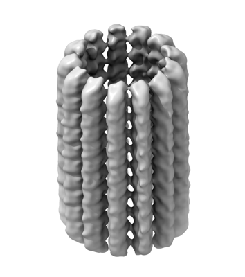
| Title | Subtomogram average of an intracellular microtubule filament | ||||||||||||
 Map data Map data | Subtomogram average of an intracellular microtubule filament | ||||||||||||
 Sample Sample |
| ||||||||||||
 Keywords Keywords | Microtubule / Cytoskeleton / STRUCTURAL PROTEIN | ||||||||||||
| Biological species |  Homo sapiens (human) Homo sapiens (human) | ||||||||||||
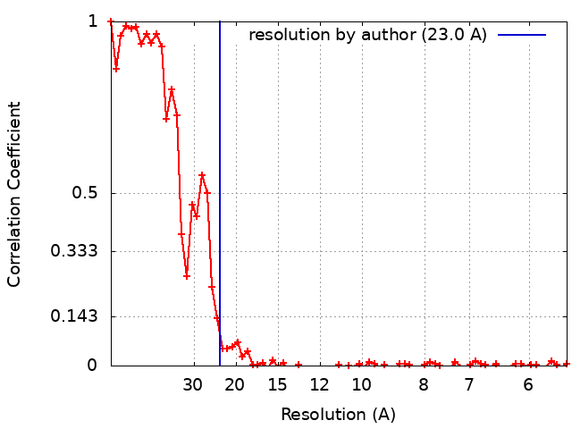
| Method | subtomogram averaging / cryo EM / Resolution: 23.0 Å | ||||||||||||
 Authors Authors | Sharp TH / Last MGF | ||||||||||||
| Funding support | European Union,  Netherlands, 3 items Netherlands, 3 items
| ||||||||||||
 Citation Citation | Journal: Sci Rep / Year: 2023 Title: Selecting optimal support grids for super-resolution cryogenic correlated light and electron microscopy. Authors: Mart G F Last / Maarten W Tuijtel / Lenard M Voortman / Thomas H Sharp /  Abstract: Cryogenic transmission electron microscopy (cryo-TEM) and super-resolution fluorescence microscopy are two popular and ever improving methods for high-resolution imaging of biological samples. In ...Cryogenic transmission electron microscopy (cryo-TEM) and super-resolution fluorescence microscopy are two popular and ever improving methods for high-resolution imaging of biological samples. In recent years, the combination of these two techniques into one correlated workflow has gained attention as a promising route towards contextualizing and enriching cryo-TEM imagery. A problem that is often encountered in the combination of these methods is that of light-induced damage to the sample during fluorescence imaging that renders the sample structure unsuitable for TEM imaging. In this paper, we describe how absorption of light by TEM sample support grids leads to sample damage, and we systematically explore the importance of parameters of grid design. We explain how, by changing the grid geometry and materials, one can increase the maximum illumination power density in fluorescence microscopy by up to an order of magnitude. Finally, we demonstrate the significant improvements in super-resolution image quality that are enabled by the selection of support grids that are optimally suited for correlated cryo-microscopy. #1: Journal: Selecting optimal support grids for super-resolution cryogenic correlated light and electron microscopyYear: 2022 Title: Selecting optimal support grids for super-resolution cryogenic correlated light and electron microscopy Authors: Sharp TH / Last MGF / Voortman LM / Tuijtel MW | ||||||||||||
| History |
|
- Structure visualization
| Supplemental images |
|---|
- Downloads & links
-EMDB archive
| Map data | emd_16307.map.gz | 5.2 MB |  EMDB map data format EMDB map data format | |
|---|---|---|---|---|
| Header (meta data) | emd-16307-v30.xmlemd-16307.xml | 16.3 KB 16.3 KB | Display Display | EMDB header |
| FSC (resolution estimation) | emd_16307_fsc.xml | 8.4 KB | Display | FSC data file |
| Images |  emd_16307.png emd_16307.png | 58.7 KB | ||
| Masks | emd_16307_msk_1.map | 22.2 MB | Mask map | |
| Filedesc metadata | emd-16307.cif.gz | 4.5 KB | ||
| Others | emd_16307_half_map_1.map.gzemd_16307_half_map_2.map.gz | 20.7 MB 20.7 MB | ||
| Archive directory |  http://ftp.pdbj.org/pub/emdb/structures/EMD-16307ftp://ftp.pdbj.org/pub/emdb/structures/EMD-16307 http://ftp.pdbj.org/pub/emdb/structures/EMD-16307ftp://ftp.pdbj.org/pub/emdb/structures/EMD-16307 | HTTPS FTP |
-Links
| EMDB pages | EMDB (EBI/PDBe) / EMDataResource |
|---|---|
| Related items in Molecule of the Month |

-Map
| File | Download / File: emd_16307.map.gz / Format: CCP4 / Size: 22.2 MB / Type: IMAGE STORED AS FLOATING POINT NUMBER (4 BYTES) | ||||||||||||||||||||||||||||||||||||
|---|---|---|---|---|---|---|---|---|---|---|---|---|---|---|---|---|---|---|---|---|---|---|---|---|---|---|---|---|---|---|---|---|---|---|---|---|---|
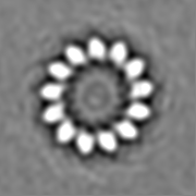
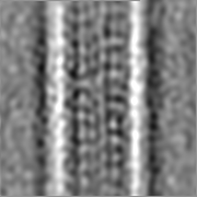
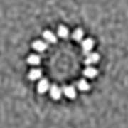
| Annotation | Subtomogram average of an intracellular microtubule filament | ||||||||||||||||||||||||||||||||||||
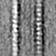
| Projections & slices | Image control
Images are generated by Spider. | ||||||||||||||||||||||||||||||||||||
| Voxel size | X=Y=Z: 2.75 Å | ||||||||||||||||||||||||||||||||||||
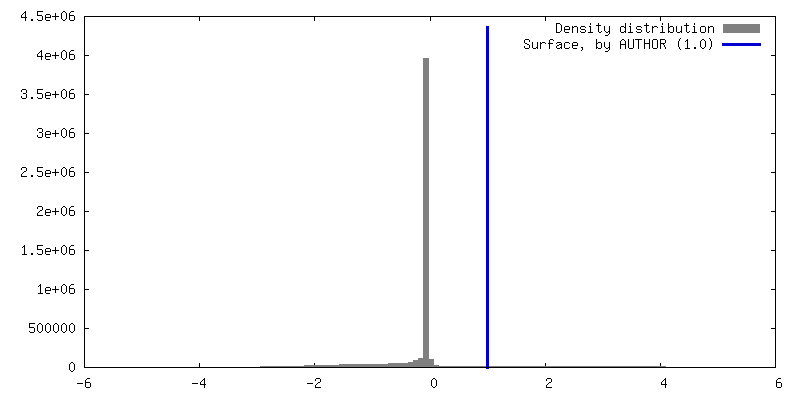
| Density |
| ||||||||||||||||||||||||||||||||||||
| Symmetry | Space group: 1 | ||||||||||||||||||||||||||||||||||||
| Details | EMDB XML:
|
 Z (Sec.)
 Y (Row.)
 X (Col.)


-Supplemental data
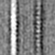
-Mask #1
| File | emd_16307_msk_1.map | ||||||||||||
|---|---|---|---|---|---|---|---|---|---|---|---|---|---|
| Projections & Slices |
| ||||||||||||
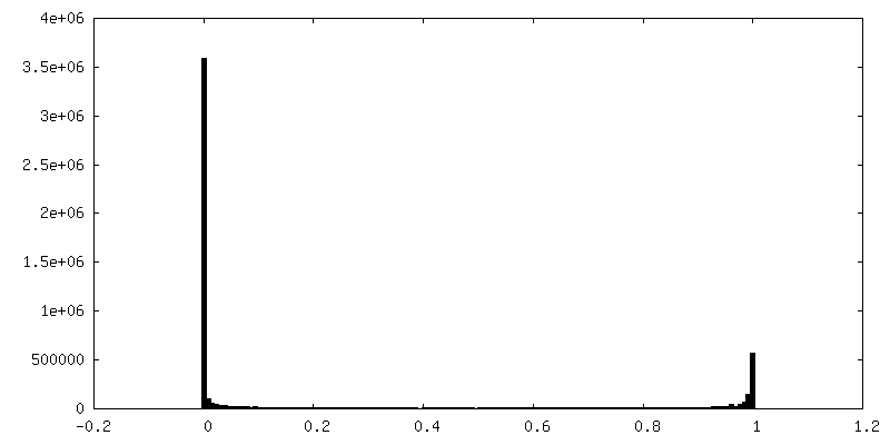
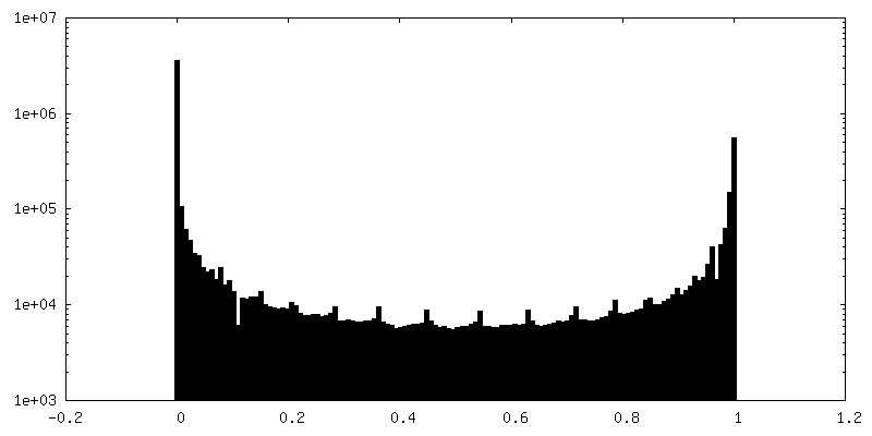
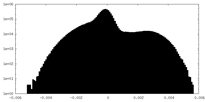
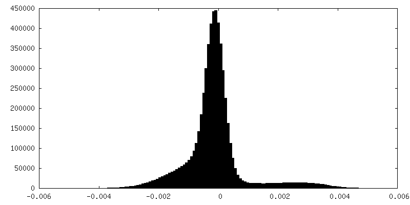
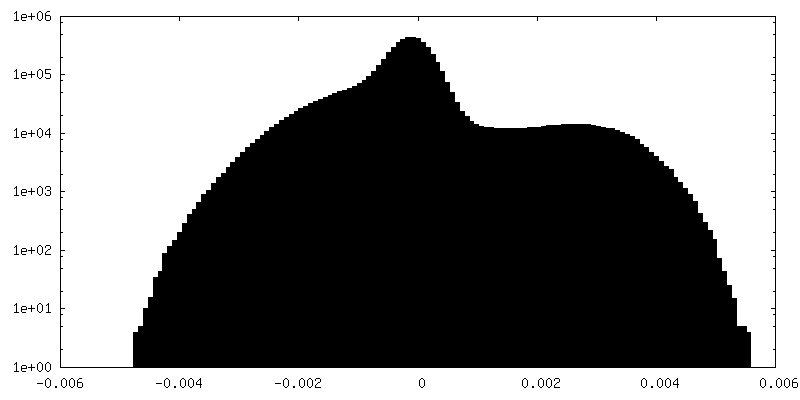
| Density Histograms |


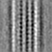
-Half map: Half map of a subtomogram average of an...
| File | emd_16307_half_map_1.map | ||||||||||||
|---|---|---|---|---|---|---|---|---|---|---|---|---|---|
| Annotation | Half map of a subtomogram average of an intracellular microtubule filament | ||||||||||||
| Projections & Slices |
| ||||||||||||
| Density Histograms |


-Half map: Half map of a subtomogram average of an...
| File | emd_16307_half_map_2.map | ||||||||||||
|---|---|---|---|---|---|---|---|---|---|---|---|---|---|
| Annotation | Half map of a subtomogram average of an intracellular microtubule filament | ||||||||||||
| Projections & Slices |
| ||||||||||||
| Density Histograms |


- Sample components
-Entire : Microtubule
| Entire | Name: Microtubule |
|---|---|
| Components |
|
-Supramolecule #1: Microtubule
| Supramolecule | Name: Microtubule / type: organelle_or_cellular_component / ID: 1 / Parent: 0 Details: Intracellular cytoskeletal filmanent composed of alpha and beta tubulin |
|---|---|
| Source (natural) | Organism: Homo sapiens (human) / Strain: U2OS |
-Experimental details
-Structure determination
| Method | cryo EM |
|---|---|
 Processing Processing | subtomogram averaging |
| Aggregation state | cell |
-Sample preparation
| Buffer | pH: 7.4 |
|---|---|
| Grid | Model: UltrAuFoil R1.2/1.3 / Material: GOLD / Mesh: 300 / Support film - Material: GOLD / Support film - topology: HOLEY ARRAY / Support film - Film thickness: 50 |
| Vitrification | Cryogen name: ETHANE / Chamber humidity: 99 % / Chamber temperature: 310 K / Instrument: LEICA EM GP |
| Details | Filaments were picked from inside vitrified cells |
- Electron microscopy
| Microscope | FEI TECNAI ARCTICA |
|---|---|
| Specialist optics | Energy filter - Name: GIF Bioquantum / Energy filter - Slit width: 20 eV |
| Image recording | Film or detector model: GATAN K3 BIOQUANTUM (6k x 4k) / Number real images: 3 / Average exposure time: 0.35 sec. / Average electron dose: 1.62 e/Å2 |
| Electron beam | Acceleration voltage: 200 kV / Electron source:  FIELD EMISSION GUN FIELD EMISSION GUN |
| Electron optics | Illumination mode: FLOOD BEAM / Imaging mode: BRIGHT FIELD / Nominal defocus max: 6.0 µm / Nominal defocus min: 2.0 µm / Nominal magnification: 31000 |
| Sample stage | Cooling holder cryogen: NITROGEN |
| Experimental equipment |  Model: Talos Arctica / Image courtesy: FEI Company |
+Image processing
-Atomic model buiding 1
| Refinement | Protocol: RIGID BODY FIT |
|---|
