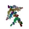
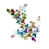
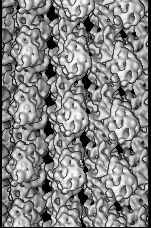
[日本語] English
- EMDB-1604: 9 Angstrom Structure of an AMPPNP Microtubule-Kinesin5 Complex -

+
データを開く


IDまたはキーワード:

読み込み中...

-
基本情報

登録情報
データベース: EMDB / ID: EMD-1604
タイトル9 Angstrom Structure of an AMPPNP Microtubule-Kinesin5 Complex
マップデータMicrotubule-kinesin5 complex
試料
  • 試料: Microtubule-KLP61F complex with AMPPNP
  • タンパク質・ペプチド: Microtubule
  • タンパク質・ペプチド: KLP61F
キーワードmicrotubule / tubulin / kinesin / kinesin-5 / KLP61F / Eg5 / mitosis
機能・相同性
機能・相同性情報


aster / plus-end directed microtubule sliding / fusome organization / fusome / COPI-dependent Golgi-to-ER retrograde traffic / Kinesins / centrosome separation / spindle elongation / mitotic spindle microtubule / microtubule bundle formation ...aster / plus-end directed microtubule sliding / fusome organization / fusome / COPI-dependent Golgi-to-ER retrograde traffic / Kinesins / centrosome separation / spindle elongation / mitotic spindle microtubule / microtubule bundle formation / plus-end-directed microtubule motor activity / positive regulation of Golgi to plasma membrane protein transport / mitotic centrosome separation / positive regulation of axon guidance / kinesin complex / microtubule associated complex / motile cilium / microtubule-based movement / mitotic spindle pole / Golgi organization / cytoskeletal motor activity / protein secretion / mitotic spindle assembly / microtubule-based process / mitotic spindle organization / spindle microtubule / structural constituent of cytoskeleton / microtubule cytoskeleton organization / spindle / neuron migration / mitotic spindle / mitotic cell cycle / microtubule cytoskeleton / microtubule binding / 加水分解酵素; 酸無水物に作用; GTPに作用・細胞または細胞小器官の運動に関与 / microtubule / hydrolase activity / protein heterodimerization activity / cell division / GTPase activity / GTP binding / endoplasmic reticulum / Golgi apparatus / ATP binding / metal ion binding / nucleus / cytoplasm
類似検索 - 分子機能
Kinesin-associated microtubule-binding domain / Kinesin-associated microtubule-binding / : / : / Kinesin motor domain signature. / Kinesin motor domain, conserved site / Kinesin motor domain / Kinesin motor domain profile. / Kinesin motor, catalytic domain. ATPase. / Kinesin motor domain ...Kinesin-associated microtubule-binding domain / Kinesin-associated microtubule-binding / : / : / Kinesin motor domain signature. / Kinesin motor domain, conserved site / Kinesin motor domain / Kinesin motor domain profile. / Kinesin motor, catalytic domain. ATPase. / Kinesin motor domain / Kinesin motor domain superfamily / Tubulin-beta mRNA autoregulation signal. / Alpha tubulin / Beta tubulin, autoregulation binding site / Beta tubulin / Tubulin / Tubulin, C-terminal / Tubulin C-terminal domain / Tubulin, conserved site / Tubulin subunits alpha, beta, and gamma signature. / Tubulin/FtsZ family, C-terminal domain / Tubulin/FtsZ-like, C-terminal domain / Tubulin/FtsZ, C-terminal / Tubulin/FtsZ, 2-layer sandwich domain / Tubulin/FtsZ family, GTPase domain / Tubulin/FtsZ family, GTPase domain / Tubulin/FtsZ, GTPase domain / Tubulin/FtsZ, GTPase domain superfamily / P-loop containing nucleoside triphosphate hydrolase
類似検索 - ドメイン・相同性
Tubulin alpha-1A chain / Tubulin beta chain / Kinesin-like protein Klp61F / Tubulin alpha-1D chain / Tubulin beta-2B chain
類似検索 - 構成要素
生物種Bos taurus (ウシ) / Drosophila melanogaster (キイロショウジョウバエ)
手法らせん対称体再構成法 / クライオ電子顕微鏡法 / 解像度: 9.4 Å
データ登録者Bodey AJ / Kikkawa M / Moores CA
引用ジャーナル: J Mol Biol / : 2009
タイトル: 9-Angström structure of a microtubule-bound mitotic motor.
著者: Andrew J Bodey / Masahide Kikkawa / Carolyn A Moores /
要旨: Kinesin-5 (K5) motors are important components of the microtubule (MT)-based cell division machinery and are targets for small-molecule inhibitors currently in cancer clinical trials. However, the ...Kinesin-5 (K5) motors are important components of the microtubule (MT)-based cell division machinery and are targets for small-molecule inhibitors currently in cancer clinical trials. However, the nature of the K5-MT interaction and the regulatory mechanisms that control it remain unclear. Using cryo-electron microscopy and image processing, we calculated the structure of a K5 motor bound to MTs at 9 A resolution, providing insight into this important interaction. Our reconstruction reveals the K5 motor domain in an ATP-like conformation in which MT binding induces the conserved nucleotide-sensing switch I and II loops to form a compact subdomain around the bound nucleotide. Our reconstruction also reveals a novel conformation for the K5-specific drug-binding loop 5, suggesting a possible role for it in switching K5s between force generation and diffusional modes of MT binding. Our data thus shed light on regulation of the interaction between spindle components important for chromosome segregation.
履歴
登録2009年2月26日-
ヘッダ(付随情報) 公開2009年3月9日-
マップ公開2009年4月1日-
更新2013年3月13日-
現状2013年3月13日処理サイト: PDBe / 状態: 公開

-
構造の表示

構造ビューアEMマップ:
SurfViewMolmilJmol/JSmol
添付画像

ダウンロードとリンク

-
マップ

ファイルダウンロード / ファイル: emd_1604.map.gz / 形式: CCP4 / 大きさ: 12.2 MB / タイプ: IMAGE STORED AS FLOATING POINT NUMBER (4 BYTES)
注釈Microtubule-kinesin5 complex
投影像・断面図

画像のコントロール

大きさ
明度
コントラスト
その他
Z (Sec.)Y (Row.)X (Col.)
1.4 Å/pix.
x 228 pix.
= 319.2 Å
1.4 Å/pix.
x 95 pix.
= 133. Å
1.4 Å/pix.
x 151 pix.
= 211.4 Å

表面

投影像

断面 (1/3)

断面 (1/2)

断面 (2/3)

画像は Spider により作成

これらの図は立方格子座標系で作成されたものです

ボクセルのサイズX=Y=Z: 1.4 Å
密度
表面レベル1: 0.5 / ムービー #1: 0
最小 - 最大-3.80928 - 5.50361
平均 (標準偏差)-0.00000000110627 (±1.0)
対称性空間群: 1
詳細

EMDB XML:

マップ形状
Axis orderXYZ
Origin-47-75-114
サイズ95151228
Spacing95151228
セルA: 211.4 Å / B: 133 Å / C: 319.2 Å
α=β=γ: 90 °

-
添付データ

-
試料の構成要素

-
全体 : Microtubule-KLP61F complex with AMPPNP

全体名称: Microtubule-KLP61F complex with AMPPNP
要素
  • 試料: Microtubule-KLP61F complex with AMPPNP
  • タンパク質・ペプチド: Microtubule
  • タンパク質・ペプチド: KLP61F

-
超分子 #1000: Microtubule-KLP61F complex with AMPPNP

超分子名称: Microtubule-KLP61F complex with AMPPNP / タイプ: sample / ID: 1000 / Number unique components: 2

-
分子 #1: Microtubule

分子名称: Microtubule / タイプ: protein_or_peptide / ID: 1 / Name.synonym: Microtubule / 組換発現: No / データベース: NCBI
由来(天然)生物種: Bos taurus (ウシ) / 別称: cattle / 組織: Brain

-
分子 #2: KLP61F

分子名称: KLP61F / タイプ: protein_or_peptide / ID: 2 / Name.synonym: KLP61F / 組換発現: Yes
由来(天然)生物種: Drosophila melanogaster (キイロショウジョウバエ)
別称: Fruit fly
分子量理論値: 42 KDa
組換発現生物種: Escherichia coli (大腸菌) / 組換プラスミド: pET-15b-TEV

-
実験情報

-
構造解析

手法クライオ電子顕微鏡法
解析らせん対称体再構成法
試料の集合状態filament

-
試料調製

緩衝液pH: 6.8
詳細: 80mM PIPES, 150mM NaCl, 7mM MgCl2, 1mM EGTA, 1mM beta-mercaptoethanol
グリッド詳細: Quantifoil grid
凍結凍結剤: ETHANE / 装置: HOMEMADE PLUNGER / 詳細: Vitrification instrument: Home made / 手法: Blot for 2-3 seconds before plunging

-
電子顕微鏡法

顕微鏡FEI TECNAI F20
アライメント法Legacy - 非点収差: Astigmatism corrected at 150,000 times magnification
撮影カテゴリ: FILM / フィルム・検出器のモデル: KODAK SO-163 FILM / デジタル化 - スキャナー: ZEISS SCAI / デジタル化 - サンプリング間隔: 7.0 µm / 実像数: 24 / 平均電子線量: 10 e/Å2 / ビット/ピクセル: 12
電子線加速電圧: 200 kV / 電子線源: FIELD EMISSION GUN
電子光学系倍率(補正後): 50000 / 照射モード: FLOOD BEAM / 撮影モード: BRIGHT FIELD / Cs: 2.0 mm / 最大 デフォーカス(公称値): 3.94 µm / 最小 デフォーカス(公称値): 1.08 µm / 倍率(公称値): 50000
試料ステージ試料ホルダー: Single tilt cryo / 試料ホルダーモデル: GATAN LIQUID NITROGEN
実験機器
モデル: Tecnai F20 / 画像提供: FEI Company

-
画像解析

最終 再構成アルゴリズム: OTHER / 解像度のタイプ: BY AUTHOR / 解像度: 9.4 Å / 解像度の算出法: FSC 0.5 CUT-OFF / ソフトウェア - 名称: Ruby-Helix
CTF補正詳細: Phase flipping, Wiener

-
原子モデル構築 1

初期モデルPDB ID:
ソフトウェア名称: UCSF Chimera
詳細Protocol: Rigid body. KLP61F, alpha-tubulin and beta-tubulin were all fitted separately
精密化空間: REAL / プロトコル: RIGID BODY FIT
得られたモデル

PDB-2wbe:
Kinesin-5-Tubulin Complex with AMPPNP

-
原子モデル構築 2

初期モデルPDB ID:
ソフトウェア名称: UCSF Chimera
詳細Protocol: Rigid body. KLP61F, alpha-tubulin and beta-tubulin were all fitted separately
精密化空間: REAL / プロトコル: RIGID BODY FIT
得られたモデル

PDB-2wbe:
Kinesin-5-Tubulin Complex with AMPPNP

-
原子モデル構築 3

初期モデルPDB ID:
ソフトウェア名称: UCSF Chimera
詳細Protocol: Rigid body. KLP61F, alpha-tubulin and beta-tubulin were all fitted separately
精密化空間: REAL / プロトコル: RIGID BODY FIT
得られたモデル

PDB-2wbe:
Kinesin-5-Tubulin Complex with AMPPNP

+
万見について

-
お知らせ

-
2022年2月9日: EMDBエントリの付随情報ファイルのフォーマットが新しくなりました

EMDBエントリの付随情報ファイルのフォーマットが新しくなりました

  • EMDBのヘッダファイルのバージョン3が、公式のフォーマットとなりました。
  • これまでは公式だったバージョン1.9は、アーカイブから削除されます。

関連情報:EMDBヘッダ

外部リンク:wwPDBはEMDBデータモデルのバージョン3へ移行します

-
2020年8月12日: 新型コロナ情報

新型コロナ情報

URL: https://pdbj.org/emnavi/covid19.php

新ページ: EM Navigatorに新型コロナウイルスの特設ページを開設しました。

関連情報:Covid-19情報 / 2020年3月5日: 新型コロナウイルスの構造データ

+
2020年3月5日: 新型コロナウイルスの構造データ

新型コロナウイルスの構造データ

関連情報:万見生物種 / 2020年8月12日: 新型コロナ情報

外部リンク:COVID-19特集ページ - PDBj / 今月の分子2020年2月:コロナウイルスプロテーアーゼ

+
2019年1月31日: EMDBのIDの桁数の変更

EMDBのIDの桁数の変更

  • EMDBエントリに付与されているアクセスコード(EMDB-ID)は4桁の数字(例、EMD-1234)でしたが、間もなく枯渇します。これまでの4桁のID番号は4桁のまま変更されませんが、4桁の数字を使い切った後に発行されるIDは5桁以上の数字(例、EMD-12345)になります。5桁のIDは2019年の春頃から発行される見通しです。
  • EM Navigator/万見では、接頭語「EMD-」は省略されています。

関連情報:Q: 「EMD」とは何ですか? / 万見/EM NavigatorにおけるID/アクセスコードの表記

外部リンク:EMDB Accession Codes are Changing Soon! / PDBjへお問い合わせ

+
2017年7月12日: PDB大規模アップデート

PDB大規模アップデート

  • 新バージョンのPDBx/mmCIF辞書形式に基づくデータがリリースされました。
  • 今回の更新はバージョン番号が4から5になる大規模なもので、全エントリデータの書き換えが行われる「Remediation」というアップデートに該当します。
  • このバージョンアップで、電子顕微鏡の実験手法に関する多くの項目の書式が改定されました(例:em_softwareなど)。
  • EM NavigatorとYorodumiでも、この改定に基づいた表示内容になります。

外部リンク:wwPDB Remediation / OneDepデータ基準に準拠した、より強化された内容のモデル構造ファイルが、PDBアーカイブで公開されました。

-
万見 (Yorodumi)

幾万の構造データを、幾万の視点から

  • 万見(Yorodumi)は、EMDB/PDB/SASBDBなどの構造データを閲覧するためのページです。
  • EM Navigatorの詳細ページの後継、Omokage検索のフロントエンドも兼ねています。

関連情報:EMDB / PDB / SASBDB / 3つのデータバンクの比較 / 万見検索 / 2016年8月31日: 新しいEM Navigatorと万見 / 万見文献 / Jmol/JSmol / 機能・相同性情報 / 新しいEM Navigatorと万見の変更点

他の情報も見る