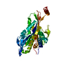
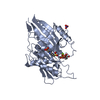
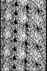
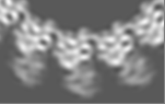
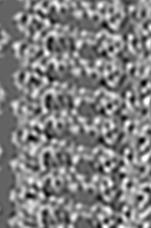
Journal: J Mol Biol / Year: 2009 Title: 9-Angström structure of a microtubule-bound mitotic motor. Authors: Andrew J Bodey / Masahide Kikkawa / Carolyn A Moores / Abstract: Kinesin-5 (K5) motors are important components of the microtubule (MT)-based cell division machinery and are targets for small-molecule inhibitors currently in cancer clinical trials. However, the ...Kinesin-5 (K5) motors are important components of the microtubule (MT)-based cell division machinery and are targets for small-molecule inhibitors currently in cancer clinical trials. However, the nature of the K5-MT interaction and the regulatory mechanisms that control it remain unclear. Using cryo-electron microscopy and image processing, we calculated the structure of a K5 motor bound to MTs at 9 A resolution, providing insight into this important interaction. Our reconstruction reveals the K5 motor domain in an ATP-like conformation in which MT binding induces the conserved nucleotide-sensing switch I and II loops to form a compact subdomain around the bound nucleotide. Our reconstruction also reveals a novel conformation for the K5-specific drug-binding loop 5, suggesting a possible role for it in switching K5s between force generation and diffusional modes of MT binding. Our data thus shed light on regulation of the interaction between spindle components important for chromosome segregation.
Cryogen name: ETHANE / Instrument: HOMEMADE PLUNGER / Details: Vitrification instrument: Home made / Method: Blot for 2-3 seconds before plunging
-
Electron microscopy
Microscope
FEI TECNAI F20
Alignment procedure
Legacy - Astigmatism: Astigmatism corrected at 150,000 times magnification
Image recording
Category: FILM / Film or detector model: KODAK SO-163 FILM / Digitization - Scanner: ZEISS SCAI / Digitization - Sampling interval: 7.0 µm / Number real images: 24 / Average electron dose: 10 e/Å2 / Bits/pixel: 12
Electron beam
Acceleration voltage: 200 kV / Electron source: FIELD EMISSION GUN
In the structure databanks used in Yorodumi, some data are registered as the other names, "COVID-19 virus" and "2019-nCoV". Here are the details of the virus and the list of structure data.
Jan 31, 2019. EMDB accession codes are about to change! (news from PDBe EMDB page)
EMDB accession codes are about to change! (news from PDBe EMDB page)
The allocation of 4 digits for EMDB accession codes will soon come to an end. Whilst these codes will remain in use, new EMDB accession codes will include an additional digit and will expand incrementally as the available range of codes is exhausted. The current 4-digit format prefixed with “EMD-” (i.e. EMD-XXXX) will advance to a 5-digit format (i.e. EMD-XXXXX), and so on. It is currently estimated that the 4-digit codes will be depleted around Spring 2019, at which point the 5-digit format will come into force.
The EM Navigator/Yorodumi systems omit the EMD- prefix.
Related info.:Q: What is EMD? / ID/Accession-code notation in Yorodumi/EM Navigator
Yorodumi is a browser for structure data from EMDB, PDB, SASBDB, etc.
This page is also the successor to EM Navigator detail page, and also detail information page/front-end page for Omokage search.
The word "yorodu" (or yorozu) is an old Japanese word meaning "ten thousand". "mi" (miru) is to see.
Related info.:EMDB / PDB / SASBDB / Comparison of 3 databanks / Yorodumi Search / Aug 31, 2016. New EM Navigator & Yorodumi / Yorodumi Papers / Jmol/JSmol / Function and homology information / Changes in new EM Navigator and Yorodumi
 Movie
Movie Controller


 Open data
 Basic information
 Map data
 Sample
 Keywords
 Function and homology information

 Authors
 Citation
 Structure visualization
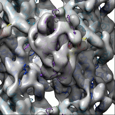
 Downloads & links
 1604.gif
 http://ftp.pdbj.org/pub/emdb/structures/EMD-1604
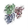


 Z (Sec.)
 Y (Row.)
 X (Col.)


 Sample components
 Processing
 Electron microscopy
 FIELD EMISSION GUN