 Movie
Movie Controller

[English] 日本語
 Yorodumi
- EMDB-16032: Structure of mouse heavy chain apoferritin solved by subtomogram ... -
+ Open data

- Basic information
| Entry |  | |||||||||
|---|---|---|---|---|---|---|---|---|---|---|
| Title | Structure of mouse heavy chain apoferritin solved by subtomogram averaging of 100 tilt series in Warp/M | |||||||||
 Map data Map data | ||||||||||
 Sample Sample |
| |||||||||
 Keywords Keywords | Apoferritin / Subtomogram averaging / STA / METAL BINDING PROTEIN | |||||||||
| Biological species |  | |||||||||
| Method | subtomogram averaging / cryo EM / Resolution: 1.6 Å | |||||||||
 Authors Authors | Obr M / Yang W / Karia D / Koh FA / Kotecha A | |||||||||
| Funding support | 1 items
| |||||||||
 Citation Citation | Journal: To Be Published Title: Structure of mouse heavy chain apoferritin solved by subtomogram averaging of 10 tilt series in Warp/M Authors: Obr M / Yang W / Karia D / Koh FA / Kotecha A | |||||||||
| History |
|
- Structure visualization
| Supplemental images |
|---|

- Downloads & links
-EMDB archive
| Map data | emd_16032.map.gz | 23 MB |  EMDB map data format EMDB map data format | |
|---|---|---|---|---|
| Header (meta data) | emd-16032-v30.xmlemd-16032.xml | 15.1 KB 15.1 KB | Display Display | EMDB header |
| FSC (resolution estimation) | emd_16032_fsc.xml | 13.5 KB | Display | FSC data file |
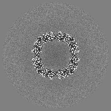
| Images | 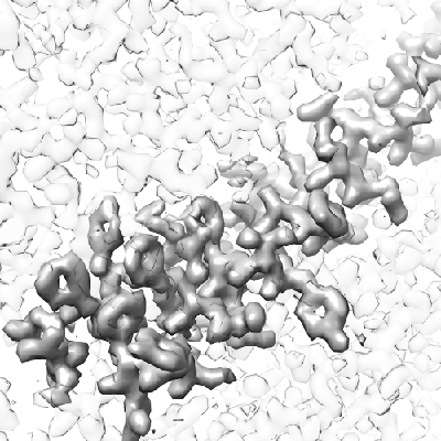 emd_16032.png emd_16032.png | 154.2 KB | ||
| Filedesc metadata | emd-16032.cif.gz | 4.4 KB | ||
| Others | emd_16032_half_map_1.map.gzemd_16032_half_map_2.map.gz | 110.9 MB 110.9 MB | ||
| Archive directory |  http://ftp.pdbj.org/pub/emdb/structures/EMD-16032ftp://ftp.pdbj.org/pub/emdb/structures/EMD-16032 http://ftp.pdbj.org/pub/emdb/structures/EMD-16032ftp://ftp.pdbj.org/pub/emdb/structures/EMD-16032 | HTTPS FTP |
-Related structure data

-Links
| EMDB pages | EMDB (EBI/PDBe) / EMDataResource |
|---|
-Map
| File | Download / File: emd_16032.map.gz / Format: CCP4 / Size: 216 MB / Type: IMAGE STORED AS FLOATING POINT NUMBER (4 BYTES) | ||||||||||||||||||||||||||||||||||||
|---|---|---|---|---|---|---|---|---|---|---|---|---|---|---|---|---|---|---|---|---|---|---|---|---|---|---|---|---|---|---|---|---|---|---|---|---|---|
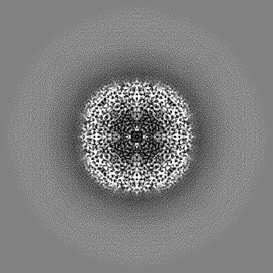
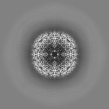
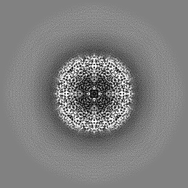
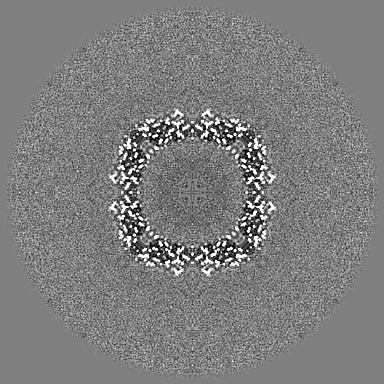
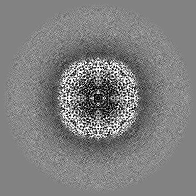
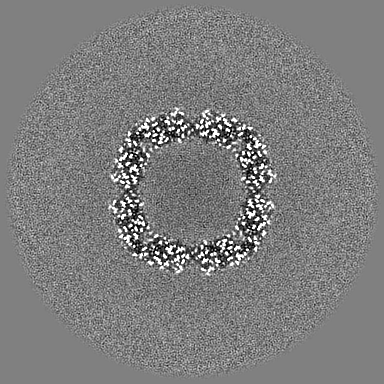
| Projections & slices | Image control
Images are generated by Spider. | ||||||||||||||||||||||||||||||||||||
| Voxel size | X=Y=Z: 0.729 Å | ||||||||||||||||||||||||||||||||||||
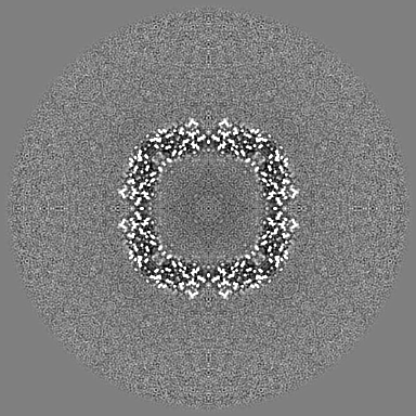
| Density |
| ||||||||||||||||||||||||||||||||||||
| Symmetry | Space group: 1 | ||||||||||||||||||||||||||||||||||||
| Details | EMDB XML:
|
 Z (Sec.)
 Y (Row.)
 X (Col.)


-Supplemental data
-Half map: #1
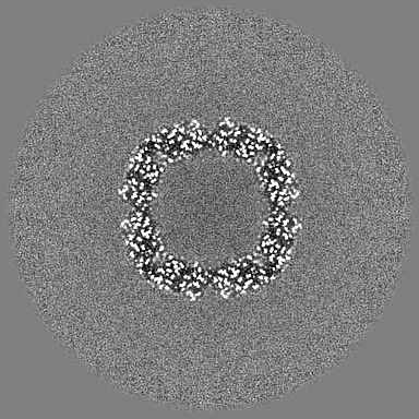
| File | emd_16032_half_map_1.map | ||||||||||||
|---|---|---|---|---|---|---|---|---|---|---|---|---|---|
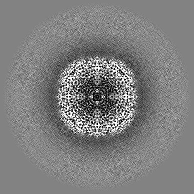
| Projections & Slices |
| ||||||||||||
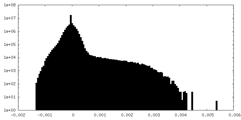
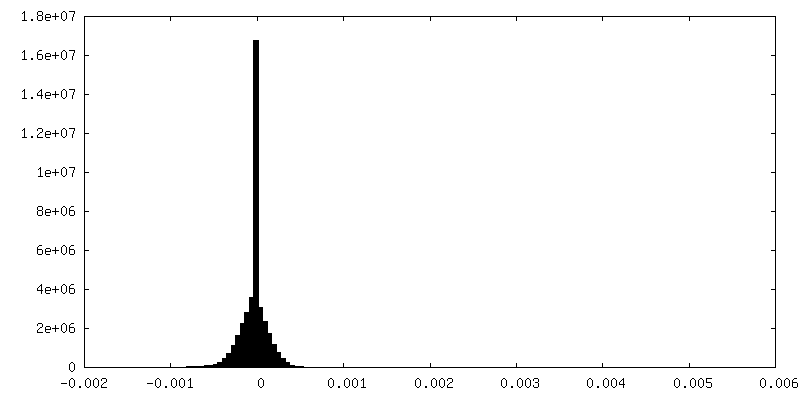
| Density Histograms |

-Half map: #2
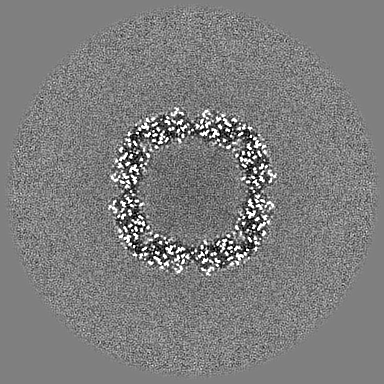
| File | emd_16032_half_map_2.map | ||||||||||||
|---|---|---|---|---|---|---|---|---|---|---|---|---|---|
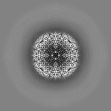
| Projections & Slices |
| ||||||||||||
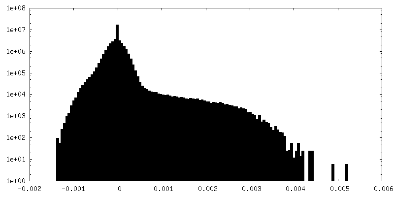
| Density Histograms |
- Sample components
-Entire : Ferritin heavy chain
| Entire | Name: Ferritin heavy chain |
|---|---|
| Components |
|
-Supramolecule #1: Ferritin heavy chain
| Supramolecule | Name: Ferritin heavy chain / type: complex / ID: 1 / Parent: 0 |
|---|---|
| Source (natural) | Organism: |
-Experimental details
-Structure determination
| Method | cryo EM |
|---|---|
 Processing Processing | subtomogram averaging |
| Aggregation state | particle |
-Sample preparation
| Concentration | 5 mg/mL | |||||||||
|---|---|---|---|---|---|---|---|---|---|---|
| Buffer | pH: 7.5 Component:
| |||||||||
| Vitrification | Cryogen name: ETHANE / Chamber humidity: 100 % / Chamber temperature: 277 K / Instrument: FEI VITROBOT MARK IV |
- Electron microscopy
| Microscope | TFS KRIOS |
|---|---|
| Specialist optics | Energy filter - Name: TFS Selectris X / Energy filter - Slit width: 10 eV |
| Details | Tilt series acquired with Hagen scheme +-48 degrees with 3 increment |
| Image recording | Film or detector model: FEI FALCON IV (4k x 4k) / Digitization - Dimensions - Width: 4096 pixel / Digitization - Dimensions - Height: 4096 pixel / Number grids imaged: 1 / Average exposure time: 0.5 sec. / Average electron dose: 3.5 e/Å2 / Details: Imaged with TFS Selectris X and Falcon 4i |
| Electron beam | Acceleration voltage: 300 kV / Electron source:  FIELD EMISSION GUN FIELD EMISSION GUN |
| Electron optics | C2 aperture diameter: 50.0 µm / Illumination mode: FLOOD BEAM / Imaging mode: BRIGHT FIELD / Cs: 2.7 mm / Nominal defocus max: 3.0 µm / Nominal defocus min: 1.0 µm / Nominal magnification: 165000 |
| Sample stage | Specimen holder model: FEI TITAN KRIOS AUTOGRID HOLDER / Cooling holder cryogen: NITROGEN |
| Experimental equipment |  Model: Titan Krios / Image courtesy: FEI Company |
-Image processing
| Final reconstruction | Applied symmetry - Point group: O (octahedral) / Algorithm: FOURIER SPACE / Resolution.type: BY AUTHOR / Resolution: 1.6 Å / Resolution method: FSC 0.143 CUT-OFF / Software - Name: Warp (ver. nb20201104) / Software - details: M / Number subtomograms used: 31478 |
|---|---|
| Extraction | Number tomograms: 100 / Number images used: 31478 / Method: Automatic 2D picking / Software: (Name: RELION (ver. 4), Warp (ver. nb20201104)) Details: Particles picked in 2D using the 0 degrees tilt image. Z coordinate was determined using ice layer fitting. |
| Final angle assignment | Type: PROJECTION MATCHING / Software - Name: Warp (ver. nb20201104) / Software - details: M |
| FSC plot (resolution estimation) | 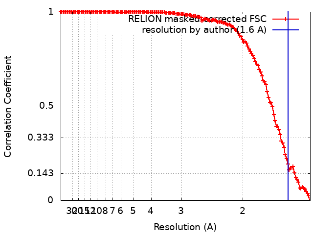 |
