 ムービー
ムービー コントローラー


+ データを開く

- 基本情報
基本情報
| 登録情報 | データベース: EMDB / ID: EMD-1597 | |||||||||
|---|---|---|---|---|---|---|---|---|---|---|
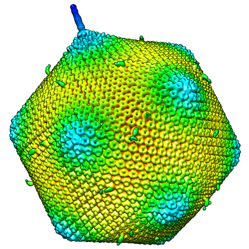
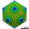
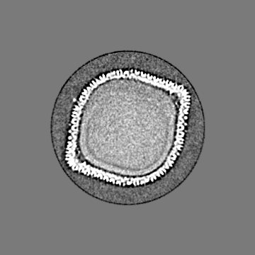
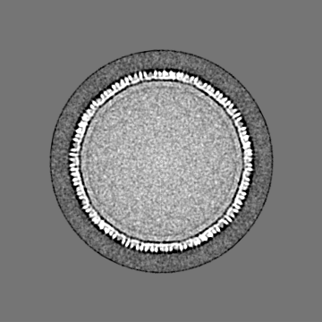
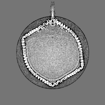
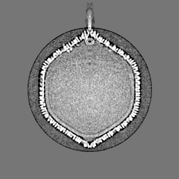
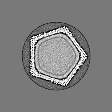
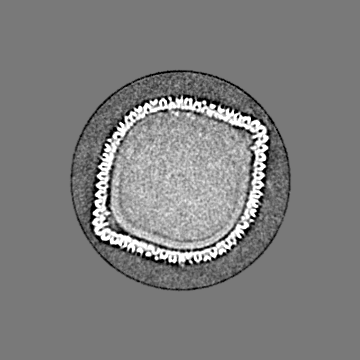
| タイトル | 5-fold averaged density map of Paramecium bursaria Chlorella virus-1 (PBCV-1). | |||||||||
 マップデータ マップデータ | 5-fold average density map of Paramecium bursaria Chlorella virus-1 (PBCV-1). | |||||||||
 試料 試料 |
| |||||||||
 キーワード キーワード | PBCV-1 / eukaryotic virus / tail / pocket / 5-fold averaged | |||||||||
| 生物種 |  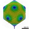 Paramecium bursaria Chlorella virus 1 (ウイルス) Paramecium bursaria Chlorella virus 1 (ウイルス) | |||||||||
| 手法 | 単粒子再構成法 / クライオ電子顕微鏡法 / 解像度: 22.0 Å | |||||||||
 データ登録者 データ登録者 | Cherrier MV / Kostyuchenko VA / Xiao C / Bowman VD / Battisti AJ / Yan X / Chipman PR / Baker TS / Van Etten JL / Rossmann MG | |||||||||
 引用 引用 | ジャーナル: Proc Natl Acad Sci U S A / 年: 2009 タイトル: An icosahedral algal virus has a complex unique vertex decorated by a spike. 著者: Mickaël V Cherrier / Victor A Kostyuchenko / Chuan Xiao / Valorie D Bowman / Anthony J Battisti / Xiaodong Yan / Paul R Chipman / Timothy S Baker / James L Van Etten / Michael G Rossmann /  要旨: Paramecium bursaria Chlorella virus-1 is an icosahedrally shaped, 1,900-A-diameter virus that infects unicellular eukaryotic green algae. A 5-fold symmetric, 3D reconstruction using cryoelectron ...Paramecium bursaria Chlorella virus-1 is an icosahedrally shaped, 1,900-A-diameter virus that infects unicellular eukaryotic green algae. A 5-fold symmetric, 3D reconstruction using cryoelectron microscopy images has now shown that the quasiicosahedral virus has a unique vertex, with a pocket on the inside and a spike structure on the outside of the capsid. The pocket might contain enzymes for use in the initial stages of infection. The unique vertex consists of virally coded proteins, some of which have been identified. Comparison of shape, size, and location of the spike with similar features in bacteriophages T4 and P22 suggests that the spike might be a cell-puncturing device. Similar asymmetric features may have been missed in previous analyses of many other viruses that had been assumed to be perfectly icosahedral. | |||||||||
| 履歴 |
|
- 構造の表示
構造の表示
| ムービー |
 ムービービューア ムービービューア |
|---|---|
| 構造ビューア | EMマップ: SurfViewMolmilJmol/JSmol |
| 添付画像 |
- ダウンロードとリンク
-EMDBアーカイブ
| マップデータ | emd_1597.map.gz | 16.3 MB | EMDBマップデータ形式 | |
|---|---|---|---|---|
| ヘッダ (付随情報) | emd-1597-v30.xmlemd-1597.xml | 11.7 KB 11.7 KB | 表示 表示 | EMDBヘッダ |
| 画像 |  EMD-1597-image.png EMD-1597-image.png | 374.6 KB | ||
| アーカイブディレクトリ |  http://ftp.pdbj.org/pub/emdb/structures/EMD-1597ftp://ftp.pdbj.org/pub/emdb/structures/EMD-1597 http://ftp.pdbj.org/pub/emdb/structures/EMD-1597ftp://ftp.pdbj.org/pub/emdb/structures/EMD-1597 | HTTPS FTP |
-検証レポート
| 文書・要旨 | emd_1597_validation.pdf.gz | 251.8 KB | 表示 | EMDB検証レポート |
|---|---|---|---|---|
| 文書・詳細版 | emd_1597_full_validation.pdf.gz | 250.9 KB | 表示 | |
| XML形式データ | emd_1597_validation.xml.gz | 6.6 KB | 表示 | |
| アーカイブディレクトリ | https://ftp.pdbj.org/pub/emdb/validation_reports/EMD-1597ftp://ftp.pdbj.org/pub/emdb/validation_reports/EMD-1597 | HTTPS FTP |
-関連構造データ
| 類似構造データ |
|---|
-リンク
| EMDBのページ | EMDB (EBI/PDBe) / EMDataResource |
|---|
-マップ
| ファイル | ダウンロード / ファイル: emd_1597.map.gz / 形式: CCP4 / 大きさ: 173.8 MB / タイプ: IMAGE STORED AS FLOATING POINT NUMBER (4 BYTES) | ||||||||||||||||||||||||||||||||||||||||||||||||||||||||||||||||||||
|---|---|---|---|---|---|---|---|---|---|---|---|---|---|---|---|---|---|---|---|---|---|---|---|---|---|---|---|---|---|---|---|---|---|---|---|---|---|---|---|---|---|---|---|---|---|---|---|---|---|---|---|---|---|---|---|---|---|---|---|---|---|---|---|---|---|---|---|---|---|
| 注釈 | 5-fold average density map of Paramecium bursaria Chlorella virus-1 (PBCV-1). | ||||||||||||||||||||||||||||||||||||||||||||||||||||||||||||||||||||
| 投影像・断面図 | 画像のコントロール
画像は Spider により作成 | ||||||||||||||||||||||||||||||||||||||||||||||||||||||||||||||||||||
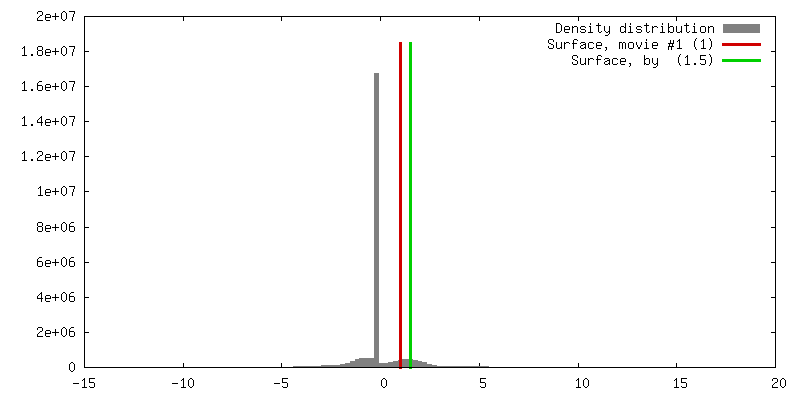
| ボクセルのサイズ | X=Y=Z: 8 Å | ||||||||||||||||||||||||||||||||||||||||||||||||||||||||||||||||||||
| 密度 |
| ||||||||||||||||||||||||||||||||||||||||||||||||||||||||||||||||||||
| 対称性 | 空間群: 1 | ||||||||||||||||||||||||||||||||||||||||||||||||||||||||||||||||||||
| 詳細 | EMDB XML:
CCP4マップ ヘッダ情報:
| ||||||||||||||||||||||||||||||||||||||||||||||||||||||||||||||||||||
 Z (Sec.)
 Y (Row.)
 X (Col.)


-添付データ
- 試料の構成要素
-全体 : Paramecium bursaria Chlorella virus-1 (PBCV-1)
| 全体 | 名称: Paramecium bursaria Chlorella virus-1 (PBCV-1) |
|---|---|
| 要素 |
|
-超分子 #1000: Paramecium bursaria Chlorella virus-1 (PBCV-1)
| 超分子 | 名称: Paramecium bursaria Chlorella virus-1 (PBCV-1) / タイプ: sample / ID: 1000 / Number unique components: 1 |
|---|
-超分子 #1: Paramecium bursaria Chlorella virus 1
| 超分子 | 名称: Paramecium bursaria Chlorella virus 1 / タイプ: virus / ID: 1 / Name.synonym: PBCV-1 / NCBI-ID: 10506 / 生物種: Paramecium bursaria Chlorella virus 1 / ウイルスタイプ: VIRION / ウイルス・単離状態: STRAIN / ウイルス・エンベロープ: Yes / ウイルス・中空状態: No / Syn species name: PBCV-1 |
|---|---|
| 宿主 | 生物種: Chlorella NC64A / 別称: ALGAE |
| ウイルス殻 | Shell ID: 1 / 名称: VP54 / 直径: 1900 Å |
-実験情報
-構造解析
| 手法 | クライオ電子顕微鏡法 |
|---|---|
 解析 解析 | 単粒子再構成法 |
| 試料の集合状態 | particle |
-試料調製
| 凍結 | 凍結剤: ETHANE / 装置: HOMEMADE PLUNGER 詳細: Vitrification instrument: Guillotine-style plunge freezing device 手法: A small vial of ethane is placed inside a larger liquid nitrogen reservoir. The grid holding a few microliters of the sample is held in place at the bottom of a plunger by the means of fine ...手法: A small vial of ethane is placed inside a larger liquid nitrogen reservoir. The grid holding a few microliters of the sample is held in place at the bottom of a plunger by the means of fine tweezers. Once the ethane in the vial is completely frozen, it needs to be slightly melted. When the liquid ethane is ready, a piece of filter paper is then pressed against the sample to blot of excess buffer, sufficient to leave a thin layer on the grid. After a predetermined time, the filter paper is removed, and the plunger is allowed to drop into the liquid ethane. Once the grid enters the liquid ethane, the sample is rapidly frozen, and the grid is transferred under liquid nitrogen to a storage box immersed liquid nitrogen for later use in the microscope. |
|---|
- 電子顕微鏡法 #1
| Microscopy ID | 1 |
|---|---|
| 顕微鏡 | FEI/PHILIPS CM200FEG |
| 温度 | 平均: 98 K |
| アライメント法 | Legacy - Electron beam tilt params: 0 |
| 撮影 | カテゴリ: FILM / フィルム・検出器のモデル: KODAK SO-163 FILM / デジタル化 - スキャナー: ZEISS SCAI / デジタル化 - サンプリング間隔: 7.0 µm / 実像数: 228 / 平均電子線量: 22 e/Å2 詳細: All particles were scaled to 4.0A per pixel, then bin twice to 8.0A per pixel. |
| Tilt angle min | 0 |
| Tilt angle max | 0 |
| 電子線 | 加速電圧: 200 kV / 電子線源:  FIELD EMISSION GUN FIELD EMISSION GUN |
| 電子光学系 | 照射モード: FLOOD BEAM / 撮影モード: BRIGHT FIELD / 最大 デフォーカス(公称値): 4.473 µm / 最小 デフォーカス(公称値): 1.302 µm / 倍率(公称値): 38000 |
| 試料ステージ | 試料ホルダー: EUCENTRIC / 試料ホルダーモデル: GATAN LIQUID NITROGEN |
-電子顕微鏡法 #2
| Microscopy ID | 2 |
|---|---|
| 顕微鏡 | FEI/PHILIPS CM300FEG/T |
| 温度 | 平均: 98 K |
| アライメント法 | Legacy - 非点収差: live FFT / Legacy - Electron beam tilt params: 0 |
| 撮影 | カテゴリ: FILM / フィルム・検出器のモデル: KODAK SO-163 FILM / デジタル化 - スキャナー: ZEISS SCAI / デジタル化 - サンプリング間隔: 7.0 µm / 実像数: 228 / 平均電子線量: 24 e/Å2 詳細: All particles were scaled to 4.0A per pixel, then bin twice to 8.0A per pixel. |
| Tilt angle min | 0 |
| Tilt angle max | 0 |
| 電子線 | 加速電圧: 300 kV / 電子線源: FIELD EMISSION GUN |
| 電子光学系 | 照射モード: FLOOD BEAM / 撮影モード: BRIGHT FIELD / Cs: 2 mm / 最大 デフォーカス(公称値): 4.728 µm / 最小 デフォーカス(公称値): 0.767 µm / 倍率(公称値): 45000 |
| 試料ステージ | 試料ホルダー: EUCENTRIC / 試料ホルダーモデル: GATAN LIQUID NITROGEN |
-画像解析
| CTF補正 | 詳細: Each particle |
|---|---|
| 最終 再構成 | 想定した対称性 - 点群: C5 (5回回転対称) / 解像度のタイプ: BY AUTHOR / 解像度: 22.0 Å / 解像度の算出法: FSC 0.5 CUT-OFF / ソフトウェア - 名称: Spider, XMIPP / 詳細: The 3D map was only 5 fold averaged. / 使用した粒子像数: 5149 |
