 Movie
Movie Controller

[English] 日本語
 Yorodumi
- EMDB-15951: Tomogram of an EBOV-infected Huh7 cell showing a late endosome wi... -
+ Open data

- Basic information
| Entry | 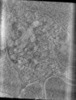 | |||||||||
|---|---|---|---|---|---|---|---|---|---|---|
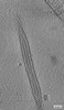
| Title | Tomogram of an EBOV-infected Huh7 cell showing a late endosome with internalized EBOV particles | |||||||||
 Map data Map data | Tomogram of a late endosome containing EBOV particles | |||||||||
 Sample Sample |
| |||||||||
 Keywords Keywords | Ebola virus / late endosome / low pH / ENDOCYTOSIS | |||||||||
| Biological species |   Ebola virus - Mayinga, Zaire, 1976 Ebola virus - Mayinga, Zaire, 1976 | |||||||||
| Method | electron tomography / cryo EM | |||||||||
 Authors Authors | Winter SL / Chlanda P | |||||||||
| Funding support |  Germany, 1 items Germany, 1 items
| |||||||||
 Citation Citation | Journal: EMBO J / Year: 2023 Title: The Ebola virus VP40 matrix layer undergoes endosomal disassembly essential for membrane fusion. Authors: Sophie L Winter / Gonen Golani / Fabio Lolicato / Melina Vallbracht / Keerthihan Thiyagarajah / Samy Sid Ahmed / Christian Lüchtenborg / Oliver T Fackler / Britta Brügger / Thomas Hoenen / ...Authors: Sophie L Winter / Gonen Golani / Fabio Lolicato / Melina Vallbracht / Keerthihan Thiyagarajah / Samy Sid Ahmed / Christian Lüchtenborg / Oliver T Fackler / Britta Brügger / Thomas Hoenen / Walter Nickel / Ulrich S Schwarz / Petr Chlanda /  Abstract: Ebola viruses (EBOVs) assemble into filamentous virions, whose shape and stability are determined by the matrix viral protein 40 (VP40). Virus entry into host cells occurs via membrane fusion in late ...Ebola viruses (EBOVs) assemble into filamentous virions, whose shape and stability are determined by the matrix viral protein 40 (VP40). Virus entry into host cells occurs via membrane fusion in late endosomes; however, the mechanism of how the remarkably long virions undergo uncoating, including virion disassembly and nucleocapsid release into the cytosol, remains unknown. Here, we investigate the structural architecture of EBOVs entering host cells and discover that the VP40 matrix disassembles prior to membrane fusion. We reveal that VP40 disassembly is caused by the weakening of VP40-lipid interactions driven by low endosomal pH that equilibrates passively across the viral envelope without a dedicated ion channel. We further show that viral membrane fusion depends on VP40 matrix integrity, and its disassembly reduces the energy barrier for fusion stalk formation. Thus, pH-driven structural remodeling of the VP40 matrix acts as a molecular switch coupling viral matrix uncoating to membrane fusion during EBOV entry. | |||||||||
| History |
|
- Structure visualization
| Supplemental images |
|---|

- Downloads & links
-EMDB archive
| Map data | emd_15951.map.gz | 348.1 MB |  EMDB map data format EMDB map data format | |
|---|---|---|---|---|
| Header (meta data) | emd-15951-v30.xmlemd-15951.xml | 8.6 KB 8.6 KB | Display Display | EMDB header |
| Images | 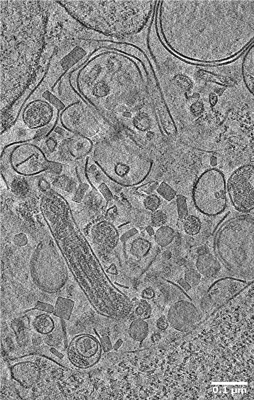 emd_15951.png emd_15951.png | 165.4 KB | ||
| Archive directory |  http://ftp.pdbj.org/pub/emdb/structures/EMD-15951ftp://ftp.pdbj.org/pub/emdb/structures/EMD-15951 http://ftp.pdbj.org/pub/emdb/structures/EMD-15951ftp://ftp.pdbj.org/pub/emdb/structures/EMD-15951 | HTTPS FTP |
-Validation report
| Summary document | emd_15951_validation.pdf.gz | 517.6 KB | Display | EMDB validaton report |
|---|---|---|---|---|
| Full document | emd_15951_full_validation.pdf.gz | 517.1 KB | Display | |
| Data in XML | emd_15951_validation.xml.gz | 4.4 KB | Display | |
| Data in CIF | emd_15951_validation.cif.gz | 5.3 KB | Display | |
| Arichive directory | https://ftp.pdbj.org/pub/emdb/validation_reports/EMD-15951ftp://ftp.pdbj.org/pub/emdb/validation_reports/EMD-15951 | HTTPS FTP |
-Related structure data
-Links
| EMDB pages | EMDB (EBI/PDBe) / EMDataResource |
|---|
-Map
| File | Download / File: emd_15951.map.gz / Format: CCP4 / Size: 455.8 MB / Type: IMAGE STORED AS SIGNED BYTE | ||||||||||||||||||||||||||||||||
|---|---|---|---|---|---|---|---|---|---|---|---|---|---|---|---|---|---|---|---|---|---|---|---|---|---|---|---|---|---|---|---|---|---|
| Annotation | Tomogram of a late endosome containing EBOV particles | ||||||||||||||||||||||||||||||||
| Projections & slices | Image control
Images are generated by Spider. generated in cubic-lattice coordinate | ||||||||||||||||||||||||||||||||
| Voxel size |
| ||||||||||||||||||||||||||||||||
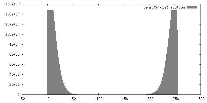
| Density |
| ||||||||||||||||||||||||||||||||
| Symmetry | Space group: 1 | ||||||||||||||||||||||||||||||||
| Details | EMDB XML:
|
 Z (Sec.)
 Y (Row.)
 X (Col.)


-Supplemental data
- Sample components
-Entire : Ebola virus - Mayinga, Zaire, 1976
| Entire | Name: Ebola virus - Mayinga, Zaire, 1976 |
|---|---|
| Components |
|
-Supramolecule #1: Ebola virus - Mayinga, Zaire, 1976
| Supramolecule | Name: Ebola virus - Mayinga, Zaire, 1976 / type: virus / ID: 1 / Parent: 0 / NCBI-ID: 128952 / Sci species name: Ebola virus - Mayinga, Zaire, 1976 / Virus type: VIRUS-LIKE PARTICLE / Virus isolate: OTHER / Virus enveloped: Yes / Virus empty: Yes |
|---|
-Experimental details
-Structure determination
| Method | cryo EM |
|---|---|
 Processing Processing | electron tomography |
| Aggregation state | particle |
-Sample preparation
| Buffer | pH: 7.4 |
|---|---|
| Vitrification | Cryogen name: ETHANE |
| Sectioning | Other: NO SECTIONING |
| Fiducial marker | Manufacturer: Aurion / Diameter: 10 nm |
- Electron microscopy
| Microscope | FEI TITAN KRIOS |
|---|---|
| Image recording | Film or detector model: GATAN K3 BIOQUANTUM (6k x 4k) / Average electron dose: 3.0 e/Å2 |
| Electron beam | Acceleration voltage: 300 kV / Electron source:  FIELD EMISSION GUN FIELD EMISSION GUN |
| Electron optics | Illumination mode: FLOOD BEAM / Imaging mode: BRIGHT FIELD / Cs: 2.7 mm / Nominal defocus max: 4.0 µm / Nominal defocus min: 4.0 µm / Nominal magnification: 33000 |
| Experimental equipment |  Model: Titan Krios / Image courtesy: FEI Company |
-Image processing
| Final reconstruction | Software - Name: IMOD / Number images used: 41 |
|---|
