 Movie
Movie Controller

[English] 日本語
 Yorodumi
- EMDB-15928: Cryo-electron tomogram of VeroE6 cells transfected with HA-GG>AA-... -
+ Open data

- Basic information
| Entry |  | |||||||||
|---|---|---|---|---|---|---|---|---|---|---|
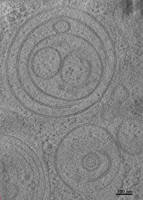
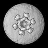
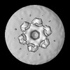
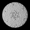
| Title | Cryo-electron tomogram of VeroE6 cells transfected with HA-GG>AA-nsp4-V5, plunge-frozen at 16 hpt and subjected to cryo-FIB milling. | |||||||||
 Map data Map data | ||||||||||
 Sample Sample |
| |||||||||
 Keywords Keywords | nsp3-4 protein / VIRAL PROTEIN | |||||||||
| Biological species |   Severe acute respiratory syndrome coronavirus 2 Severe acute respiratory syndrome coronavirus 2 | |||||||||
| Method | electron tomography / cryo EM | |||||||||
 Authors Authors | Chlanda P / Zimmermann L | |||||||||
| Funding support |  Germany, 1 items Germany, 1 items
| |||||||||
 Citation Citation | Journal: Nat Commun / Year: 2023 Title: SARS-CoV-2 nsp3 and nsp4 are minimal constituents of a pore spanning replication organelle. Authors: Liv Zimmermann / Xiaohan Zhao / Jana Makroczyova / Moritz Wachsmuth-Melm / Vibhu Prasad / Zach Hensel / Ralf Bartenschlager / Petr Chlanda /  Abstract: Coronavirus replication is associated with the remodeling of cellular membranes, resulting in the formation of double-membrane vesicles (DMVs). A DMV-spanning pore was identified as a putative portal ...Coronavirus replication is associated with the remodeling of cellular membranes, resulting in the formation of double-membrane vesicles (DMVs). A DMV-spanning pore was identified as a putative portal for viral RNA. However, the exact components and the structure of the SARS-CoV-2 DMV pore remain to be determined. Here, we investigate the structure of the DMV pore by in situ cryo-electron tomography combined with subtomogram averaging. We identify non-structural protein (nsp) 3 and 4 as minimal components required for the formation of a DMV-spanning pore, which is dependent on nsp3-4 proteolytic cleavage. In addition, we show that Mac2-Mac3-DPUP-Ubl2 domains are critical for nsp3 oligomerization and crown integrity which influences membrane curvature required for biogenesis of DMVs. Altogether, SARS-CoV-2 nsp3-4 have a dual role by driving the biogenesis of replication organelles and assembly of DMV-spanning pores which we propose here to term replicopores. | |||||||||
| History |
|
- Structure visualization
| Supplemental images |
|---|
- Downloads & links
-EMDB archive
| Map data | emd_15928.map.gz | 693.2 MB |  EMDB map data format EMDB map data format | |
|---|---|---|---|---|
| Header (meta data) | emd-15928-v30.xmlemd-15928.xml | 9.4 KB 9.4 KB | Display Display | EMDB header |
| Images |  emd_15928.png emd_15928.png | 135.5 KB | ||
| Filedesc metadata | emd-15928.cif.gz | 3.8 KB | ||
| Archive directory |  http://ftp.pdbj.org/pub/emdb/structures/EMD-15928ftp://ftp.pdbj.org/pub/emdb/structures/EMD-15928 http://ftp.pdbj.org/pub/emdb/structures/EMD-15928ftp://ftp.pdbj.org/pub/emdb/structures/EMD-15928 | HTTPS FTP |
-Related structure data

-Links
| EMDB pages | EMDB (EBI/PDBe) / EMDataResource |
|---|
-Map
| File | Download / File: emd_15928.map.gz / Format: CCP4 / Size: 821.7 MB / Type: IMAGE STORED AS SIGNED BYTE | ||||||||||||||||||||||||||||||||
|---|---|---|---|---|---|---|---|---|---|---|---|---|---|---|---|---|---|---|---|---|---|---|---|---|---|---|---|---|---|---|---|---|---|
| Projections & slices | Image control
Images are generated by Spider. generated in cubic-lattice coordinate | ||||||||||||||||||||||||||||||||
| Voxel size | X=Y=Z: 6.468 Å | ||||||||||||||||||||||||||||||||
| Density |
| ||||||||||||||||||||||||||||||||
| Symmetry | Space group: 1 | ||||||||||||||||||||||||||||||||
| Details | EMDB XML:
|
 Z (Sec.)
 Y (Row.)
 X (Col.)


-Supplemental data
- Sample components
-Entire : VeroE6 cells transfected with SARS-CoV-2 nsp3-4 truncation: pCDNA...
| Entire | Name: VeroE6 cells transfected with SARS-CoV-2 nsp3-4 truncation: pCDNA3.1-HA-nsp3-deltaUbl1-Mac1-nsp4-V5 |
|---|---|
| Components |
|
-Supramolecule #1: VeroE6 cells transfected with SARS-CoV-2 nsp3-4 truncation: pCDNA...
| Supramolecule | Name: VeroE6 cells transfected with SARS-CoV-2 nsp3-4 truncation: pCDNA3.1-HA-nsp3-deltaUbl1-Mac1-nsp4-V5 type: cell / ID: 1 / Parent: 0 |
|---|---|
| Source (natural) | Organism: Severe acute respiratory syndrome coronavirus 2 |
-Experimental details
-Structure determination
| Method | cryo EM |
|---|---|
 Processing Processing | electron tomography |
| Aggregation state | cell |
-Sample preparation
| Buffer | pH: 7.4 |
|---|---|
| Vitrification | Cryogen name: ETHANE |
| Cryo protectant | no |
| Sectioning | Focused ion beam - Instrument: OTHER / Focused ion beam - Ion: OTHER / Focused ion beam - Voltage: 30 / Focused ion beam - Current: 0.03 / Focused ion beam - Duration: 200 / Focused ion beam - Temperature: 90 K / Focused ion beam - Initial thickness: 1000 / Focused ion beam - Final thickness: 200 Focused ion beam - Details: The value given for _em_focused_ion_beam.instrument is Aquilos cryo-FIB-SEM (Thermo Fisher Scientific). This is not in a list of allowed values {'DB235', 'OTHER'} so OTHER ...Focused ion beam - Details: The value given for _em_focused_ion_beam.instrument is Aquilos cryo-FIB-SEM (Thermo Fisher Scientific). This is not in a list of allowed values {'DB235', 'OTHER'} so OTHER is written into the XML file. |
- Electron microscopy
| Microscope | FEI TITAN KRIOS |
|---|---|
| Specialist optics | Energy filter - Name: GIF Quantum LS / Energy filter - Slit width: 20 eV |
| Image recording | Film or detector model: GATAN K3 (6k x 4k) / Average electron dose: 3.0 e/Å2 |
| Electron beam | Acceleration voltage: 300 kV / Electron source: LAB6 |
| Electron optics | Illumination mode: FLOOD BEAM / Imaging mode: BRIGHT FIELD / Nominal defocus max: 4.0 µm / Nominal defocus min: 2.5 µm / Nominal magnification: 42000 |
| Sample stage | Cooling holder cryogen: NITROGEN |
| Experimental equipment |  Model: Titan Krios / Image courtesy: FEI Company |
-Image processing
| Final reconstruction | Software - Name: SerialEM / Number images used: 41 |
|---|
