 ムービー
ムービー コントローラー


+ データを開く

- 基本情報
基本情報
| 登録情報 | データベース: EMDB / ID: EMD-1590 | |||||||||
|---|---|---|---|---|---|---|---|---|---|---|
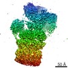
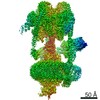
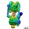
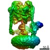
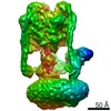
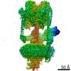
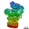
| タイトル | Structure of the Manduca sexta V-ATPase by cryo-electron microscopy | |||||||||
 マップデータ マップデータ | Manduca sexta vacuolar ATPase | |||||||||
 試料 試料 |
| |||||||||
 キーワード キーワード | H-ATPase / V-ATPase / cryo-electron microscopy / vacuolar membrane | |||||||||
| 生物種 |  Manduca sexta (蝶・蛾) Manduca sexta (蝶・蛾) | |||||||||
| 手法 | 単粒子再構成法 / クライオ電子顕微鏡法 / 解像度: 17.0 Å | |||||||||
 データ登録者 データ登録者 | Muench SP / Huss M / Phillips C / Song CF / Wieczorek H / Trinick J / Harrison MA | |||||||||
 引用 引用 | ジャーナル: J Mol Biol / 年: 2009 タイトル: Cryo-electron microscopy of the vacuolar ATPase motor reveals its mechanical and regulatory complexity. 著者: Stephen P Muench / Markus Huss / Chun Feng Song / Clair Phillips / Helmut Wieczorek / John Trinick / Michael A Harrison /  要旨: The vacuolar H+-ATPase (V-ATPase) is an ATP-driven rotary molecular motor that is a transmembrane proton pump in all eukaryotic cells. Although its activity is fundamental to many physiological ...The vacuolar H+-ATPase (V-ATPase) is an ATP-driven rotary molecular motor that is a transmembrane proton pump in all eukaryotic cells. Although its activity is fundamental to many physiological processes, our understanding of the structure and mechanism of the V-ATPase is poor. Using cryo-electron microscopy of the tobacco hornworm (Manduca sexta) enzyme, we have calculated the first 3D reconstruction of the intact pump in its native state. The resolution of 16.5 A is significantly higher than that of previous cryo-electron microscopy models of either V-ATPase or the related F1F0-ATPase. A network of four stalk structures connecting the V1 catalytic domain and the V0 membrane domain is now fully resolved, demonstrating substantially greater complexity than that found in the F-ATPase. Three peripheral stator stalks connect these domains to a horizontal collar that partly encircles the region between V1 and V0. The fourth stalk is a central axle that connects to V0 but makes minimal contact with V1. Several subunit crystal structures can be fit accurately into the reconstruction. The model thus provides new insights into the organisation of key components involved in mechanical coupling between the domains and regulation of activity. | |||||||||
| 履歴 |
|
- 構造の表示
構造の表示
| ムービー |
 ムービービューア ムービービューア |
|---|---|
| 構造ビューア | EMマップ: SurfViewMolmilJmol/JSmol |
| 添付画像 |

- ダウンロードとリンク
-EMDBアーカイブ
| マップデータ | emd_1590.map.gz | 3.5 MB | EMDBマップデータ形式 | |
|---|---|---|---|---|
| ヘッダ (付随情報) | emd-1590-v30.xmlemd-1590.xml | 9.4 KB 9.4 KB | 表示 表示 | EMDBヘッダ |
| 画像 |  1590.gif 1590.gif | 50.5 KB | ||
| アーカイブディレクトリ |  http://ftp.pdbj.org/pub/emdb/structures/EMD-1590ftp://ftp.pdbj.org/pub/emdb/structures/EMD-1590 http://ftp.pdbj.org/pub/emdb/structures/EMD-1590ftp://ftp.pdbj.org/pub/emdb/structures/EMD-1590 | HTTPS FTP |
-関連構造データ

-リンク
| EMDBのページ | EMDB (EBI/PDBe) / EMDataResource |
|---|
-マップ
| ファイル | ダウンロード / ファイル: emd_1590.map.gz / 形式: CCP4 / 大きさ: 3.7 MB / タイプ: IMAGE STORED AS FLOATING POINT NUMBER (4 BYTES) | ||||||||||||||||||||||||||||||||||||||||||||||||||||||||||||||||||||
|---|---|---|---|---|---|---|---|---|---|---|---|---|---|---|---|---|---|---|---|---|---|---|---|---|---|---|---|---|---|---|---|---|---|---|---|---|---|---|---|---|---|---|---|---|---|---|---|---|---|---|---|---|---|---|---|---|---|---|---|---|---|---|---|---|---|---|---|---|---|
| 注釈 | Manduca sexta vacuolar ATPase | ||||||||||||||||||||||||||||||||||||||||||||||||||||||||||||||||||||
| 投影像・断面図 | 画像のコントロール
画像は Spider により作成 | ||||||||||||||||||||||||||||||||||||||||||||||||||||||||||||||||||||
| ボクセルのサイズ | X=Y=Z: 4.36 Å | ||||||||||||||||||||||||||||||||||||||||||||||||||||||||||||||||||||
| 密度 |
| ||||||||||||||||||||||||||||||||||||||||||||||||||||||||||||||||||||
| 対称性 | 空間群: 1 | ||||||||||||||||||||||||||||||||||||||||||||||||||||||||||||||||||||
| 詳細 | EMDB XML:
CCP4マップ ヘッダ情報:
| ||||||||||||||||||||||||||||||||||||||||||||||||||||||||||||||||||||
 Z (Sec.)
 Y (Row.)
 X (Col.)


-添付データ
- 試料の構成要素
-全体 : Manduca sexta vacuolar ATPase complex
| 全体 | 名称: Manduca sexta vacuolar ATPase complex |
|---|---|
| 要素 |
|
-超分子 #1000: Manduca sexta vacuolar ATPase complex
| 超分子 | 名称: Manduca sexta vacuolar ATPase complex / タイプ: sample / ID: 1000 / 詳細: The sample was monodisperse / 集合状態: monomer / Number unique components: 1 |
|---|---|
| 分子量 | 実験値: 900 KDa / 理論値: 900 KDa |
-超分子 #1: membrane proton pump
| 超分子 | 名称: membrane proton pump / タイプ: organelle_or_cellular_component / ID: 1 / Name.synonym: V-ATPase / 集合状態: monomer / 組換発現: No |
|---|---|
| 由来(天然) | 生物種: Manduca sexta (蝶・蛾) / 別称: tobacco hornworm / 組織: midgut |
-実験情報
-構造解析
| 手法 | クライオ電子顕微鏡法 |
|---|---|
 解析 解析 | 単粒子再構成法 |
| 試料の集合状態 | particle |
-試料調製
| 濃度 | 2.0 mg/mL |
|---|---|
| 緩衝液 | pH: 8.1 詳細: 150 mM NaCl, 9.6 mM B mercaptoethanol, 20 mM TrisHCl. Solubilised in C12E10 detergent |
| グリッド | 詳細: 300 mesh Cu Lacey grid |
| 凍結 | 凍結剤: ETHANE / チャンバー内温度: 22 K / 装置: HOMEMADE PLUNGER 詳細: Vitrification instrument: Computer controlled blotting device 手法: A small vial of ethane was placed inside a larger liquid nitrogen reservoir. 3ul of protein sample was then applied to a lacey grid which had been glow discharged for 30 seconds prior to use. ...手法: A small vial of ethane was placed inside a larger liquid nitrogen reservoir. 3ul of protein sample was then applied to a lacey grid which had been glow discharged for 30 seconds prior to use. The grid was then blotted (1.6 seconds) and quickly frozen in liquid ethane using a computer operated device as described in White et al., 2003, J. Struct, Biol 144 246-252 |
- 電子顕微鏡法
| 顕微鏡 | FEI TECNAI F20 |
|---|---|
| 温度 | 平均: 22 K |
| アライメント法 | Legacy - 非点収差: astigmatism corrected at 100,000 magnification |
| 日付 | 2007年11月10日 |
| 撮影 | カテゴリ: CCD フィルム・検出器のモデル: GENERIC GATAN (4k x 4k) デジタル化 - サンプリング間隔: 15.0 µm / 実像数: 320 / 平均電子線量: 15 e/Å2 |
| Tilt angle min | 0 |
| Tilt angle max | 0 |
| 電子線 | 加速電圧: 200 kV / 電子線源:  FIELD EMISSION GUN FIELD EMISSION GUN |
| 電子光学系 | 倍率(補正後): 69000 / 照射モード: FLOOD BEAM / 撮影モード: BRIGHT FIELD / Cs: 2.0 mm / 最大 デフォーカス(公称値): 4.5 µm / 最小 デフォーカス(公称値): 1.5 µm / 倍率(公称値): 50000 |
| 試料ステージ | 試料ホルダー: Gatan side entry cryo holder / 試料ホルダーモデル: GATAN LIQUID NITROGEN |
| 実験機器 |  モデル: Tecnai F20 / 画像提供: FEI Company |
-画像解析
| 詳細 | Particles were picked using BOXER |
|---|---|
| CTF補正 | 詳細: phase flipping each particle |
| 最終 再構成 | 想定した対称性 - 点群: C1 (非対称) / アルゴリズム: OTHER / 解像度のタイプ: BY AUTHOR / 解像度: 17.0 Å / 解像度の算出法: FSC 0.5 CUT-OFF / ソフトウェア - 名称: Imagic Eman / 使用した粒子像数: 11742 |
