Ministry of Education, Culture, Sports, Science and Technology (Japan)
日本
引用
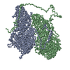
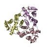
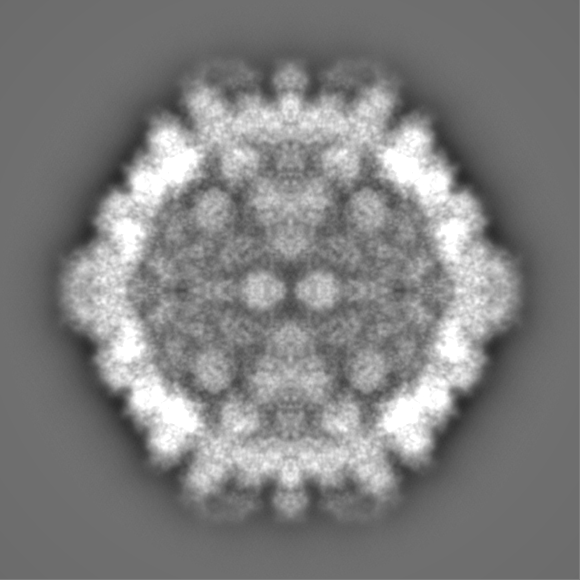
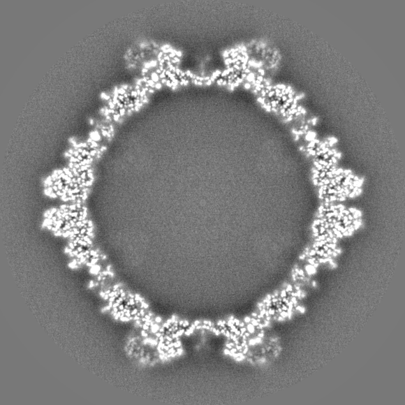
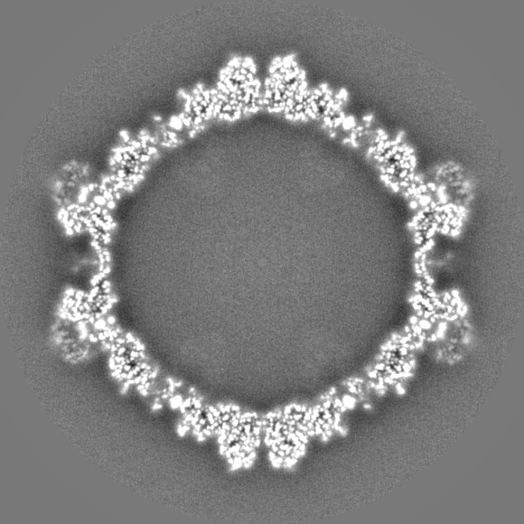
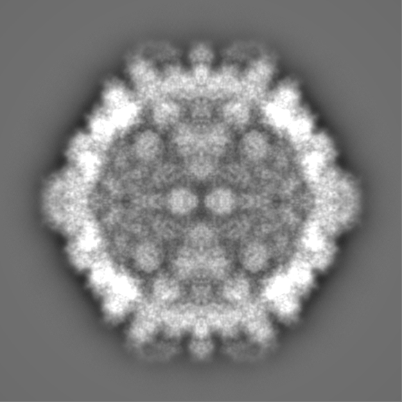
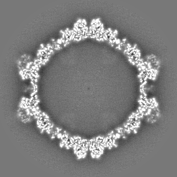
ジャーナル: PLoS Pathog / 年: 2023 タイトル: Capsid structure of a fungal dsRNA megabirnavirus reveals its previously unidentified surface architecture. 著者: Han Wang / Lakha Salaipeth / Naoyuki Miyazaki / Nobuhiro Suzuki / Kenta Okamoto / 要旨: Rosellinia necatrix megabirnavirus 1-W779 (RnMBV1) is a non-enveloped icosahedral double-stranded (ds)RNA virus that infects the ascomycete fungus Rosellinia necatrix, a causative agent that induces ...Rosellinia necatrix megabirnavirus 1-W779 (RnMBV1) is a non-enveloped icosahedral double-stranded (ds)RNA virus that infects the ascomycete fungus Rosellinia necatrix, a causative agent that induces a lethal plant disease white root rot. Herein, we have first resolved the atomic structure of the RnMBV1 capsid at 3.2 Å resolution using cryo-electron microscopy (cryo-EM) single-particle analysis. Compared with other non-enveloped icosahedral dsRNA viruses, the RnMBV1 capsid protein structure exhibits an extra-long C-terminal arm and a surface protrusion domain. In addition, the previously unrecognized crown proteins are identified in a symmetry-expanded cryo-EM model and are present over the 3-fold axes. These exclusive structural features of the RnMBV1 capsid could have been acquired for playing essential roles in transmission and/or particle assembly of the megabirnaviruses. Our findings, therefore, will reinforce the understanding of how the structural and molecular machineries of the megabirnaviruses influence the virulence of the disease-related ascomycete fungus.
 ムービー
ムービー コントローラー


 データを開く
 基本情報
基本情報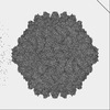
 マップデータ
 試料
試料 Rosellinia necatrix megabirnavirus 1/W779 (ウイルス)
 データ登録者
 スウェーデン,
  日本, 2件
  引用
引用 構造の表示
構造の表示
 ダウンロードとリンク
 EMDBマップデータ形式
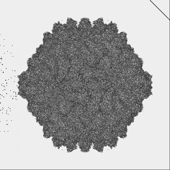
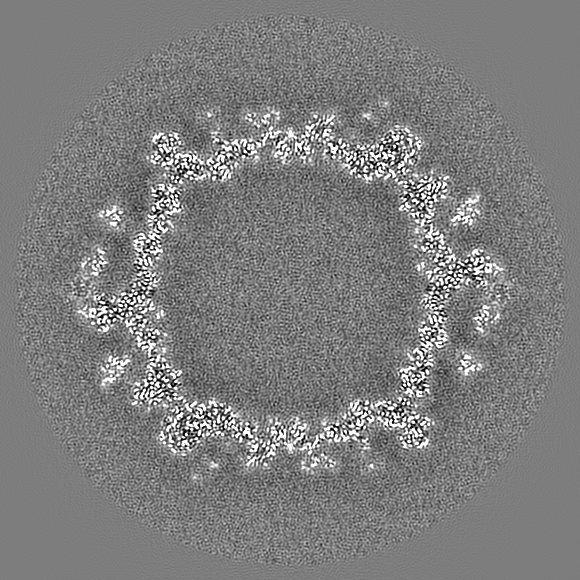
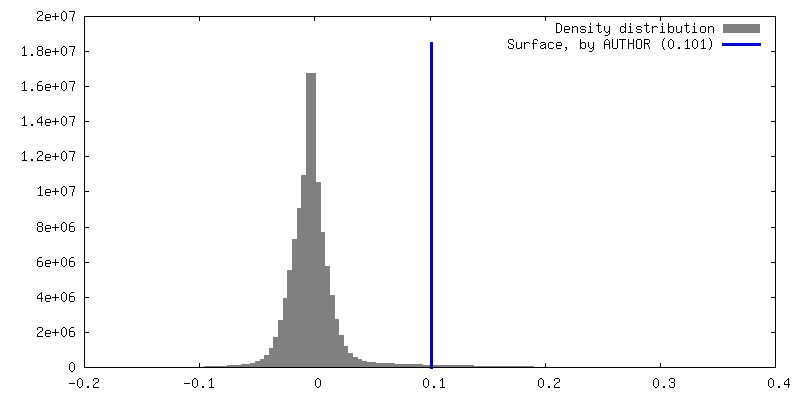
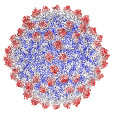 emd_15857.png
 http://ftp.pdbj.org/pub/emdb/structures/EMD-15857
 Z (Sec.)
 Y (Row.)
 X (Col.)


 試料の構成要素
 解析
解析 電子顕微鏡法
 FIELD EMISSION GUN
