 Movie
Movie Controller

[English] 日本語
 Yorodumi
- EMDB-1582: Three-dimensional structure of the 70S E. coli ribosome in its na... -
+ Open data

- Basic information
| Entry | Database: EMDB / ID: EMD-1582 | |||||||||
|---|---|---|---|---|---|---|---|---|---|---|
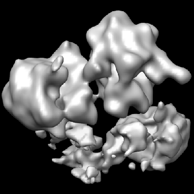
| Title | Three-dimensional structure of the 70S E. coli ribosome in its native 3D organization in polysomes. | |||||||||
 Map data Map data | This is a 3D density of class t-t ribosomes obtained by averaging of polysomal particles. | |||||||||
 Sample Sample |
| |||||||||
 Keywords Keywords | 70S ribosome / polysome / translation / protein folding / cryoelectron tomography | |||||||||
| Biological species |  | |||||||||
| Method | subtomogram averaging / cryo EM / Resolution: 30.0 Å | |||||||||
 Authors Authors | Brandt F / Elcock AH / Etchells SA / Ortiz JO / Hartl FU / Baumeister W | |||||||||
 Citation Citation | Journal: Cell / Year: 2009 Title: The native 3D organization of bacterial polysomes. Authors: Florian Brandt / Stephanie A Etchells / Julio O Ortiz / Adrian H Elcock / F Ulrich Hartl / Wolfgang Baumeister /  Abstract: Recent advances have led to insights into the structure of the bacterial ribosome, but little is known about the 3D organization of ribosomes in the context of translating polysomes. We employed ...Recent advances have led to insights into the structure of the bacterial ribosome, but little is known about the 3D organization of ribosomes in the context of translating polysomes. We employed cryoelectron tomography and a template-matching approach to map 70S ribosomes in vitrified bacterial translation extracts and in lysates of active E. coli spheroplasts. In these preparations, polysomal arrangements were observed in which neighboring ribosomes are densely packed and exhibit preferred orientations. Analysis of characteristic examples of polysomes reveals a staggered or pseudohelical organization of ribosomes along the mRNA trace, with the transcript being sequestered on the inside, the tRNA entrance sites being accessible, and the polypeptide exit sites facing the cytosol. Modeling of elongating nascent polypeptide chains suggests that this arrangement maximizes the distance between nascent chains on adjacent ribosomes, thereby reducing the probability of intermolecular interactions that would give rise to aggregation and limit productive folding. | |||||||||
| History |
|
- Structure visualization
| Movie |
 Movie viewer Movie viewer |
|---|---|
| Structure viewer | EM map: SurfViewMolmilJmol/JSmol |
| Supplemental images |
- Downloads & links
-EMDB archive
| Map data | emd_1582.map.gz | 7.2 MB | EMDB map data format | |
|---|---|---|---|---|
| Header (meta data) | emd-1582-v30.xmlemd-1582.xml | 11.4 KB 11.4 KB | Display Display | EMDB header |
| Images |  1582.gif 1582.gif | 86.7 KB | ||
| Archive directory |  http://ftp.pdbj.org/pub/emdb/structures/EMD-1582ftp://ftp.pdbj.org/pub/emdb/structures/EMD-1582 http://ftp.pdbj.org/pub/emdb/structures/EMD-1582ftp://ftp.pdbj.org/pub/emdb/structures/EMD-1582 | HTTPS FTP |
-Validation report
| Summary document | emd_1582_validation.pdf.gz | 213.5 KB | Display | EMDB validaton report |
|---|---|---|---|---|
| Full document | emd_1582_full_validation.pdf.gz | 212.6 KB | Display | |
| Data in XML | emd_1582_validation.xml.gz | 4.5 KB | Display | |
| Arichive directory | https://ftp.pdbj.org/pub/emdb/validation_reports/EMD-1582ftp://ftp.pdbj.org/pub/emdb/validation_reports/EMD-1582 | HTTPS FTP |
-Links
| EMDB pages | EMDB (EBI/PDBe) / EMDataResource |
|---|---|
| Related items in Molecule of the Month |

-Map
| File | Download / File: emd_1582.map.gz / Format: CCP4 / Size: 7.8 MB / Type: IMAGE STORED AS FLOATING POINT NUMBER (4 BYTES) | ||||||||||||||||||||||||||||||||||||||||||||||||||||||||||||||||||||
|---|---|---|---|---|---|---|---|---|---|---|---|---|---|---|---|---|---|---|---|---|---|---|---|---|---|---|---|---|---|---|---|---|---|---|---|---|---|---|---|---|---|---|---|---|---|---|---|---|---|---|---|---|---|---|---|---|---|---|---|---|---|---|---|---|---|---|---|---|---|
| Annotation | This is a 3D density of class t-t ribosomes obtained by averaging of polysomal particles. | ||||||||||||||||||||||||||||||||||||||||||||||||||||||||||||||||||||
| Voxel size | X=Y=Z: 5.6 Å | ||||||||||||||||||||||||||||||||||||||||||||||||||||||||||||||||||||
| Density |
| ||||||||||||||||||||||||||||||||||||||||||||||||||||||||||||||||||||
| Symmetry | Space group: 1 | ||||||||||||||||||||||||||||||||||||||||||||||||||||||||||||||||||||
| Details | EMDB XML:
CCP4 map header:
| ||||||||||||||||||||||||||||||||||||||||||||||||||||||||||||||||||||
-Supplemental data
- Sample components
-Entire : 70S ribosome in a native polysome with a neighbor ribosome at the...
| Entire | Name: 70S ribosome in a native polysome with a neighbor ribosome at the 5' end oriented in a tt-class |
|---|---|
| Components |
|
-Supramolecule #1000: 70S ribosome in a native polysome with a neighbor ribosome at the...
| Supramolecule | Name: 70S ribosome in a native polysome with a neighbor ribosome at the 5' end oriented in a tt-class type: sample / ID: 1000 Details: The sample is indeed heterogeneous, a prokaryotic lysate containing many cellular components Oligomeric state: ribosomes forming polysomes / Number unique components: 1 |
|---|---|
| Molecular weight | Experimental: 2.7 MDa / Theoretical: 2.7 MDa / Method: Sedimentation |
-Supramolecule #1: 70S ribosome
| Supramolecule | Name: 70S ribosome / type: complex / ID: 1 / Name.synonym: 70S ribosome Details: The ribosomes under scrutiny are part of large polysomal arrangements Recombinant expression: No / Database: NCBI / Ribosome-details: ribosome-prokaryote: ALL |
|---|---|
| Source (natural) | Organism: |
| Molecular weight | Experimental: 2.7 MDa / Theoretical: 2.7 MDa |
-Experimental details
-Structure determination
| Method | cryo EM |
|---|---|
 Processing Processing | subtomogram averaging |
| Aggregation state | particle |
-Sample preparation
| Concentration | 20 mg/mL |
|---|---|
| Buffer | pH: 7.5 Details: Roche reconstitution buffer from the RTS kit, ATP,GTP, 8-15 mM Mg2, 100-250 nm K,NH4,DTT, protease inhibitors,NaN3 |
| Grid | Details: 400 mesh gold grid |
| Vitrification | Cryogen name: ETHANE / Chamber temperature: 77 K / Instrument: HOMEMADE PLUNGER Details: Vitrification instrument: plunger. Vitrification carried out in air Method: Blot for 1 s before plunging |
- Electron microscopy
| Microscope | FEI/PHILIPS CM200FEG |
|---|---|
| Temperature | Min: 70 K / Max: 90 K / Average: 77 K |
| Alignment procedure | Legacy - Astigmatism: Objective astigmatism was corrected using a quadrupole stigmator at 50,000 times magnification Legacy - Electron beam tilt params: -4 |
| Date | Dec 1, 2006 |
| Image recording | Category: CCD / Film or detector model: GENERIC TVIPS (4k x 4k) / Average electron dose: 50 e/Å2 |
| Electron beam | Acceleration voltage: 160 kV / Electron source:  FIELD EMISSION GUN FIELD EMISSION GUN |
| Electron optics | Calibrated magnification: 53960 / Illumination mode: FLOOD BEAM / Imaging mode: BRIGHT FIELD / Cs: 2 mm / Nominal defocus max: 4.0 µm / Nominal defocus min: 3.0 µm / Nominal magnification: 27500 |
| Sample stage | Specimen holder: Side entry liquid nitrogen- / Specimen holder model: GATAN LIQUID NITROGEN / Tilt series - Axis1 - Min angle: -60 ° / Tilt series - Axis1 - Max angle: 60 ° |
-Image processing
| Details | Average number of projections used in the 3D reconstructions: 391. |
|---|---|
| Final reconstruction | Algorithm: OTHER / Resolution.type: BY AUTHOR / Resolution: 30.0 Å / Resolution method: FSC 0.5 CUT-OFF / Software - Name: TOM ToolBox / Details: CET |
-Atomic model buiding 1
| Initial model | PDB ID:  2aw7 |
|---|---|
| Software | Name: Chimera |
| Details | Protocol: Rigid Body |
| Refinement | Space: REAL / Protocol: RIGID BODY FIT |
-Atomic model buiding 2
| Initial model | PDB ID: 2awb |
|---|---|
| Software | Name: Chimera |
| Details | Protocol: Rigid Body |
| Refinement | Space: REAL / Protocol: RIGID BODY FIT |
