 Movie
Movie Controller

[English] 日本語
 Yorodumi
- EMDB-15775: Cryo-EM structure of the Tripartite ATP-independent Periplasmic (... -
+ Open data

- Basic information
| Entry |  | |||||||||
|---|---|---|---|---|---|---|---|---|---|---|
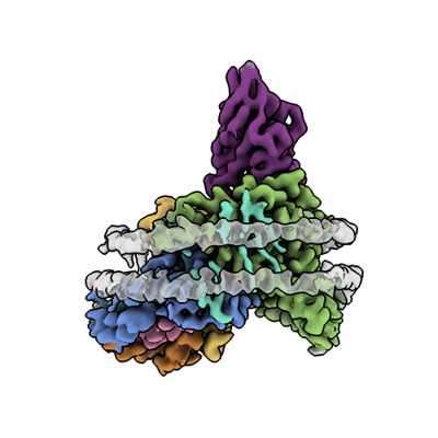
| Title | Cryo-EM structure of the Tripartite ATP-independent Periplasmic (TRAP) transporter SiaQM from Photobacterium profundum in a nanodisc | |||||||||
 Map data Map data | ||||||||||
 Sample Sample |
| |||||||||
 Keywords Keywords | TRAP / elevator / secondary transporter / sialic acid / TRANSPORT PROTEIN | |||||||||
| Function / homology |  Function and homology information Function and homology informationC4-dicarboxylate transport / transmembrane transporter activity / plasma membrane Similarity search - Function | |||||||||
| Biological species |  Photobacterium profundum (bacteria) / Photobacterium profundum (bacteria) /  Helicobacter pylori (bacteria) / Photobacterium profundum SS9 (bacteria) / synthetic construct (others) Helicobacter pylori (bacteria) / Photobacterium profundum SS9 (bacteria) / synthetic construct (others) | |||||||||
| Method | single particle reconstruction / cryo EM / Resolution: 3.03 Å | |||||||||
 Authors Authors | Davies JS / North RA / Dobson RCJ | |||||||||
| Funding support |  New Zealand, 2 items New Zealand, 2 items
| |||||||||
 Citation Citation | Journal: Nat Commun / Year: 2023 Title: Structure and mechanism of a tripartite ATP-independent periplasmic TRAP transporter. Authors: James S Davies / Michael J Currie / Rachel A North / Mariafrancesca Scalise / Joshua D Wright / Jack M Copping / Daniela M Remus / Ashutosh Gulati / Dustin R Morado / Sam A Jamieson / ...Authors: James S Davies / Michael J Currie / Rachel A North / Mariafrancesca Scalise / Joshua D Wright / Jack M Copping / Daniela M Remus / Ashutosh Gulati / Dustin R Morado / Sam A Jamieson / Michael C Newton-Vesty / Gayan S Abeysekera / Subramanian Ramaswamy / Rosmarie Friemann / Soichi Wakatsuki / Jane R Allison / Cesare Indiveri / David Drew / Peter D Mace / Renwick C J Dobson /     Abstract: In bacteria and archaea, tripartite ATP-independent periplasmic (TRAP) transporters uptake essential nutrients. TRAP transporters receive their substrates via a secreted soluble substrate-binding ...In bacteria and archaea, tripartite ATP-independent periplasmic (TRAP) transporters uptake essential nutrients. TRAP transporters receive their substrates via a secreted soluble substrate-binding protein. How a sodium ion-driven secondary active transporter is strictly coupled to a substrate-binding protein is poorly understood. Here we report the cryo-EM structure of the sialic acid TRAP transporter SiaQM from Photobacterium profundum at 2.97 Å resolution. SiaM comprises a "transport" domain and a "scaffold" domain, with the transport domain consisting of helical hairpins as seen in the sodium ion-coupled elevator transporter VcINDY. The SiaQ protein forms intimate contacts with SiaM to extend the size of the scaffold domain, suggesting that TRAP transporters may operate as monomers, rather than the typically observed oligomers for elevator-type transporters. We identify the Na and sialic acid binding sites in SiaM and demonstrate a strict dependence on the substrate-binding protein SiaP for uptake. We report the SiaP crystal structure that, together with docking studies, suggest the molecular basis for how sialic acid is delivered to the SiaQM transporter complex. We thus propose a model for substrate transport by TRAP proteins, which we describe herein as an 'elevator-with-an-operator' mechanism. | |||||||||
| History |
|
- Structure visualization
| Supplemental images |
|---|
- Downloads & links
-EMDB archive
| Map data | emd_15775.map.gz | 51.4 MB | EMDB map data format | |
|---|---|---|---|---|
| Header (meta data) | emd-15775-v30.xmlemd-15775.xml | 21.6 KB 21.6 KB | Display Display | EMDB header |
| FSC (resolution estimation) | emd_15775_fsc.xml | 9.9 KB | Display | FSC data file |
| Images |  emd_15775.png emd_15775.png | 92.9 KB | ||
| Filedesc metadata | emd-15775.cif.gz | 6.7 KB | ||
| Others | emd_15775_half_map_1.map.gzemd_15775_half_map_2.map.gz | 95.7 MB 95.7 MB | ||
| Archive directory |  http://ftp.pdbj.org/pub/emdb/structures/EMD-15775ftp://ftp.pdbj.org/pub/emdb/structures/EMD-15775 http://ftp.pdbj.org/pub/emdb/structures/EMD-15775ftp://ftp.pdbj.org/pub/emdb/structures/EMD-15775 | HTTPS FTP |
-Validation report
| Summary document | emd_15775_validation.pdf.gz | 919.1 KB | Display | EMDB validaton report |
|---|---|---|---|---|
| Full document | emd_15775_full_validation.pdf.gz | 918.7 KB | Display | |
| Data in XML | emd_15775_validation.xml.gz | 18.2 KB | Display | |
| Data in CIF | emd_15775_validation.cif.gz | 23.6 KB | Display | |
| Arichive directory | https://ftp.pdbj.org/pub/emdb/validation_reports/EMD-15775ftp://ftp.pdbj.org/pub/emdb/validation_reports/EMD-15775 | HTTPS FTP |
-Related structure data
| Related structure data |  8b01MC  7qhaC  7t3eC C: citing same article ( M: atomic model generated by this map |
|---|---|
| Similar structure data |
-Links
| EMDB pages | EMDB (EBI/PDBe) / EMDataResource |
|---|
-Map
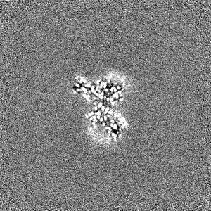
| File | Download / File: emd_15775.map.gz / Format: CCP4 / Size: 103 MB / Type: IMAGE STORED AS FLOATING POINT NUMBER (4 BYTES) | ||||||||||||||||||||||||||||||||||||
|---|---|---|---|---|---|---|---|---|---|---|---|---|---|---|---|---|---|---|---|---|---|---|---|---|---|---|---|---|---|---|---|---|---|---|---|---|---|
| Projections & slices | Image control
Images are generated by Spider. | ||||||||||||||||||||||||||||||||||||
| Voxel size | X=Y=Z: 0.886 Å | ||||||||||||||||||||||||||||||||||||
| Density |
| ||||||||||||||||||||||||||||||||||||
| Symmetry | Space group: 1 | ||||||||||||||||||||||||||||||||||||
| Details | EMDB XML:
|
 Z (Sec.)
 Y (Row.)
 X (Col.)


-Supplemental data
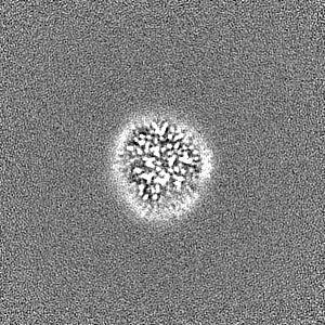
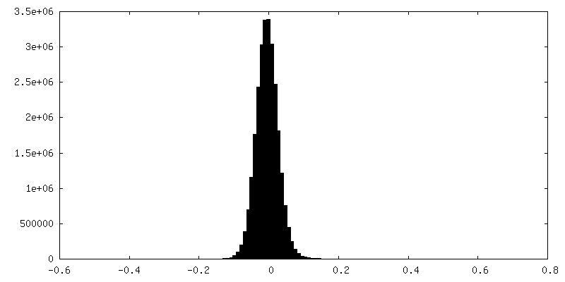
-Half map: #2
| File | emd_15775_half_map_1.map | ||||||||||||
|---|---|---|---|---|---|---|---|---|---|---|---|---|---|
| Projections & Slices |
| ||||||||||||
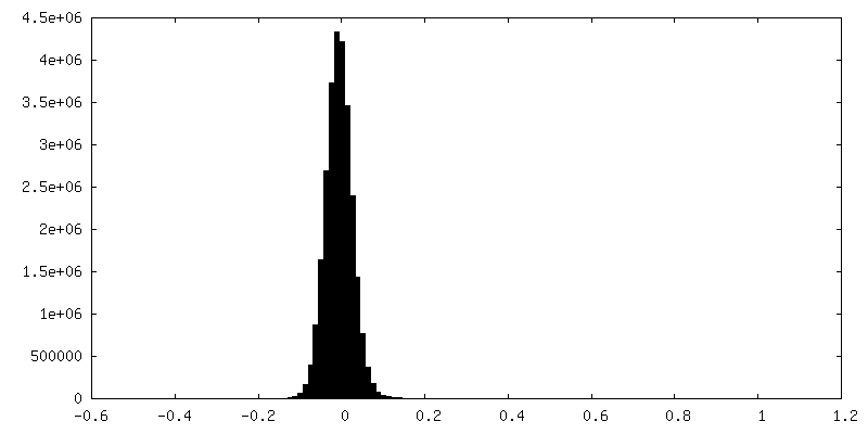
| Density Histograms |


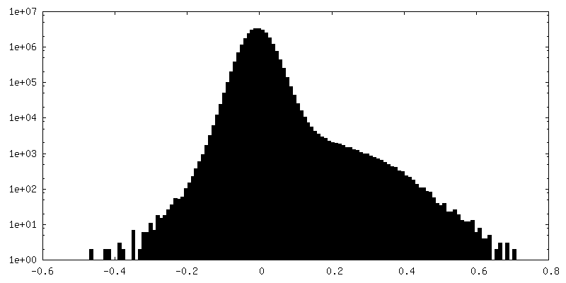
-Half map: #1
| File | emd_15775_half_map_2.map | ||||||||||||
|---|---|---|---|---|---|---|---|---|---|---|---|---|---|
| Projections & Slices |
| ||||||||||||
| Density Histograms |

- Sample components
+Entire : SiaQM with megabody
+Supramolecule #1: SiaQM with megabody
+Supramolecule #2: SiaQM
+Supramolecule #3: MegaBody
+Macromolecule #1: Putative TRAP-type C4-dicarboxylate transport system, small perme...
+Macromolecule #2: Putative TRAP-type C4-dicarboxylate transport system, large perme...
+Macromolecule #3: Megabody c7HopQ
+Macromolecule #4: N-OCTANE

+Macromolecule #5: PHOSPHATIDYLETHANOLAMINE

+Macromolecule #6: DOCOSANE
+Macromolecule #7: DECANE

+Macromolecule #8: SODIUM ION
+Macromolecule #9: TRIDECANE
+Macromolecule #10: HEXANE
-Experimental details
-Structure determination
| Method | cryo EM |
|---|---|
 Processing Processing | single particle reconstruction |
| Aggregation state | particle |
-Sample preparation
| Concentration | 1 mg/mL |
|---|---|
| Buffer | pH: 8 |
| Vitrification | Cryogen name: ETHANE / Chamber humidity: 100 % / Chamber temperature: 277 K / Instrument: FEI VITROBOT MARK IV |
- Electron microscopy
| Microscope | FEI TITAN KRIOS |
|---|---|
| Image recording | Film or detector model: GATAN K3 BIOQUANTUM (6k x 4k) / Number grids imaged: 1 / Number real images: 8127 / Average exposure time: 2.0 sec. / Average electron dose: 70.9 e/Å2 |
| Electron beam | Acceleration voltage: 300 kV / Electron source:  FIELD EMISSION GUN FIELD EMISSION GUN |
| Electron optics | Illumination mode: FLOOD BEAM / Imaging mode: BRIGHT FIELD / Nominal defocus max: 2.0 µm / Nominal defocus min: 0.4 µm |
| Experimental equipment |  Model: Titan Krios / Image courtesy: FEI Company |

