 ムービー
ムービー コントローラー


+ データを開く

- 基本情報
基本情報
| 登録情報 | データベース: EMDB / ID: EMD-1576 | |||||||||
|---|---|---|---|---|---|---|---|---|---|---|
| タイトル | Structural analysis of the interaction of human adenovirus 5 with erythrocytes. | |||||||||
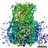
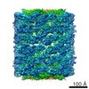
 マップデータ マップデータ | This is a map of the adenovirus fivefold vertex interacting directly with the surface of a human red blood cell ghost. | |||||||||
 試料 試料 |
| |||||||||
 キーワード キーワード | Adenovirus / erythrocyte / complement receptor / coxsackie-adenovirus receptor / penton base | |||||||||
| 生物種 |  Homo sapiens (ヒト) / Adenovirus serotype 5 Homo sapiens (ヒト) / Adenovirus serotype 5 | |||||||||
| 手法 | 単粒子再構成法 / クライオ電子顕微鏡法 / 解像度: 40.0 Å | |||||||||
 データ登録者 データ登録者 | Carlisle RC / Di Y / Cerny AM / Sonnen AF-P / Sim RB / Green NK / Subr V / Ulbrich K / Gilbert RJC / Fisher KD ...Carlisle RC / Di Y / Cerny AM / Sonnen AF-P / Sim RB / Green NK / Subr V / Ulbrich K / Gilbert RJC / Fisher KD / Finberg RW / Seymour LW | |||||||||
 引用 引用 | ジャーナル: Blood / 年: 2009 タイトル: Human erythrocytes bind and inactivate type 5 adenovirus by presenting Coxsackie virus-adenovirus receptor and complement receptor 1. 著者: Robert C Carlisle / Ying Di / Anna M Cerny / Andreas F-P Sonnen / Robert B Sim / Nicola K Green / Vladimir Subr / Karel Ulbrich / Robert J C Gilbert / Kerry D Fisher / Robert W Finberg / Leonard W Seymour /  要旨: Type 5 adenovirus (Ad5) is a human pathogen that has been widely developed for therapeutic uses, with only limited success to date. We report here the novel finding that human erythrocytes present ...Type 5 adenovirus (Ad5) is a human pathogen that has been widely developed for therapeutic uses, with only limited success to date. We report here the novel finding that human erythrocytes present Coxsackie virus-adenovirus receptor (CAR) providing an Ad5 sequestration mechanism that protects against systemic infection. Interestingly, erythrocytes from neither mice nor rhesus macaques present CAR. Excess Ad5 fiber protein or anti-CAR antibody inhibits the binding of Ad5 to human erythrocytes and cryo-electron microscopy shows attachment via the fiber protein of Ad5, leading to close juxtaposition with the erythrocyte membrane. Human, but not murine, erythrocytes also present complement receptor (CR1), which binds Ad5 in the presence of antibodies and complement. Transplantation of human erythrocytes into nonobese diabetic/severe combined immunodeficiency mice extends blood circulation of intravenous Ad5 but decreases its extravasation into human xenograft tumors. Ad5 also shows extended circulation in transgenic mice presenting CAR on their erythrocytes, although it clears rapidly in transgenic mice presenting erythrocyte CR1. Hepatic infection is inhibited in both transgenic models. Erythrocytes may therefore restrict Ad5 infection (natural and therapeutic) in humans, independent of antibody status, presenting a formidable challenge to Ad5 therapeutics. "Stealthing" of Ad5 using hydrophilic polymers may enable circumvention of these natural virus traps. | |||||||||
| 履歴 |
|
- 構造の表示
構造の表示
| ムービー |
 ムービービューア ムービービューア |
|---|---|
| 構造ビューア | EMマップ: SurfViewMolmilJmol/JSmol |
| 添付画像 |


- ダウンロードとリンク
-EMDBアーカイブ
| マップデータ | emd_1576.map.gz | 2 MB | EMDBマップデータ形式 | |
|---|---|---|---|---|
| ヘッダ (付随情報) | emd-1576-v30.xmlemd-1576.xml | 10.1 KB 10.1 KB | 表示 表示 | EMDBヘッダ |
| 画像 |  1576.gif 1576.gif | 90.8 KB | ||
| その他 | IMAGE_DETAILSemd_1576_1.tifemd_1576_2.tifemd_1576_3.tifemd_1576_4.tifemd_1576_5.tifemd_1576_6.tifemd_1576_7.tif | 771 B 807.1 KB 688.3 KB 833.2 KB 805.4 KB 818 KB 703.4 KB 88 KB | ||
| アーカイブディレクトリ |  http://ftp.pdbj.org/pub/emdb/structures/EMD-1576ftp://ftp.pdbj.org/pub/emdb/structures/EMD-1576 http://ftp.pdbj.org/pub/emdb/structures/EMD-1576ftp://ftp.pdbj.org/pub/emdb/structures/EMD-1576 | HTTPS FTP |
-検証レポート
| 文書・要旨 | emd_1576_validation.pdf.gz | 233.8 KB | 表示 | EMDB検証レポート |
|---|---|---|---|---|
| 文書・詳細版 | emd_1576_full_validation.pdf.gz | 233 KB | 表示 | |
| XML形式データ | emd_1576_validation.xml.gz | 6.1 KB | 表示 | |
| アーカイブディレクトリ | https://ftp.pdbj.org/pub/emdb/validation_reports/EMD-1576ftp://ftp.pdbj.org/pub/emdb/validation_reports/EMD-1576 | HTTPS FTP |
-関連構造データ
| 類似構造データ |
|---|
-リンク
| EMDBのページ | EMDB (EBI/PDBe) / EMDataResource |
|---|
-マップ
| ファイル | ダウンロード / ファイル: emd_1576.map.gz / 形式: CCP4 / 大きさ: 29.8 MB / タイプ: IMAGE STORED AS FLOATING POINT NUMBER (4 BYTES) | ||||||||||||||||||||||||||||||||||||||||||||||||||||||||||||||||||||
|---|---|---|---|---|---|---|---|---|---|---|---|---|---|---|---|---|---|---|---|---|---|---|---|---|---|---|---|---|---|---|---|---|---|---|---|---|---|---|---|---|---|---|---|---|---|---|---|---|---|---|---|---|---|---|---|---|---|---|---|---|---|---|---|---|---|---|---|---|---|
| 注釈 | This is a map of the adenovirus fivefold vertex interacting directly with the surface of a human red blood cell ghost. | ||||||||||||||||||||||||||||||||||||||||||||||||||||||||||||||||||||
| 投影像・断面図 | 画像のコントロール
画像は Spider により作成 | ||||||||||||||||||||||||||||||||||||||||||||||||||||||||||||||||||||
| ボクセルのサイズ | X=Y=Z: 2.6 Å | ||||||||||||||||||||||||||||||||||||||||||||||||||||||||||||||||||||
| 密度 |
| ||||||||||||||||||||||||||||||||||||||||||||||||||||||||||||||||||||
| 対称性 | 空間群: 1 | ||||||||||||||||||||||||||||||||||||||||||||||||||||||||||||||||||||
| 詳細 | EMDB XML:
CCP4マップ ヘッダ情報:
| ||||||||||||||||||||||||||||||||||||||||||||||||||||||||||||||||||||
 Z (Sec.)
 X (Row.)
 Y (Col.)


-添付データ
-その他
| 詳細 | [IMAGE_DETAILS] First six images are of the interaction, and will be published in the Blood manuscript with the reconstruction. The last one is an aligned average, showing the virus, membrane and fibre attaching. The images are of Ad5 interacting with erythrocyte ghost membranes via its fibre that projects from the fivefold vertex represent the stage in virus-membrane interaction prior to the one reconstructed in the deposited map. The interaction is via the coxsackie-adenovirus receptor (CAR), which binds to the end of the fibre. The main focus of the manuscript describing this work was the demonstration that CAR is found on human erythrocytes, along with complement receptor 1 (CR1) and that this has major implications for the use of Ad5 as a vector in immunisation. |
|---|---|
| 画像 | |
| 画像 | |
| 画像 | |
| 画像 | |
| 画像 | |
| 画像 | |
| 画像 |
- 試料の構成要素
-全体 : Human adenovirus serotype 5 interacting with the surface of human...
| 全体 | 名称: Human adenovirus serotype 5 interacting with the surface of human erythrocyte-derived ghosts. |
|---|---|
| 要素 |
|
-超分子 #1000: Human adenovirus serotype 5 interacting with the surface of human...
| 超分子 | 名称: Human adenovirus serotype 5 interacting with the surface of human erythrocyte-derived ghosts. タイプ: sample / ID: 1000 集合状態: One penton vertex binds to the erythrocyte surface Number unique components: 2 |
|---|
-超分子 #1: Adenovirus serotype 5
| 超分子 | 名称: Adenovirus serotype 5 / タイプ: virus / ID: 1 / Name.synonym: Adenovirus 詳細: The adenovirus was mixed with erythrocyte ghosts, to which they bound. 生物種: Adenovirus serotype 5 / ウイルスタイプ: VIRION / ウイルス・単離状態: SEROTYPE / ウイルス・エンベロープ: No / ウイルス・中空状態: Yes / Syn species name: Adenovirus |
|---|---|
| 宿主 | 生物種: Homo sapiens (ヒト) / 別称: VERTEBRATES |
-超分子 #2: Erythrocyte ghost
| 超分子 | 名称: Erythrocyte ghost / タイプ: organelle_or_cellular_component / ID: 2 / Name.synonym: red blood cell ghost 詳細: The ghosts were prepared from fresh human blood using osmotic shock and sonication. 組換発現: No |
|---|---|
| 由来(天然) | 生物種: Homo sapiens (ヒト) / 別称: Human / 組織: Blood / 細胞: Erythrocyte / 細胞中の位置: Plasma membrane |
-実験情報
-構造解析
| 手法 | クライオ電子顕微鏡法 |
|---|---|
 解析 解析 | 単粒子再構成法 |
| 試料の集合状態 | particle |
-試料調製
| 緩衝液 | pH: 7.4 / 詳細: PBS |
|---|---|
| グリッド | 詳細: 300 mesh copper grid with lacey carbon film |
| 凍結 | 凍結剤: ETHANE / 装置: HOMEMADE PLUNGER / 詳細: Vitrification instrument: Homemade plunger / 手法: Blot for 1-2 seconds before plunging. |
- 電子顕微鏡法
| 顕微鏡 | FEI TECNAI F30 |
|---|---|
| 撮影 | カテゴリ: FILM / フィルム・検出器のモデル: KODAK SO-163 FILM / デジタル化 - スキャナー: ZEISS SCAI / デジタル化 - サンプリング間隔: 7 µm / 実像数: 4 / ビット/ピクセル: 8 |
| 電子線 | 加速電圧: 300 kV / 電子線源:  FIELD EMISSION GUN FIELD EMISSION GUN |
| 電子光学系 | 照射モード: FLOOD BEAM / 撮影モード: BRIGHT FIELD / Cs: 2 mm / 最大 デフォーカス(公称値): 5.0 µm / 最小 デフォーカス(公称値): 1.0 µm / 倍率(公称値): 27000 |
| 試料ステージ | 試料ホルダー: Eucentric / 試料ホルダーモデル: GATAN LIQUID NITROGEN |
| 実験機器 |  モデル: Tecnai F30 / 画像提供: FEI Company |
-画像解析
| CTF補正 | 詳細: Per micrograph |
|---|---|
| 最終 再構成 | 想定した対称性 - 点群: C1 (非対称) / アルゴリズム: OTHER / 解像度のタイプ: BY AUTHOR / 解像度: 40.0 Å / 解像度の算出法: FSC 0.5 CUT-OFF / ソフトウェア - 名称: SPIDER 詳細: Final map was calculated using imposed fivefold symmetry orthogonal to the axis of view. 使用した粒子像数: 27 |
| 最終 角度割当 | 詳細: Angles around y axis, since viewed perpendicular to fivefold vertex, using Euler convention of SPIDER and XPLOR |
