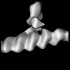
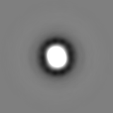
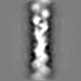
Journal: Commun Biol / Year: 2022 Title: A network of mixed actin polarity in the leading edge of spreading cells. Authors: Wen-Lu Chung / Matthias Eibauer / Wenhong Li / Rajaa Boujemaa-Paterski / Benjamin Geiger / Ohad Medalia / Abstract: Physical interactions of cells with the underlying extracellular matrix (ECM) play key roles in multiple cellular processes. The actin cytoskeleton is a central driver and regulator of cellular ...Physical interactions of cells with the underlying extracellular matrix (ECM) play key roles in multiple cellular processes. The actin cytoskeleton is a central driver and regulator of cellular dynamics, that produces membrane-protrusions such as lamellipodia and filopodia. Here, we examined actin organization in expanding lamellipodia during early stages of cell spreading. To gain insight into the 3D actin organization, we plated fibroblasts on galectin-8 coated EM grids, an ECM protein presents in disease states. We then combined cryo-electron tomography with advanced image processing tools for reconstructing the structure of F-actin in the lamellipodia. This approach enabled us to resolve the polarity and orientation of filaments, and the structure of the Arp2/3 complexes associated with F-actin branches. We show that F-actin in lamellipodial protrusions forms a dense network with three distinct sub-domains. One consists primarily of radial filaments, with their barbed ends pointing towards the membrane, the other is enriched with parallel filaments that run between the radial fibers, in addition to an intermediate sub-domain. Surprisingly, a minor, yet significant (~10%) population of actin filaments, are oriented with their barbed-ends towards the cell center. Our results provide structural insights into F-actin assembly and dynamic reorganization in the leading edge of spreading cells.
In the structure databanks used in Yorodumi, some data are registered as the other names, "COVID-19 virus" and "2019-nCoV". Here are the details of the virus and the list of structure data.
Jan 31, 2019. EMDB accession codes are about to change! (news from PDBe EMDB page)
EMDB accession codes are about to change! (news from PDBe EMDB page)
The allocation of 4 digits for EMDB accession codes will soon come to an end. Whilst these codes will remain in use, new EMDB accession codes will include an additional digit and will expand incrementally as the available range of codes is exhausted. The current 4-digit format prefixed with “EMD-” (i.e. EMD-XXXX) will advance to a 5-digit format (i.e. EMD-XXXXX), and so on. It is currently estimated that the 4-digit codes will be depleted around Spring 2019, at which point the 5-digit format will come into force.
The EM Navigator/Yorodumi systems omit the EMD- prefix.
Related info.:Q: What is EMD? / ID/Accession-code notation in Yorodumi/EM Navigator
Yorodumi is a browser for structure data from EMDB, PDB, SASBDB, etc.
This page is also the successor to EM Navigator detail page, and also detail information page/front-end page for Omokage search.
The word "yorodu" (or yorozu) is an old Japanese word meaning "ten thousand". "mi" (miru) is to see.
Related info.:EMDB / PDB / SASBDB / Comparison of 3 databanks / Yorodumi Search / Aug 31, 2016. New EM Navigator & Yorodumi / Yorodumi Papers / Jmol/JSmol / Function and homology information / Changes in new EM Navigator and Yorodumi
 Movie
Movie Controller

 Yorodumi
 Open data
 Basic information
 Map data
 Sample
 Keywords
 Authors
 Switzerland, European Union, 2 items
  Citation
 Structure visualization
 Downloads & links
 EMDB map data format
 emd_15666.png
 http://ftp.pdbj.org/pub/emdb/structures/EMD-15666
 Z (Sec.)
 Y (Row.)
 X (Col.)


 Sample components
 Processing
 Electron microscopy
 FIELD EMISSION GUN
