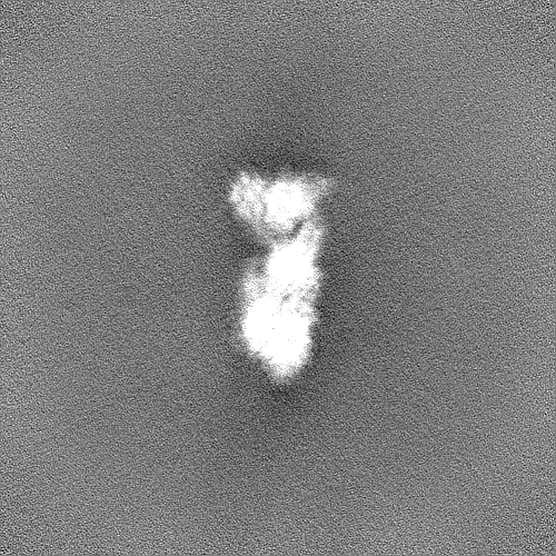
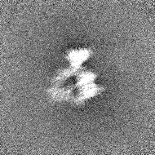
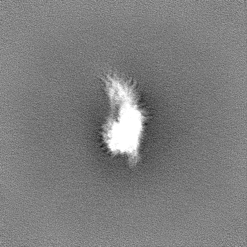
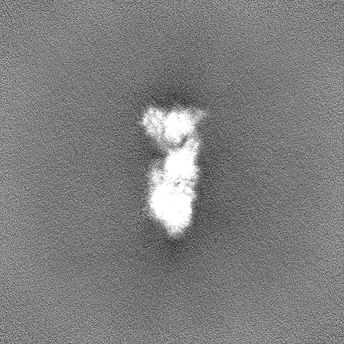
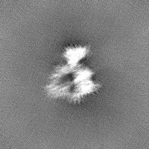
- EMDB-15353: structure of the human beta-cardiac myosin folded-back off state -
+
Open data
ID or keywords:
Loading...
-
Basic information
Entry
Database: EMDB / ID: EMD-15353
Title
structure of the human beta-cardiac myosin folded-back off state
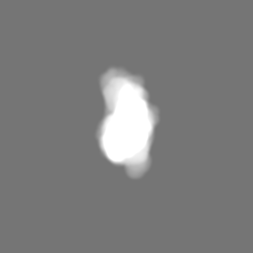
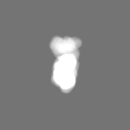
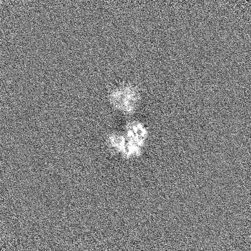
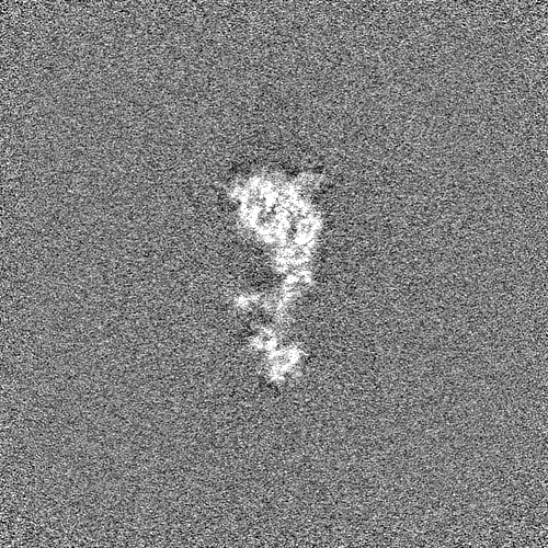
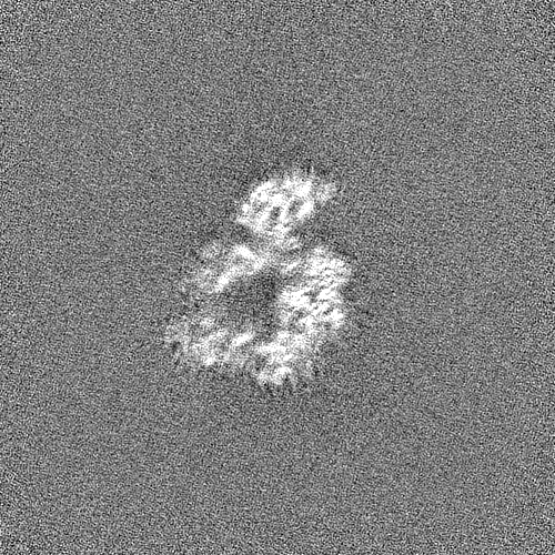
Map data
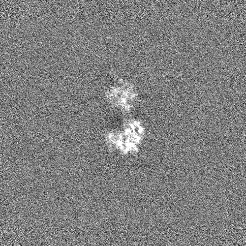
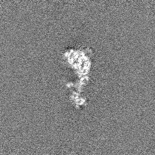
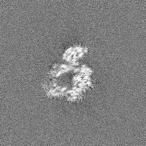
density map of the IHMof b-cardiac myosin
Sample
Complex: human beta-cardiac myosin folded-back off state
Protein or peptide: Myosin-7
Protein or peptide: Myosin light chain 3
Protein or peptide: Myosin regulatory light chain 2, ventricular/cardiac muscle isoform
Ligand: ADENOSINE-5'-DIPHOSPHATE
Ligand: PHOSPHATE ION
Ligand: MAGNESIUM ION
Keywords
Cardiac Myosin / Myosin / Human / folded-back off state / CONTRACTILE PROTEIN
Function / homology
Function and homology information
myosin II heavy chain binding / muscle cell fate specification / regulation of slow-twitch skeletal muscle fiber contraction / regulation of the force of skeletal muscle contraction / regulation of striated muscle contraction / cardiac myofibril / A band / muscle myosin complex / cardiac myofibril assembly / regulation of the force of heart contraction ...myosin II heavy chain binding / muscle cell fate specification / regulation of slow-twitch skeletal muscle fiber contraction / regulation of the force of skeletal muscle contraction / regulation of striated muscle contraction / cardiac myofibril / A band / muscle myosin complex / cardiac myofibril assembly / regulation of the force of heart contraction / transition between fast and slow fiber / myosin filament / Striated Muscle Contraction / cardiac muscle hypertrophy in response to stress / muscle filament sliding / myosin complex / adult heart development / myosin II complex / I band / structural constituent of muscle / ventricular cardiac muscle tissue morphogenesis / microfilament motor activity / myosin heavy chain binding / heart contraction / positive regulation of the force of heart contraction / myofibril / cytoskeletal motor activity / actin monomer binding / skeletal muscle tissue development / ATP metabolic process / striated muscle contraction / skeletal muscle contraction / cardiac muscle contraction / stress fiber / regulation of heart rate / muscle contraction / post-embryonic development / sarcomere / negative regulation of cell growth / Z disc / actin filament binding / heart development / cytoskeleton / calmodulin binding / calcium ion binding / ATP binding / cytoplasm / cytosol Similarity search - Function
: / DNA repair protein XRCC4-like, C-terminal / Myosin tail / Myosin tail / Myosin N-terminal SH3-like domain / Myosin S1 fragment, N-terminal / Myosin, N-terminal, SH3-like / Myosin N-terminal SH3-like domain profile. / Short calmodulin-binding motif containing conserved Ile and Gln residues. / IQ motif, EF-hand binding site ...: / DNA repair protein XRCC4-like, C-terminal / Myosin tail / Myosin tail / Myosin N-terminal SH3-like domain / Myosin S1 fragment, N-terminal / Myosin, N-terminal, SH3-like / Myosin N-terminal SH3-like domain profile. / Short calmodulin-binding motif containing conserved Ile and Gln residues. / IQ motif, EF-hand binding site / Myosin motor domain profile. / Myosin head, motor domain / Myosin head (motor domain) / Myosin. Large ATPases. / IQ motif profile. / Kinesin motor domain superfamily / : / EF-hand domain pair / EF-hand, calcium binding motif / EF-Hand 1, calcium-binding site / EF-hand calcium-binding domain. / EF-hand calcium-binding domain profile. / EF-hand domain / EF-hand domain pair / P-loop containing nucleoside triphosphate hydrolase Similarity search - Domain/homology
National Institutes of Health/National Institute of General Medical Sciences (NIH/NIGMS)
NIH-RM1GM131981-01
United States
Agence Nationale de la Recherche (ANR)
ANR-21-CE11-0022-01
France
National Institutes of Health/National Institute of General Medical Sciences (NIH/NIGMS)
NIH-R01GM33289
United States
Citation
Journal: Nat Commun / Year: 2023 Title: Cryo-EM structure of the folded-back state of human β-cardiac myosin. Authors: Alessandro Grinzato / Daniel Auguin / Carlos Kikuti / Neha Nandwani / Dihia Moussaoui / Divya Pathak / Eaazhisai Kandiah / Kathleen M Ruppel / James A Spudich / Anne Houdusse / Julien Robert-Paganin / Abstract: To save energy and precisely regulate cardiac contractility, cardiac muscle myosin heads are sequestered in an 'off' state that can be converted to an 'on' state when exertion is increased. The 'off' ...To save energy and precisely regulate cardiac contractility, cardiac muscle myosin heads are sequestered in an 'off' state that can be converted to an 'on' state when exertion is increased. The 'off' state is equated with a folded-back structure known as the interacting-heads motif (IHM), which is a regulatory feature of all class-2 muscle and non-muscle myosins. We report here the human β-cardiac myosin IHM structure determined by cryo-electron microscopy to 3.6 Å resolution, providing details of all the interfaces stabilizing the 'off' state. The structure shows that these interfaces are hot spots of hypertrophic cardiomyopathy mutations that are thought to cause hypercontractility by destabilizing the 'off' state. Importantly, the cardiac and smooth muscle myosin IHM structures dramatically differ, providing structural evidence for the divergent physiological regulation of these muscle types. The cardiac IHM structure will facilitate development of clinically useful new molecules that modulate IHM stability.
Type of model: OTHER Details: The coiled-coil S2 region was fitted from the crystal structure (Blankenfeldt et al., 2006; PDB code 2FXM). The IQ2/RLC region was obtained by homology modeling based on the SmMyo2 IHM ...Details: The coiled-coil S2 region was fitted from the crystal structure (Blankenfeldt et al., 2006; PDB code 2FXM). The IQ2/RLC region was obtained by homology modeling based on the SmMyo2 IHM (Heissler et al., 2021; PDB code 7MF3).
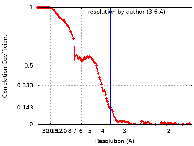
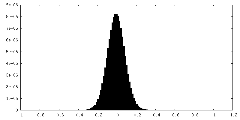
Final reconstruction
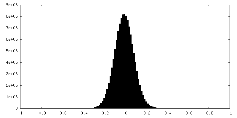
Resolution.type: BY AUTHOR / Resolution: 3.6 Å / Resolution method: FSC 0.143 CUT-OFF / Number images used: 213596
Initial angle assignment
Type: MAXIMUM LIKELIHOOD
Final angle assignment
Type: MAXIMUM LIKELIHOOD
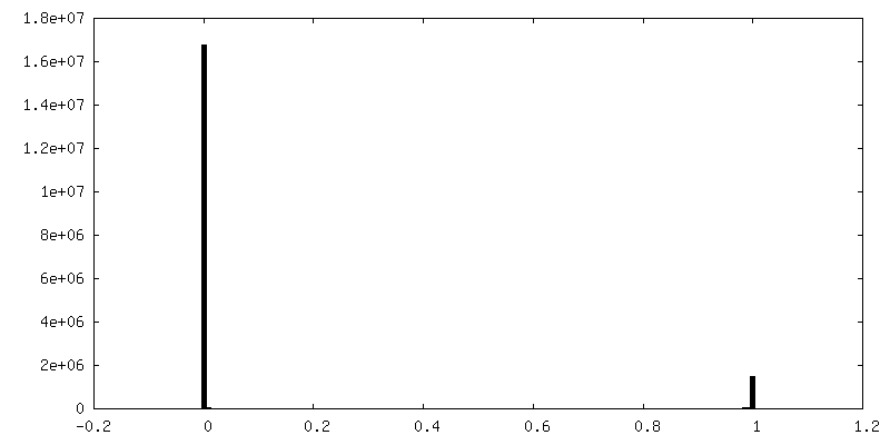
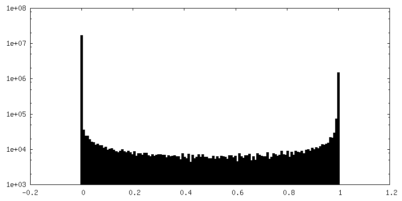
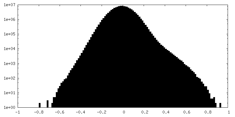
FSC plot (resolution estimation)
+
About Yorodumi
-
News
-
Feb 9, 2022. New format data for meta-information of EMDB entries
New format data for meta-information of EMDB entries
Version 3 of the EMDB header file is now the official format.
The previous official version 1.9 will be removed from the archive.
In the structure databanks used in Yorodumi, some data are registered as the other names, "COVID-19 virus" and "2019-nCoV". Here are the details of the virus and the list of structure data.
Jan 31, 2019. EMDB accession codes are about to change! (news from PDBe EMDB page)
EMDB accession codes are about to change! (news from PDBe EMDB page)
The allocation of 4 digits for EMDB accession codes will soon come to an end. Whilst these codes will remain in use, new EMDB accession codes will include an additional digit and will expand incrementally as the available range of codes is exhausted. The current 4-digit format prefixed with “EMD-” (i.e. EMD-XXXX) will advance to a 5-digit format (i.e. EMD-XXXXX), and so on. It is currently estimated that the 4-digit codes will be depleted around Spring 2019, at which point the 5-digit format will come into force.
The EM Navigator/Yorodumi systems omit the EMD- prefix.
Related info.:Q: What is EMD? / ID/Accession-code notation in Yorodumi/EM Navigator
Yorodumi is a browser for structure data from EMDB, PDB, SASBDB, etc.
This page is also the successor to EM Navigator detail page, and also detail information page/front-end page for Omokage search.
The word "yorodu" (or yorozu) is an old Japanese word meaning "ten thousand". "mi" (miru) is to see.
Related info.:EMDB / PDB / SASBDB / Comparison of 3 databanks / Yorodumi Search / Aug 31, 2016. New EM Navigator & Yorodumi / Yorodumi Papers / Jmol/JSmol / Function and homology information / Changes in new EM Navigator and Yorodumi
 Movie
Movie Controller

 Yorodumi
 Open data
 Basic information
 Map data
 Sample
 Keywords
 Function and homology information
 Homo sapiens (human)
 Authors
 United States,
  France, 3 items
  Citation
 Structure visualization
 Downloads & links
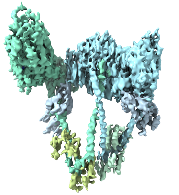 emd_15353.png
 http://ftp.pdbj.org/pub/emdb/structures/EMD-15353


 Z (Sec.)
 Y (Row.)
 X (Col.)


 Sample components

 Processing
 Electron microscopy
 FIELD EMISSION GUN