 Movie
Movie Controller

[English] 日本語
 Yorodumi
- EMDB-15274: Single Particle cryo-EM of the lipid binding protein P116 (MPN213... -
+ Open data

- Basic information
| Entry |  | ||||||||||||
|---|---|---|---|---|---|---|---|---|---|---|---|---|---|
| Title | Single Particle cryo-EM of the lipid binding protein P116 (MPN213) from Mycoplasma pneumoniae at 3.3 Angstrom resolution. | ||||||||||||
 Map data Map data | |||||||||||||
 Sample Sample |
| ||||||||||||
 Keywords Keywords | Lipid binding protein P116 (MPN213) from Mycoplasma pneumoniae / LIPID BINDING / LIPID BINDING PROTEIN | ||||||||||||
| Function / homology | : / : / P116-like protein / membrane / Uncharacterized protein MG075 homolog Function and homology information Function and homology information | ||||||||||||
| Biological species |  Mycoplasma pneumoniae M129 (bacteria) Mycoplasma pneumoniae M129 (bacteria) | ||||||||||||
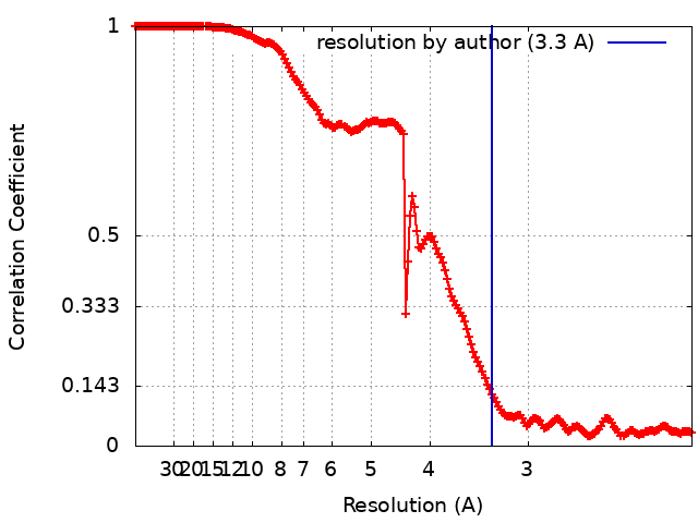
| Method | single particle reconstruction / cryo EM / Resolution: 3.3 Å | ||||||||||||
 Authors Authors | Sprankel L / Vizarraga D | ||||||||||||
| Funding support |  Germany, 3 items Germany, 3 items
| ||||||||||||
 Citation Citation | Journal: Nat Struct Mol Biol / Year: 2023 Title: Essential protein P116 extracts cholesterol and other indispensable lipids for Mycoplasmas. Authors: Lasse Sprankel / David Vizarraga / Jesús Martín / Sina Manger / Jakob Meier-Credo / Marina Marcos / Josep Julve / Noemi Rotllan / Margot P Scheffer / Joan Carles Escolà-Gil / Julian D ...Authors: Lasse Sprankel / David Vizarraga / Jesús Martín / Sina Manger / Jakob Meier-Credo / Marina Marcos / Josep Julve / Noemi Rotllan / Margot P Scheffer / Joan Carles Escolà-Gil / Julian D Langer / Jaume Piñol / Ignacio Fita / Achilleas S Frangakis /  Abstract: Mycoplasma pneumoniae, responsible for approximately 30% of community-acquired human pneumonia, needs to extract lipids from the host environment for survival and proliferation. Here, we report a ...Mycoplasma pneumoniae, responsible for approximately 30% of community-acquired human pneumonia, needs to extract lipids from the host environment for survival and proliferation. Here, we report a comprehensive structural and functional analysis of the previously uncharacterized protein P116 (MPN_213). Single-particle cryo-electron microscopy of P116 reveals a homodimer presenting a previously unseen fold, forming a huge hydrophobic cavity, which is fully accessible to solvent. Lipidomics analysis shows that P116 specifically extracts lipids such as phosphatidylcholine, sphingomyelin and cholesterol. Structures of different conformational states reveal the mechanism by which lipids are extracted. This finding immediately suggests a way to control Mycoplasma infection by interfering with lipid uptake. | ||||||||||||
| History |
|
- Structure visualization
| Supplemental images |
|---|

- Downloads & links
-EMDB archive
| Map data | emd_15274.map.gz | 484 MB | EMDB map data format | |
|---|---|---|---|---|
| Header (meta data) | emd-15274-v30.xmlemd-15274.xml | 21.9 KB 21.9 KB | Display Display | EMDB header |
| FSC (resolution estimation) | emd_15274_fsc.xml | 17.7 KB | Display | FSC data file |
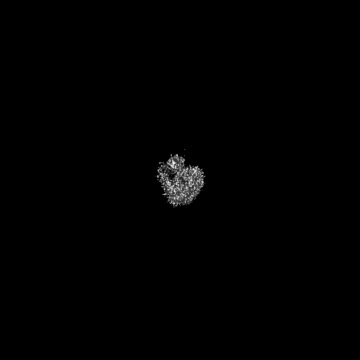
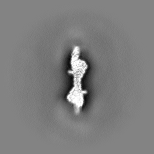
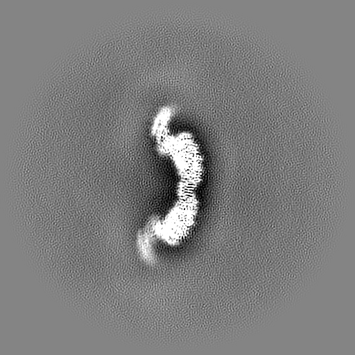
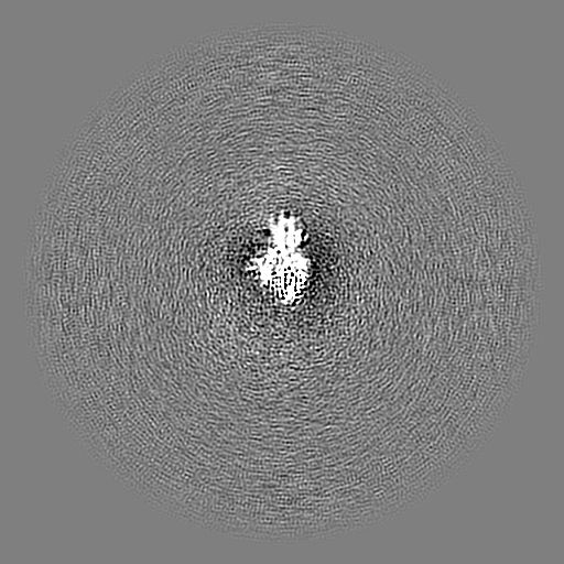
| Images |  emd_15274.png emd_15274.png | 44.3 KB | ||
| Masks | emd_15274_msk_1.map | 512 MB | Mask map | |
| Filedesc metadata | emd-15274.cif.gz | 7.1 KB | ||
| Others | emd_15274_half_map_1.map.gzemd_15274_half_map_2.map.gz | 475.7 MB 475.7 MB | ||
| Archive directory |  http://ftp.pdbj.org/pub/emdb/structures/EMD-15274ftp://ftp.pdbj.org/pub/emdb/structures/EMD-15274 http://ftp.pdbj.org/pub/emdb/structures/EMD-15274ftp://ftp.pdbj.org/pub/emdb/structures/EMD-15274 | HTTPS FTP |
-Related structure data
| Related structure data |  8a9aMC  8a9bC M: atomic model generated by this map C: citing same article ( |
|---|---|
| Similar structure data |


-Links
| EMDB pages | EMDB (EBI/PDBe) / EMDataResource |
|---|
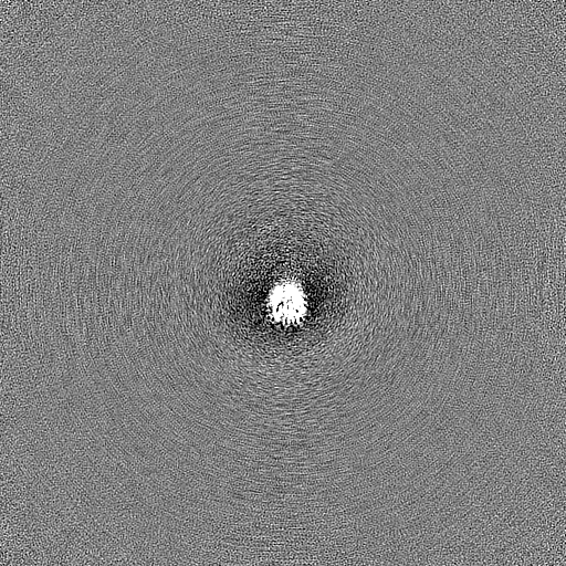
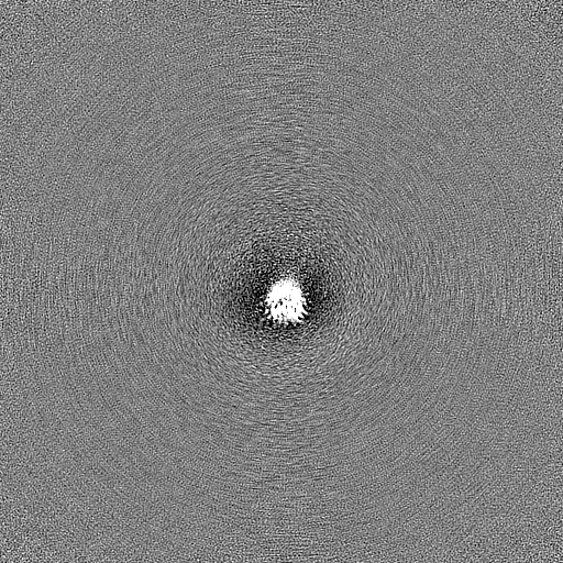
-Map
| File | Download / File: emd_15274.map.gz / Format: CCP4 / Size: 512 MB / Type: IMAGE STORED AS FLOATING POINT NUMBER (4 BYTES) | ||||||||||||||||||||||||||||||||||||
|---|---|---|---|---|---|---|---|---|---|---|---|---|---|---|---|---|---|---|---|---|---|---|---|---|---|---|---|---|---|---|---|---|---|---|---|---|---|
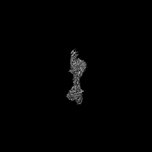
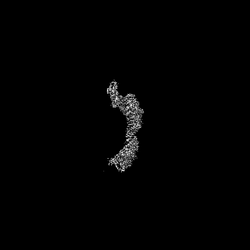
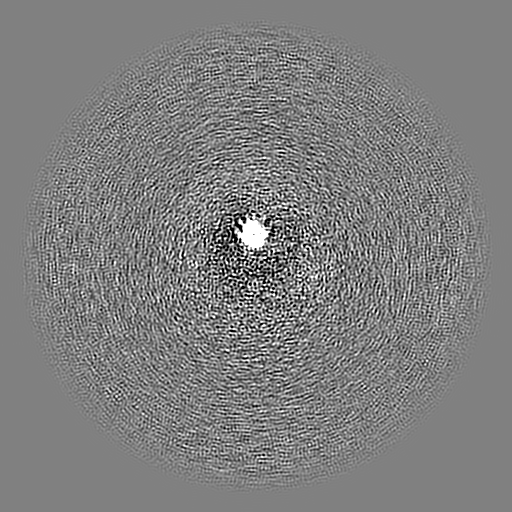
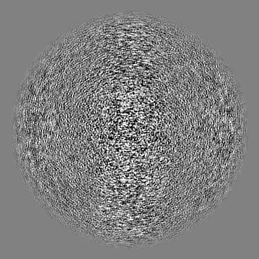
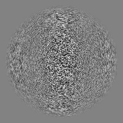
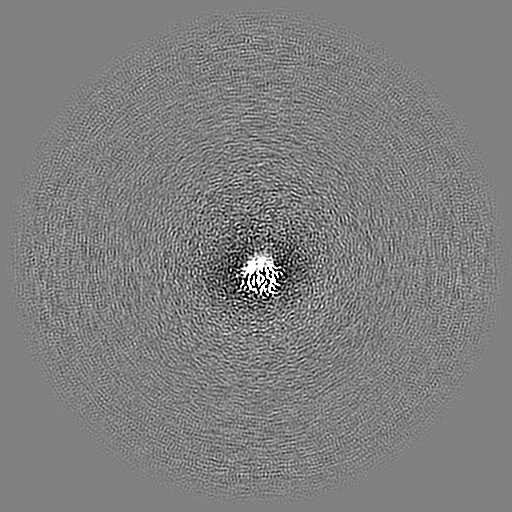
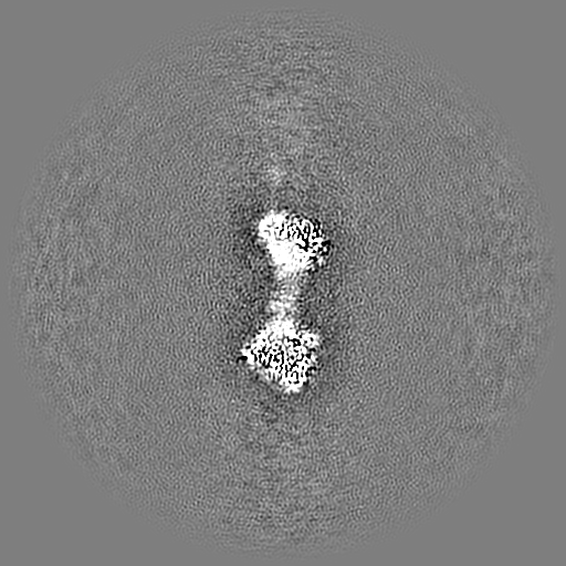
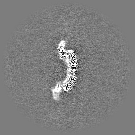
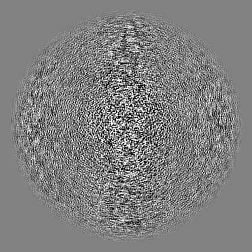
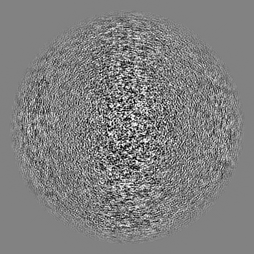
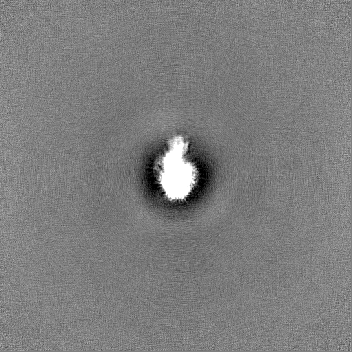
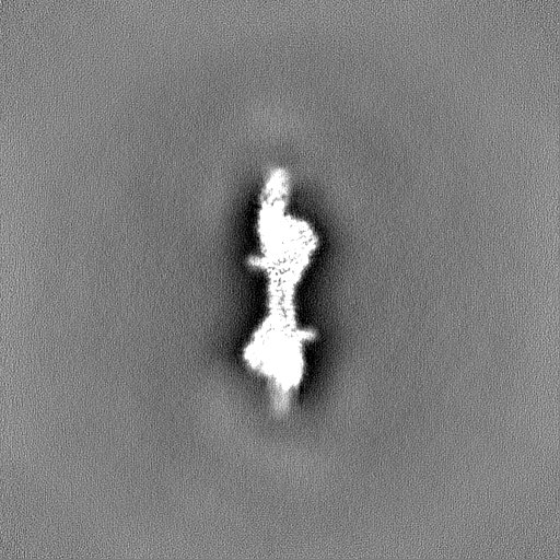
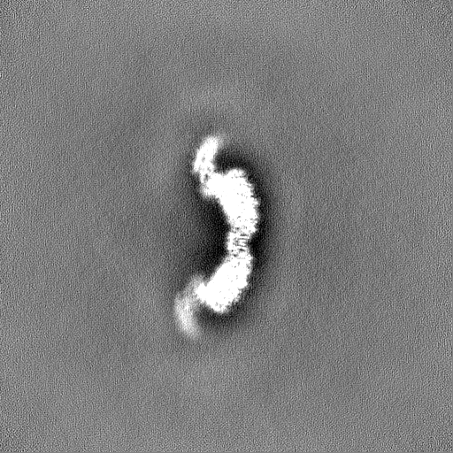
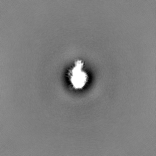
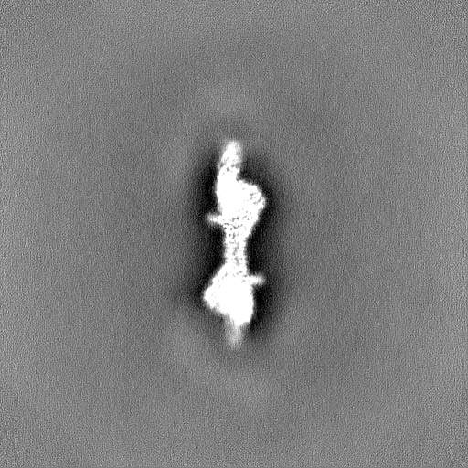
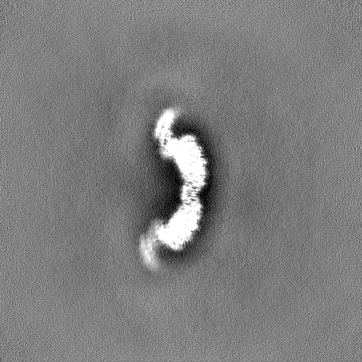
| Projections & slices | Image control
Images are generated by Spider. | ||||||||||||||||||||||||||||||||||||
| Voxel size | X=Y=Z: 1.05 Å | ||||||||||||||||||||||||||||||||||||
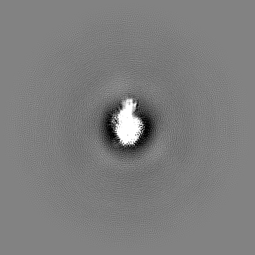
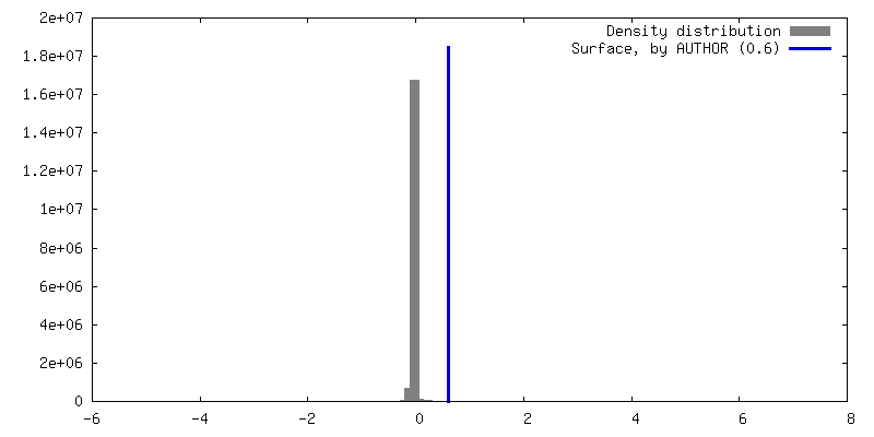
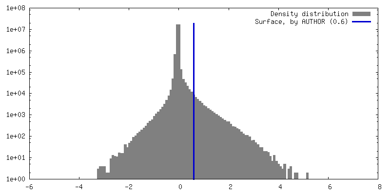
| Density |
| ||||||||||||||||||||||||||||||||||||
| Symmetry | Space group: 1 | ||||||||||||||||||||||||||||||||||||
| Details | EMDB XML:
|
 Z (Sec.)
 Y (Row.)
 X (Col.)


-Supplemental data
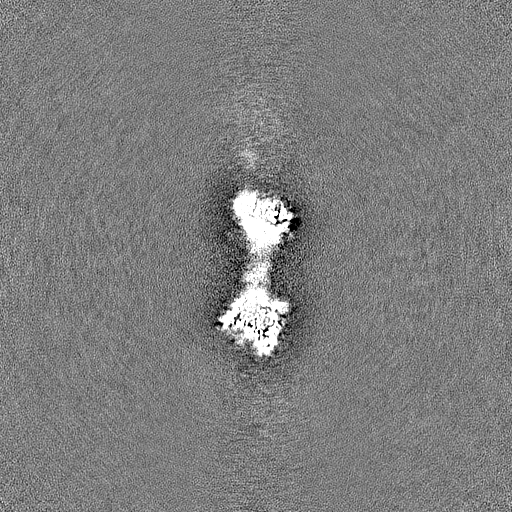
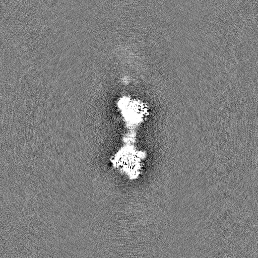
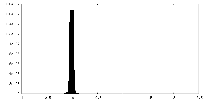
-Mask #1
| File | emd_15274_msk_1.map | ||||||||||||
|---|---|---|---|---|---|---|---|---|---|---|---|---|---|
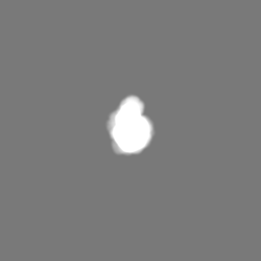
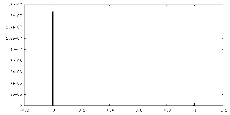
| Projections & Slices |
| ||||||||||||
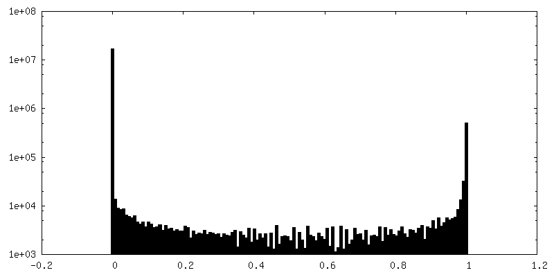
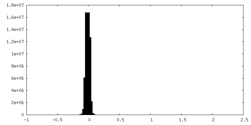
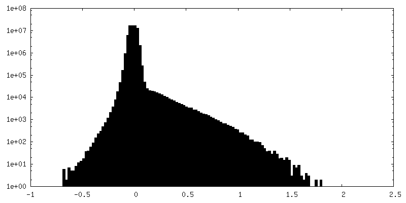
| Density Histograms |


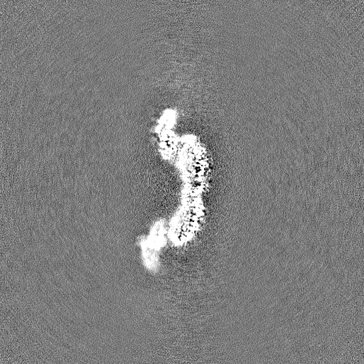
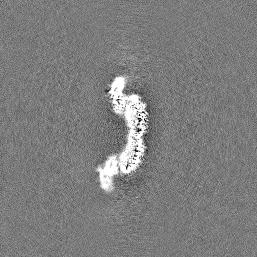
-Half map: #2
| File | emd_15274_half_map_1.map | ||||||||||||
|---|---|---|---|---|---|---|---|---|---|---|---|---|---|
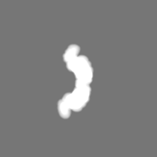
| Projections & Slices |
| ||||||||||||
| Density Histograms |
-Half map: #1
| File | emd_15274_half_map_2.map | ||||||||||||
|---|---|---|---|---|---|---|---|---|---|---|---|---|---|
| Projections & Slices |
| ||||||||||||
| Density Histograms |

- Sample components
-Entire : P116 homodimer
| Entire | Name: P116 homodimer |
|---|---|
| Components |
|
-Supramolecule #1: P116 homodimer
| Supramolecule | Name: P116 homodimer / type: complex / ID: 1 / Parent: 0 / Macromolecule list: all |
|---|---|
| Source (natural) | Organism: Mycoplasma pneumoniae M129 (bacteria) |
-Macromolecule #1: Lipid binding protein P116 (MPN213)
| Macromolecule | Name: Lipid binding protein P116 (MPN213) / type: protein_or_peptide / ID: 1 / Number of copies: 2 / Enantiomer: LEVO |
|---|---|
| Source (natural) | Organism: Mycoplasma pneumoniae M129 (bacteria) / Strain: ATCC 29342 / M129 |
| Molecular weight | Theoretical: 114.149852 KDa |
| Recombinant expression | Organism: |
| Sequence | String: NKTHQVEHES EQSDFQDIRF GLNSVKLPKA QPAAATRITV ENGTDKLVNY KSSPQQLFLA KNALKDKLQG EFDKFLSDAK AFPALTADL QEWVDQQLFN PNQSFFDLSA PRSNFTLSSD KKASLDFIFR FTNFTESVQL LKLPEGVSVV VDSKQSFDYY V NASAQKLL ...String: NKTHQVEHES EQSDFQDIRF GLNSVKLPKA QPAAATRITV ENGTDKLVNY KSSPQQLFLA KNALKDKLQG EFDKFLSDAK AFPALTADL QEWVDQQLFN PNQSFFDLSA PRSNFTLSSD KKASLDFIFR FTNFTESVQL LKLPEGVSVV VDSKQSFDYY V NASAQKLL VLPLSLPDYT LGLNYMFDHI TLNGKVVNKF SFNPFKTNLN LAFSNVYNGV DVFEAQKNLV GKGKYLNTHV KA EDVKKDV NANIKNQFDI AKIIAELMGK ALKEFGNQQE GQPLSFLKVM DKVKEDFEKL FNLVRPGLGK FVKDLIQSSS QAE NKITVY KLIFDNKKTI LNLLKELSIP ELNSSLGLVD VLFDGITDSD GLYERLQSFK DLIVPAVKTN EKTAALSPLI EELL TQKDT YVFDLIQKHK GILTNLLKNF LADFQKSTPF MADQVAIFTE LFDNEGAFDL FGEADFVDKI AELFLTKRTV KNGEK IETK DSLLVTSLKS LLGEKVAALG DLLDSYIFKN ELLNRSVEVA KAEAKDTKGA TDYKKEQAKA LKKLFKHIGE NTLSKT NLD KITLKEVKNT ENVELEETET TLKVKKLDVE YKVELGNFEI KNGLIKAMLE FLPDTKDLET TLDKLLFKGE SYKAMKD KY IKEGFPGYGW AKGVVPGAFE SIENTFKSAI DKTKSIRDLF GDMLFGNDLS SVKETDSFIT LGGSFDIKYG GENLNVLP A YYSLINSEIG YQIIGVDTTI DATKVKVELK NKEYKGKSPA INGQVKLSQS FFNVWTNMFD SITKQIFQKK YEFKDNIQV FARNEDNTSR LELDISDPEQ RVIPFAFVDG FGIQLKAVDK NITKEAGNTE PKSPVIQLYE ALNKEKDQKQ QSKQSPKQLD TKTQLGYLL KLGDNWSKDD YKSLIDDTII NNNYLEASFN SKITVDRLGI PIDLWLFKIW PKFNLEIPMQ GSLQLYSSSV I FPYGIYDT SVQDAAKIVK RLNFTDMGFK LNDPKPNFWF VGFKHHHHH UniProtKB: Uncharacterized protein MG075 homolog |
-Experimental details
-Structure determination
| Method | cryo EM |
|---|---|
 Processing Processing | single particle reconstruction |
| Aggregation state | particle |
-Sample preparation
| Concentration | 0.6 mg/mL | ||||||
|---|---|---|---|---|---|---|---|
| Buffer | pH: 7.4 / Component:
| ||||||
| Grid | Model: C-flat-1.2/1.3 / Material: COPPER / Mesh: 300 / Pretreatment - Type: GLOW DISCHARGE / Pretreatment - Time: 45 sec. / Pretreatment - Atmosphere: AIR | ||||||
| Vitrification | Cryogen name: ETHANE / Chamber humidity: 100 % / Chamber temperature: 277.15 K / Instrument: FEI VITROBOT MARK IV |
- Electron microscopy
| Microscope | FEI TITAN KRIOS |
|---|---|
| Specialist optics | Energy filter - Name: GIF Quantum SE / Energy filter - Slit width: 20 eV |
| Image recording | Film or detector model: GATAN K2 SUMMIT (4k x 4k) / Detector mode: COUNTING / Digitization - Dimensions - Width: 3710 pixel / Digitization - Dimensions - Height: 3838 pixel / Number grids imaged: 1 / Number real images: 4376 / Average electron dose: 50.0 e/Å2 |
| Electron beam | Acceleration voltage: 300 kV / Electron source:  FIELD EMISSION GUN FIELD EMISSION GUN |
| Electron optics | C2 aperture diameter: 70.0 µm / Calibrated defocus max: 3.5 µm / Calibrated defocus min: 1.0 µm / Calibrated magnification: 130000 / Illumination mode: FLOOD BEAM / Imaging mode: BRIGHT FIELD / Cs: 2.7 mm / Nominal defocus max: 3.5 µm / Nominal defocus min: 1.0 µm / Nominal magnification: 130000 |
| Sample stage | Specimen holder model: FEI TITAN KRIOS AUTOGRID HOLDER / Cooling holder cryogen: NITROGEN |
| Experimental equipment |  Model: Titan Krios / Image courtesy: FEI Company |
+Image processing
-Atomic model buiding 1
| Refinement | Space: REAL / Protocol: AB INITIO MODEL |
|---|---|
| Output model | PDB-8a9a: |
