 ムービー
ムービー コントローラー


+ データを開く

- 基本情報
基本情報
| 登録情報 |  | ||||||||||||||||||||||||
|---|---|---|---|---|---|---|---|---|---|---|---|---|---|---|---|---|---|---|---|---|---|---|---|---|---|
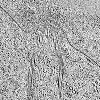
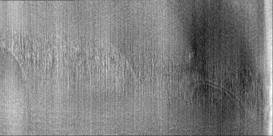
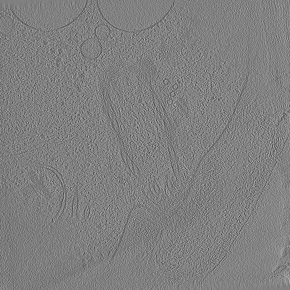
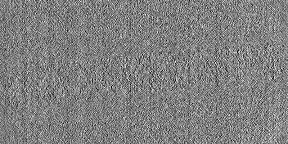
| タイトル | Tomogram #3 of the C. reinhardtii ciliary transition zone | ||||||||||||||||||||||||
 マップデータ マップデータ | Tomogram at bin4 deconvoluted using ad-hoc deconvolution filter (tom_deconv.m) | ||||||||||||||||||||||||
 試料 試料 |
| ||||||||||||||||||||||||
 キーワード キーワード | cilia / transition zone / flagella / ciliary pore / intraflagellar transport / motility / signaling / basal body / microtubules / chlamydomonas / STRUCTURAL PROTEIN | ||||||||||||||||||||||||
| 生物種 |   Chlamydomonas reinhardtii (クラミドモナス) Chlamydomonas reinhardtii (クラミドモナス) | ||||||||||||||||||||||||
| 手法 | 電子線トモグラフィー法 / クライオ電子顕微鏡法 | ||||||||||||||||||||||||
 データ登録者 データ登録者 | van den Hoek HG / Schaffer M / Erdmann PS / Wan WN / Plitzko JM / Baumeister W / Engel BD | ||||||||||||||||||||||||
| 資金援助 | European Union,  ドイツ, ドイツ,  スイス, 7件 スイス, 7件
| ||||||||||||||||||||||||
 引用 引用 | ジャーナル: Science / 年: 2022 タイトル: In situ architecture of the ciliary base reveals the stepwise assembly of intraflagellar transport trains. 著者: Hugo van den Hoek / Nikolai Klena / Mareike A Jordan / Gonzalo Alvarez Viar / Ricardo D Righetto / Miroslava Schaffer / Philipp S Erdmann / William Wan / Stefan Geimer / Jürgen M Plitzko / ...著者: Hugo van den Hoek / Nikolai Klena / Mareike A Jordan / Gonzalo Alvarez Viar / Ricardo D Righetto / Miroslava Schaffer / Philipp S Erdmann / William Wan / Stefan Geimer / Jürgen M Plitzko / Wolfgang Baumeister / Gaia Pigino / Virginie Hamel / Paul Guichard / Benjamin D Engel /   要旨: The cilium is an antenna-like organelle that performs numerous cellular functions, including motility, sensing, and signaling. The base of the cilium contains a selective barrier that regulates the ...The cilium is an antenna-like organelle that performs numerous cellular functions, including motility, sensing, and signaling. The base of the cilium contains a selective barrier that regulates the entry of large intraflagellar transport (IFT) trains, which carry cargo proteins required for ciliary assembly and maintenance. However, the native architecture of the ciliary base and the process of IFT train assembly remain unresolved. In this work, we used in situ cryo-electron tomography to reveal native structures of the transition zone region and assembling IFT trains at the ciliary base in . We combined this direct cellular visualization with ultrastructure expansion microscopy to describe the front-to-back stepwise assembly of IFT trains: IFT-B forms the backbone, onto which bind IFT-A, dynein-1b, and finally kinesin-2 before entry into the cilium. | ||||||||||||||||||||||||
| 履歴 |
|
- 構造の表示
構造の表示
| 添付画像 |
|---|
- ダウンロードとリンク
-EMDBアーカイブ
| マップデータ | emd_15262.map.gz | 385.5 MB |  EMDBマップデータ形式 EMDBマップデータ形式 | |
|---|---|---|---|---|
| ヘッダ (付随情報) | emd-15262-v30.xmlemd-15262.xml | 14.1 KB 14.1 KB | 表示 表示 | EMDBヘッダ |
| 画像 |  emd_15262.png emd_15262.png | 295 KB | ||
| Filedesc metadata | emd-15262.cif.gz | 4.6 KB | ||
| その他 | emd_15262_additional_1.map.gz | 380.2 MB | ||
| アーカイブディレクトリ |  http://ftp.pdbj.org/pub/emdb/structures/EMD-15262ftp://ftp.pdbj.org/pub/emdb/structures/EMD-15262 http://ftp.pdbj.org/pub/emdb/structures/EMD-15262ftp://ftp.pdbj.org/pub/emdb/structures/EMD-15262 | HTTPS FTP |
-関連構造データ
| 関連構造データ | C: 同じ文献を引用 ( |
|---|---|
| 電子顕微鏡画像生データ | EMPIAR-11078 (タイトル: In situ cryo-electron tomography of the C. reinhardtii ciliary transition zone Data size: 302.0 Data #1: Deconvoluted bin4 tomograms for visualization [reconstructed volumes] Data #2: CTF-corrected bin4 tomograms [reconstructed volumes] Data #3: CTF-corrected bin2 tomograms [reconstructed volumes] Data #4: Subtomograms at bin2 [subtomograms] / Data #5: Aligned tilt series stacks [tilt series] Data #6: Unaligned tilt series raw frames [micrographs - multiframe]) |


-リンク
| EMDBのページ | EMDB (EBI/PDBe) / EMDataResource |
|---|
-マップ
| ファイル | ダウンロード / ファイル: emd_15262.map.gz / 形式: CCP4 / 大きさ: 762.2 MB / タイプ: IMAGE STORED AS SIGNED INTEGER (2 BYTES) | ||||||||||||||||||||||||||||||||
|---|---|---|---|---|---|---|---|---|---|---|---|---|---|---|---|---|---|---|---|---|---|---|---|---|---|---|---|---|---|---|---|---|---|
| 注釈 | Tomogram at bin4 deconvoluted using ad-hoc deconvolution filter (tom_deconv.m) | ||||||||||||||||||||||||||||||||
| 投影像・断面図 | 画像のコントロール
画像は Spider により作成 これらの図は立方格子座標系で作成されたものです | ||||||||||||||||||||||||||||||||
| ボクセルのサイズ | X=Y=Z: 13.68 Å | ||||||||||||||||||||||||||||||||
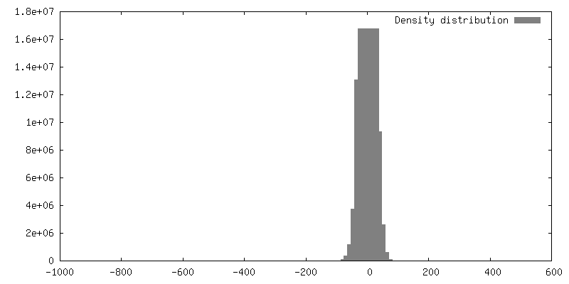
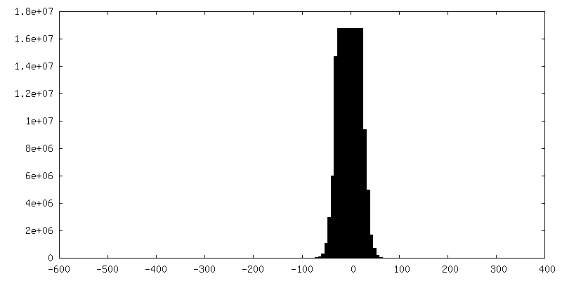
| 密度 |
| ||||||||||||||||||||||||||||||||
| 対称性 | 空間群: 1 | ||||||||||||||||||||||||||||||||
| 詳細 | EMDB XML:
|
 Z (Sec.)
 Y (Row.)
 X (Col.)


-添付データ
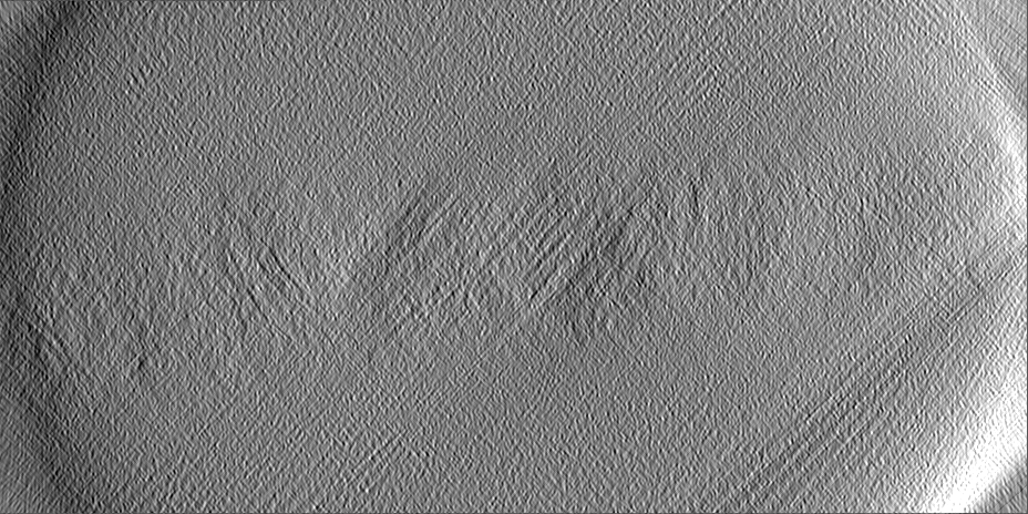
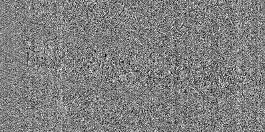
-追加マップ: Tomogram reconstructed with IMOD at bin4
| ファイル | emd_15262_additional_1.map | ||||||||||||
|---|---|---|---|---|---|---|---|---|---|---|---|---|---|
| 注釈 | Tomogram reconstructed with IMOD at bin4 | ||||||||||||
| 投影像・断面図 |
| ||||||||||||
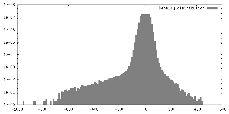
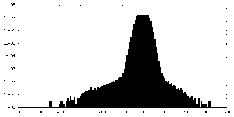
| 密度ヒストグラム |

- 試料の構成要素
-全体 : C. reinhardtii ciliary transition zone
| 全体 | 名称: C. reinhardtii ciliary transition zone |
|---|---|
| 要素 |
|
-超分子 #1: C. reinhardtii ciliary transition zone
| 超分子 | 名称: C. reinhardtii ciliary transition zone / タイプ: cell / ID: 1 / 親要素: 0 |
|---|---|
| 由来(天然) | 生物種: Chlamydomonas reinhardtii (クラミドモナス) 株: CC-3994 |
-実験情報
-構造解析
| 手法 | クライオ電子顕微鏡法 |
|---|---|
 解析 解析 | 電子線トモグラフィー法 |
| 試料の集合状態 | cell |
-試料調製
| 緩衝液 | pH: 7 |
|---|---|
| グリッド | モデル: Quantifoil R2/1 / 材質: COPPER / 支持フィルム - 材質: CARBON / 支持フィルム - トポロジー: CONTINUOUS / 前処理 - タイプ: GLOW DISCHARGE |
| 凍結 | 凍結剤: ETHANE-PROPANE / 装置: FEI VITROBOT MARK IV |
| 切片作成 | 集束イオンビーム - 装置: OTHER / 集束イオンビーム - イオン: OTHER / 集束イオンビーム - 電圧: 30 / 集束イオンビーム - 電流: 0.03 / 集束イオンビーム - 時間: 2700 / 集束イオンビーム - 温度: 91 K / 集束イオンビーム - Initial thickness: 6000 / 集束イオンビーム - 最終 厚さ: 180 集束イオンビーム - 詳細: The value given for _em_focused_ion_beam.instrument is FEI Quanta FIB. This is not in a list of allowed values {'DB235', 'OTHER'} so OTHER is written into the XML file. |
- 電子顕微鏡法
| 顕微鏡 | FEI TITAN KRIOS |
|---|---|
| 特殊光学系 | エネルギーフィルター - 名称: GIF Quantum LS / エネルギーフィルター - スリット幅: 20 eV |
| 撮影 | フィルム・検出器のモデル: GATAN K2 SUMMIT (4k x 4k) 検出モード: COUNTING / 平均電子線量: 100.0 e/Å2 |
| 電子線 | 加速電圧: 300 kV / 電子線源:  FIELD EMISSION GUN FIELD EMISSION GUN |
| 電子光学系 | 照射モード: FLOOD BEAM / 撮影モード: BRIGHT FIELD / Cs: 2.7 mm / 最大 デフォーカス(公称値): 6.0 µm / 最小 デフォーカス(公称値): 4.0 µm / 倍率(公称値): 42000 |
| 試料ステージ | 試料ホルダーモデル: FEI TITAN KRIOS AUTOGRID HOLDER ホルダー冷却材: NITROGEN |
| 実験機器 |  モデル: Titan Krios / 画像提供: FEI Company |
-画像解析
| 最終 再構成 | アルゴリズム: BACK PROJECTION / ソフトウェア - 名称: STOPGAP (ver. 0.3) / 使用した粒子像数: 60 |
|---|
