 ムービー
ムービー コントローラー


+ データを開く

- 基本情報
基本情報
| 登録情報 |  | |||||||||
|---|---|---|---|---|---|---|---|---|---|---|
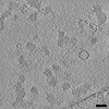
| タイトル | Whole cell cryo-electron tomogram of Apilactobacillus kunkeei and secreted nanoparticles (TS_04.2) | |||||||||
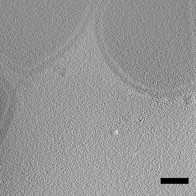
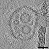
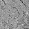
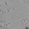
 マップデータ マップデータ | Cryo-electron tomogram of A. kunkeei strain A1401 showing the release of extracellular particles. This tomogram is a bit faint but shows the release of several particles from the cell surface. | |||||||||
 試料 試料 |
| |||||||||
 キーワード キーワード | Membrane vesicles / extracellular protein complexes / UNKNOWN FUNCTION | |||||||||
| 生物種 |  Apilactobacillus kunkeei (バクテリア) Apilactobacillus kunkeei (バクテリア) | |||||||||
| 手法 | 電子線トモグラフィー法 / クライオ電子顕微鏡法 / 解像度: 8.452 Å | |||||||||
 データ登録者 データ登録者 | Seeger C / Andersson SGE | |||||||||
| 資金援助 |  スウェーデン, 1件 スウェーデン, 1件
| |||||||||
 引用 引用 | ジャーナル: To Be Published タイトル: Cryo-eletron tomogram of secreted particles from Apilactobacillus kunkeei (TS_01) 著者: Seeger C / Andersson SGE | |||||||||
| 履歴 |
|
- 構造の表示
構造の表示
| 添付画像 |
|---|
- ダウンロードとリンク
-EMDBアーカイブ
| マップデータ | emd_15200.map.gz | 331.6 MB |  EMDBマップデータ形式 EMDBマップデータ形式 | |
|---|---|---|---|---|
| ヘッダ (付随情報) | emd-15200-v30.xmlemd-15200.xml | 10.5 KB 10.5 KB | 表示 表示 | EMDBヘッダ |
| 画像 |  emd_15200.png emd_15200.png | 277.4 KB | ||
| アーカイブディレクトリ |  http://ftp.pdbj.org/pub/emdb/structures/EMD-15200ftp://ftp.pdbj.org/pub/emdb/structures/EMD-15200 http://ftp.pdbj.org/pub/emdb/structures/EMD-15200ftp://ftp.pdbj.org/pub/emdb/structures/EMD-15200 | HTTPS FTP |
-検証レポート
| 文書・要旨 | emd_15200_validation.pdf.gz | 518.9 KB | 表示 | EMDB検証レポート |
|---|---|---|---|---|
| 文書・詳細版 | emd_15200_full_validation.pdf.gz | 518.5 KB | 表示 | |
| XML形式データ | emd_15200_validation.xml.gz | 4.8 KB | 表示 | |
| CIF形式データ | emd_15200_validation.cif.gz | 5.9 KB | 表示 | |
| アーカイブディレクトリ | https://ftp.pdbj.org/pub/emdb/validation_reports/EMD-15200ftp://ftp.pdbj.org/pub/emdb/validation_reports/EMD-15200 | HTTPS FTP |
-関連構造データ


-リンク
| EMDBのページ | EMDB (EBI/PDBe) / EMDataResource |
|---|
-マップ
| ファイル | ダウンロード / ファイル: emd_15200.map.gz / 形式: CCP4 / 大きさ: 492.2 MB / タイプ: IMAGE STORED AS SIGNED BYTE | ||||||||||||||||||||
|---|---|---|---|---|---|---|---|---|---|---|---|---|---|---|---|---|---|---|---|---|---|
| 注釈 | Cryo-electron tomogram of A. kunkeei strain A1401 showing the release of extracellular particles. This tomogram is a bit faint but shows the release of several particles from the cell surface. | ||||||||||||||||||||
| ボクセルのサイズ | X=Y=Z: 8.452 Å | ||||||||||||||||||||
| 密度 |
| ||||||||||||||||||||
| 対称性 | 空間群: 1 | ||||||||||||||||||||
| 詳細 | EMDB XML:
|
-添付データ
- 試料の構成要素
-全体 : Extracellular nanoparticles (membrane vesicles, protein complexes...
| 全体 | 名称: Extracellular nanoparticles (membrane vesicles, protein complexes) secreted from the cell surface of Apilactobacillus kunkeei. |
|---|---|
| 要素 |
|
-超分子 #1: Extracellular nanoparticles (membrane vesicles, protein complexes...
| 超分子 | 名称: Extracellular nanoparticles (membrane vesicles, protein complexes) secreted from the cell surface of Apilactobacillus kunkeei. タイプ: cell / ID: 1 / 親要素: 0 |
|---|---|
| 由来(天然) | 生物種: Apilactobacillus kunkeei (バクテリア) / 株: A1401 |
-実験情報
-構造解析
| 手法 | クライオ電子顕微鏡法 |
|---|---|
 解析 解析 | 電子線トモグラフィー法 |
| 試料の集合状態 | cell |
-試料調製
| 緩衝液 | pH: 7.4 / 構成要素 - 濃度: 1.0 1 / 構成要素 - 名称: HyClone / 詳細: HyClone |
|---|---|
| グリッド | モデル: Quantifoil R2/2 / 材質: COPPER / メッシュ: 200 / 支持フィルム - 材質: CARBON / 支持フィルム - トポロジー: HOLEY / 支持フィルム - Film thickness: 2 / 前処理 - タイプ: GLOW DISCHARGE / 前処理 - 時間: 60 sec. / 前処理 - 雰囲気: AIR / 前処理 - 気圧: 101.325 kPa |
| 凍結 | 凍結剤: ETHANE / チャンバー内湿度: 100 % / チャンバー内温度: 277 K / 装置: FEI VITROBOT MARK IV / 詳細: manual back-side blotting of 5 seconds. |
| 詳細 | Cells were cultivated until log-phase (OD approx. 0.3), gently pelleted (500 x g, 2 min, RT) and re-suspended in HyClone to a final concentration of OD600 approx. 1. |
| 切片作成 | その他: NO SECTIONING |
| 位置合わせマーカー | Manufacturer: Aurion / 直径: 10 nm |
- 電子顕微鏡法
| 顕微鏡 | FEI TITAN KRIOS |
|---|---|
| 特殊光学系 | エネルギーフィルター - スリット幅: 20 eV |
| 撮影 | フィルム・検出器のモデル: GATAN K3 (6k x 4k) / 実像数: 61 / 平均露光時間: 0.4 sec. / 平均電子線量: 85.0 e/Å2 詳細: Datasets were collected using a Titan Krios G3i microscope (FEI/Thermo Fisher Scientific) outfitted with a K3 detector and BioQuantum imaging filter (Gatan) operated at 300 kV in nanoprobe ...詳細: Datasets were collected using a Titan Krios G3i microscope (FEI/Thermo Fisher Scientific) outfitted with a K3 detector and BioQuantum imaging filter (Gatan) operated at 300 kV in nanoprobe and EF-TEM mode, a C2 aperture size of 50 micron, an objective aperture size of 100 micron, and an energy filter slit width of 20 eV in Zero-Loss mode. SerialEM software was used to acquire each tilt-series using a dose-symmetric tilt scheme with a range of +/-60deg, 2deg angular increment and a target defocus of -3 to -6 micron. Each tilt-series of 61 10-frame movies was recorded in counting mode with a pixel size of 2.11angstrom at a dose rate of 15 e-/px/s for 0.4 s and a total dose per tilt-series of ~83e-/A2. |
| 電子線 | 加速電圧: 300 kV / 電子線源: OTHER |
| 電子光学系 | C2レンズ絞り径: 50.0 µm / 照射モード: OTHER / 撮影モード: OTHER / 最大 デフォーカス(公称値): 3.0 µm / 最小 デフォーカス(公称値): 6.0 µm |
| 試料ステージ | ホルダー冷却材: NITROGEN |
| 実験機器 |  モデル: Titan Krios / 画像提供: FEI Company |
-画像解析
| 詳細 | The initial raw movies were aligned and dose-weight filtered using alignframes from the IMOD package. Tilt-series were aligned using the gold fiducial markers, and tomograms were reconstructed by weighted back-projection using programs within within IMOD v4.11.6 |
|---|---|
| 最終 再構成 | アルゴリズム: BACK PROJECTION / 解像度のタイプ: BY AUTHOR / 解像度: 8.452 Å / ソフトウェア - 名称:  IMOD (ver. v4.11.6) / 使用した粒子像数: 61 IMOD (ver. v4.11.6) / 使用した粒子像数: 61 |
