 Movie
Movie Controller


+ Open data

- Basic information
| Entry |  | |||||||||
|---|---|---|---|---|---|---|---|---|---|---|
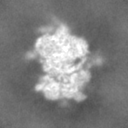
| Title | Translating 80S ribosome from HeLa cell lysate | |||||||||
 Map data Map data | ||||||||||
 Sample Sample |
| |||||||||
 Keywords Keywords | eukaryotic polyribosome ribosomal translation complex / RIBOSOME | |||||||||
| Biological species |  Homo sapiens (human) Homo sapiens (human) | |||||||||
| Method | single particle reconstruction / cryo EM / Resolution: 7.7 Å | |||||||||
 Authors Authors | Baymukhametov TN / Chesnokov YM / Afonina ZA | |||||||||
| Funding support |  Russian Federation, 1 items Russian Federation, 1 items
| |||||||||
 Citation Citation | Journal: Nucleic Acids Res / Year: 2023 Title: Polyribosomes of circular topology are prevalent in mammalian cells. Authors: Timur N Baymukhametov / Dmitry N Lyabin / Yury M Chesnokov / Ivan I Sorokin / Evgeniya V Pechnikova / Alexander L Vasiliev / Zhanna A Afonina / Abstract: Polyribosomes, the groups of ribosomes simultaneously translating a single mRNA molecule, are very common in both, prokaryotic and eukaryotic cells. Even in early EM studies, polyribosomes have been ...Polyribosomes, the groups of ribosomes simultaneously translating a single mRNA molecule, are very common in both, prokaryotic and eukaryotic cells. Even in early EM studies, polyribosomes have been shown to possess various spatial conformations, including a ring-shaped configuration which was considered to be functionally important. However, a recent in situ cryo-ET analysis of predominant regular inter-ribosome contacts did not confirm the abundance of ring-shaped polyribosomes in a cell cytoplasm. To address this discrepancy, here we analyzed the cryo-ET structure of polyribosomes in diluted lysates of HeLa cells. It was shown that the vast majority of the ribosomes were combined into polysomes and were proven to be translationally active. Tomogram analysis revealed that circular polyribosomes are indeed very common in the cytoplasm, but they mostly possess pseudo-regular structures without specific inter-ribosomal contacts. Although the size of polyribosomes varied widely, most circular polysomes were relatively small in size (4-8 ribosomes). Our results confirm the recent data that it is cellular mRNAs with short ORF that most commonly form circular structures providing an enhancement of translation. | |||||||||
| History |
|
- Structure visualization
| Supplemental images |
|---|

- Downloads & links
-EMDB archive
| Map data | emd_15018.map.gz | 713.4 KB |  EMDB map data format EMDB map data format | |
|---|---|---|---|---|
| Header (meta data) | emd-15018-v30.xmlemd-15018.xml | 15.4 KB 15.4 KB | Display Display | EMDB header |
| FSC (resolution estimation) | emd_15018_fsc.xml | 4.4 KB | Display | FSC data file |
| Images | 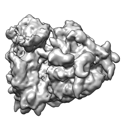 emd_15018.png emd_15018.png | 96.3 KB | ||
| Masks | emd_15018_msk_1.map | 8 MB | Mask map | |
| Filedesc metadata | emd-15018.cif.gz | 4.5 KB | ||
| Others | emd_15018_half_map_1.map.gzemd_15018_half_map_2.map.gz | 7.4 MB 7.4 MB | ||
| Archive directory |  http://ftp.pdbj.org/pub/emdb/structures/EMD-15018ftp://ftp.pdbj.org/pub/emdb/structures/EMD-15018 http://ftp.pdbj.org/pub/emdb/structures/EMD-15018ftp://ftp.pdbj.org/pub/emdb/structures/EMD-15018 | HTTPS FTP |
-Related structure data
-Links
| EMDB pages | EMDB (EBI/PDBe) / EMDataResource |
|---|
-Map
| File | Download / File: emd_15018.map.gz / Format: CCP4 / Size: 8 MB / Type: IMAGE STORED AS FLOATING POINT NUMBER (4 BYTES) | ||||||||||||||||||||||||||||||||||||
|---|---|---|---|---|---|---|---|---|---|---|---|---|---|---|---|---|---|---|---|---|---|---|---|---|---|---|---|---|---|---|---|---|---|---|---|---|---|
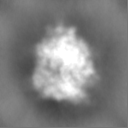
| Projections & slices | Image control
Images are generated by Spider. | ||||||||||||||||||||||||||||||||||||
| Voxel size | X=Y=Z: 3.7 Å | ||||||||||||||||||||||||||||||||||||
| Density |
| ||||||||||||||||||||||||||||||||||||
| Symmetry | Space group: 1 | ||||||||||||||||||||||||||||||||||||
| Details | EMDB XML:
|
 Z (Sec.)
 Y (Row.)
 X (Col.)


-Supplemental data
-Mask #1
| File | emd_15018_msk_1.map | ||||||||||||
|---|---|---|---|---|---|---|---|---|---|---|---|---|---|
| Projections & Slices |
| ||||||||||||
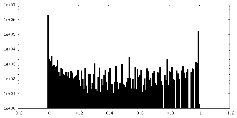
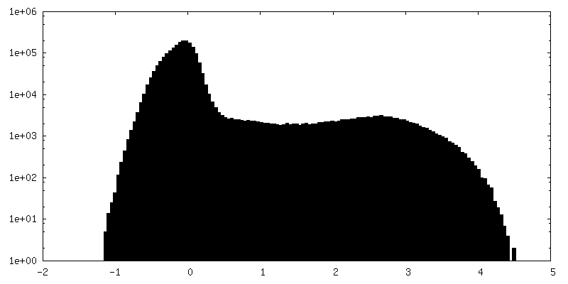
| Density Histograms |


-Half map: #1
| File | emd_15018_half_map_1.map | ||||||||||||
|---|---|---|---|---|---|---|---|---|---|---|---|---|---|
| Projections & Slices |
| ||||||||||||
| Density Histograms |


-Half map: #2
| File | emd_15018_half_map_2.map | ||||||||||||
|---|---|---|---|---|---|---|---|---|---|---|---|---|---|
| Projections & Slices |
| ||||||||||||
| Density Histograms |


- Sample components
-Entire : Polyribosomes from HeLa cytoplasm
| Entire | Name: Polyribosomes from HeLa cytoplasm |
|---|---|
| Components |
|
-Supramolecule #1: Polyribosomes from HeLa cytoplasm
| Supramolecule | Name: Polyribosomes from HeLa cytoplasm / type: complex / ID: 1 / Parent: 0 / Macromolecule list: #1 Details: Polysomes in cell lysate obtained by osmotic lysis of HeLa cells |
|---|---|
| Source (natural) | Organism: Homo sapiens (human) / Strain: HeLa / Location in cell: cytoplasm |
-Experimental details
-Structure determination
| Method | cryo EM |
|---|---|
 Processing Processing | single particle reconstruction |
| Aggregation state | particle |
-Sample preparation
| Buffer | pH: 7.6 |
|---|---|
| Grid | Model: Quantifoil R2/2 / Material: COPPER / Mesh: 300 / Support film - Material: CARBON / Support film - topology: HOLEY / Pretreatment - Type: GLOW DISCHARGE / Pretreatment - Time: 30 sec. / Pretreatment - Atmosphere: AIR / Pretreatment - Pressure: 26.0 kPa |
| Vitrification | Cryogen name: ETHANE / Chamber humidity: 100 % / Chamber temperature: 293 K / Instrument: FEI VITROBOT MARK IV |
- Electron microscopy
| Microscope | FEI TITAN KRIOS |
|---|---|
| Specialist optics | Spherical aberration corrector: Cs image corrector (CEOS, Germany) |
| Image recording | Film or detector model: FEI FALCON II (4k x 4k) / Detector mode: INTEGRATING / Digitization - Dimensions - Width: 4096 pixel / Digitization - Dimensions - Height: 4096 pixel / Digitization - Frames/image: 1-24 / Number grids imaged: 1 / Number real images: 500 / Average electron dose: 61.0 e/Å2 |
| Electron beam | Acceleration voltage: 300 kV / Electron source:  FIELD EMISSION GUN FIELD EMISSION GUN |
| Electron optics | C2 aperture diameter: 100.0 µm / Calibrated magnification: 3783 / Illumination mode: FLOOD BEAM / Imaging mode: BRIGHT FIELD / Cs: 0.01 mm / Nominal defocus max: 3.5 µm / Nominal defocus min: 1.5 µm / Nominal magnification: 18000 |
| Sample stage | Specimen holder model: FEI TITAN KRIOS AUTOGRID HOLDER / Cooling holder cryogen: NITROGEN |
| Experimental equipment |  Model: Titan Krios / Image courtesy: FEI Company |

