 ムービー
ムービー コントローラー


+ データを開く

- 基本情報
基本情報
| 登録情報 |  | |||||||||
|---|---|---|---|---|---|---|---|---|---|---|
| タイトル | Map2 from paper entitled: Structure of a cholinergic cell membrane | |||||||||
 マップデータ マップデータ | Density map of acetylcholine receptors in native lipid bilayer obtained by helical reconstruction of tubular vesicles from Torpedo | |||||||||
 試料 試料 |
| |||||||||
 キーワード キーワード | cholesterol / phospholipid / lipid bilayer / acetylcholine receptor / membrane protein | |||||||||
| 生物種 |  | |||||||||
| 手法 | らせん対称体再構成法 / クライオ電子顕微鏡法 / 解像度: 5.5 Å | |||||||||
 データ登録者 データ登録者 | Unwin N | |||||||||
| 資金援助 |  英国, 1件 英国, 1件
| |||||||||
 引用 引用 | ジャーナル: Proc Natl Acad Sci U S A / 年: 2022 タイトル: Structure of a cholinergic cell membrane. 著者: Nigel Unwin / 要旨: Cell membranes are complex assemblies of proteins and lipids making transient or long-term associations that have yet to be characterized at a molecular level. Here, cryo-electron microscopy is ...Cell membranes are complex assemblies of proteins and lipids making transient or long-term associations that have yet to be characterized at a molecular level. Here, cryo-electron microscopy is applied to determine how phospholipids and cholesterol arrange between neighboring proteins (nicotinic acetylcholine receptors) of cholinergic membrane. The lipids exhibit distinct properties in the two leaflets of the bilayer, influenced by the protein surfaces and by differences in cholesterol concentration. In the outer leaflet, the lipids show no consistent motif away from the protein surfaces, in keeping with their assumed fluidity. In the inner leaflet, where the cholesterol concentration is higher, the lipids organize into extensive close-packed linear arrays. These arrays are built from the sterol groups of cholesterol and the initial saturated portions of the phospholipid hydrocarbon chains. Together, they create an ordered ∼7 Å-thick "skin" within the hydrophobic core of the bilayer. The packing of lipids in the arrays appears to bear a close relationship to the linear cholesterol arrays that form crystalline monolayers at the air-water interface. | |||||||||
| 履歴 |
|
- 構造の表示
構造の表示
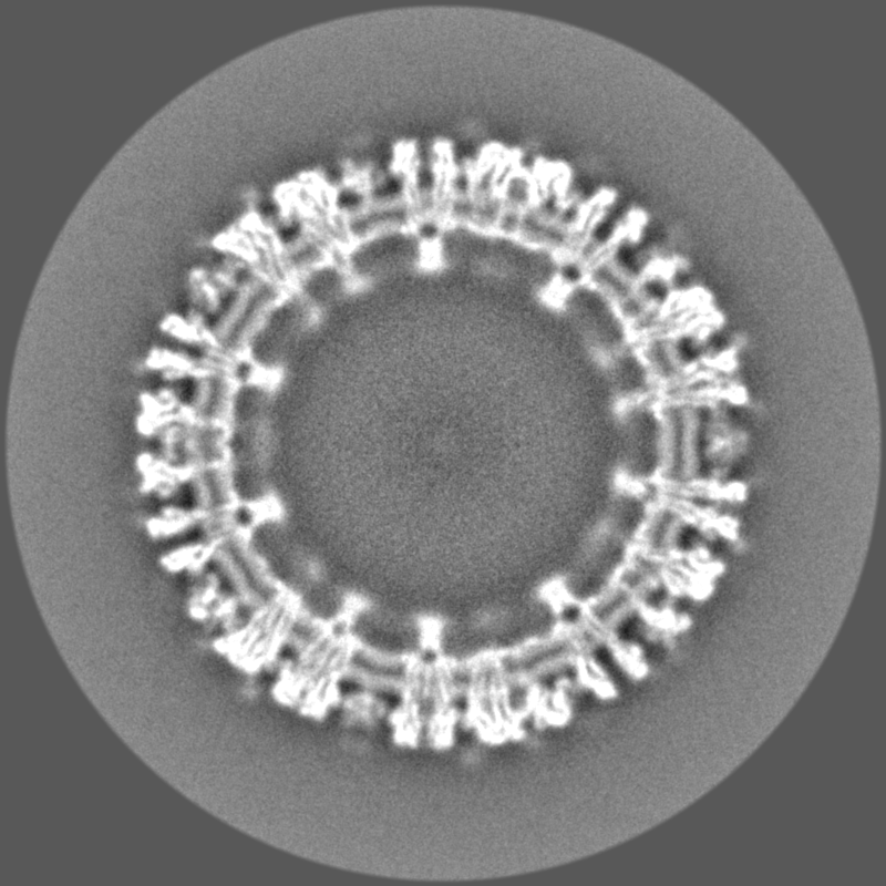
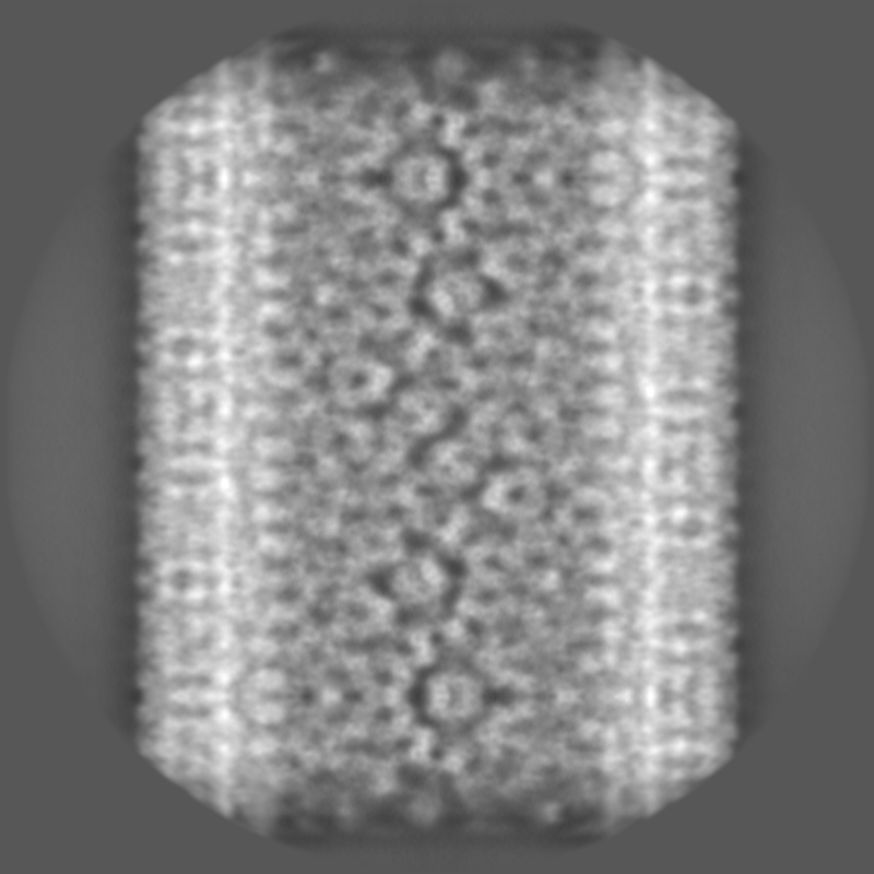
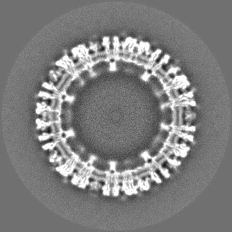
| 添付画像 |
|---|

- ダウンロードとリンク
-EMDBアーカイブ
| マップデータ | emd_14946.map.gz | 475.2 MB |  EMDBマップデータ形式 EMDBマップデータ形式 | |
|---|---|---|---|---|
| ヘッダ (付随情報) | emd-14946-v30.xmlemd-14946.xml | 17.1 KB 17.1 KB | 表示 表示 | EMDBヘッダ |
| 画像 | 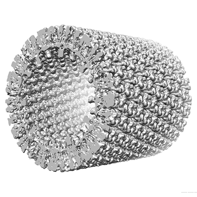 emd_14946.png emd_14946.png | 162.8 KB | ||
| Filedesc metadata | emd-14946.cif.gz | 4.9 KB | ||
| その他 | emd_14946_half_map_1.map.gzemd_14946_half_map_2.map.gz | 925.5 MB 965.3 MB | ||
| アーカイブディレクトリ |  http://ftp.pdbj.org/pub/emdb/structures/EMD-14946ftp://ftp.pdbj.org/pub/emdb/structures/EMD-14946 http://ftp.pdbj.org/pub/emdb/structures/EMD-14946ftp://ftp.pdbj.org/pub/emdb/structures/EMD-14946 | HTTPS FTP |
-検証レポート
| 文書・要旨 | emd_14946_validation.pdf.gz | 985.7 KB | 表示 | EMDB検証レポート |
|---|---|---|---|---|
| 文書・詳細版 | emd_14946_full_validation.pdf.gz | 985.3 KB | 表示 | |
| XML形式データ | emd_14946_validation.xml.gz | 25.9 KB | 表示 | |
| CIF形式データ | emd_14946_validation.cif.gz | 30.8 KB | 表示 | |
| アーカイブディレクトリ | https://ftp.pdbj.org/pub/emdb/validation_reports/EMD-14946ftp://ftp.pdbj.org/pub/emdb/validation_reports/EMD-14946 | HTTPS FTP |
-関連構造データ
-リンク
| EMDBのページ | EMDB (EBI/PDBe) / EMDataResource |
|---|
-マップ
| ファイル | ダウンロード / ファイル: emd_14946.map.gz / 形式: CCP4 / 大きさ: 1.9 GB / タイプ: IMAGE STORED AS FLOATING POINT NUMBER (4 BYTES) | ||||||||||||||||||||
|---|---|---|---|---|---|---|---|---|---|---|---|---|---|---|---|---|---|---|---|---|---|
| 注釈 | Density map of acetylcholine receptors in native lipid bilayer obtained by helical reconstruction of tubular vesicles from Torpedo | ||||||||||||||||||||
| ボクセルのサイズ | X=Y=Z: 1.34 Å | ||||||||||||||||||||
| 密度 |
| ||||||||||||||||||||
| 対称性 | 空間群: 1 | ||||||||||||||||||||
| 詳細 | EMDB XML:
|
-添付データ
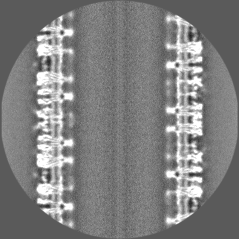
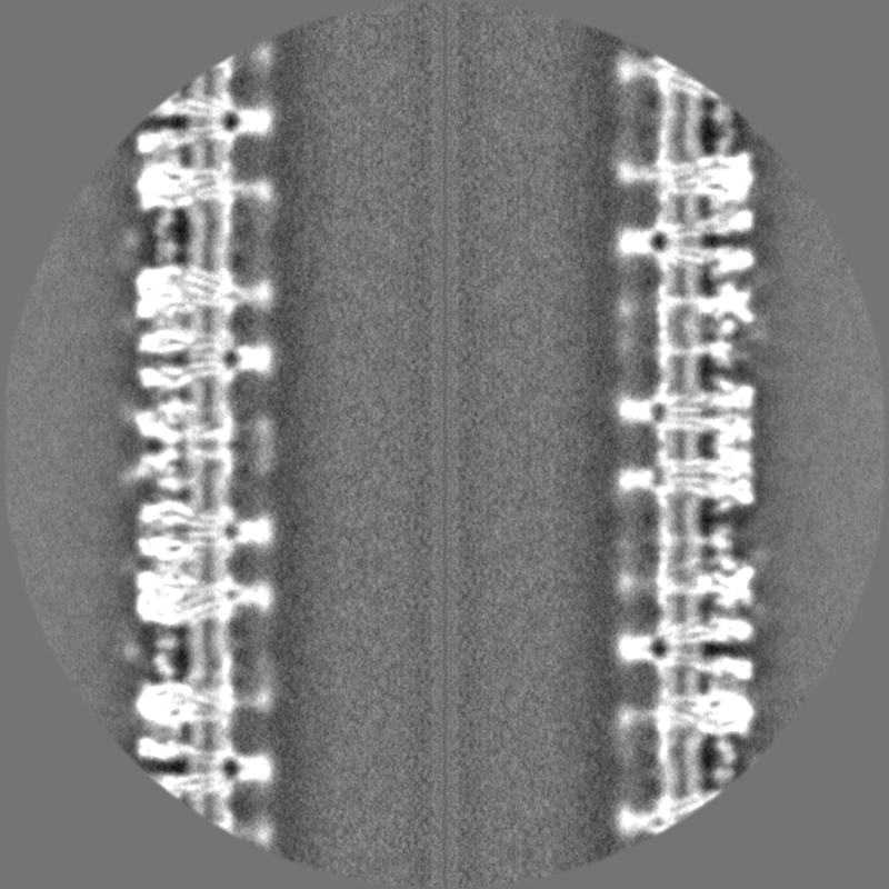
-ハーフマップ: First half-map
| ファイル | emd_14946_half_map_1.map | ||||||||||||
|---|---|---|---|---|---|---|---|---|---|---|---|---|---|
| 注釈 | First half-map | ||||||||||||
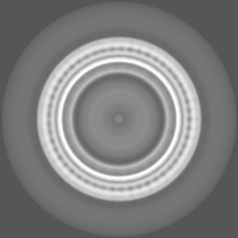
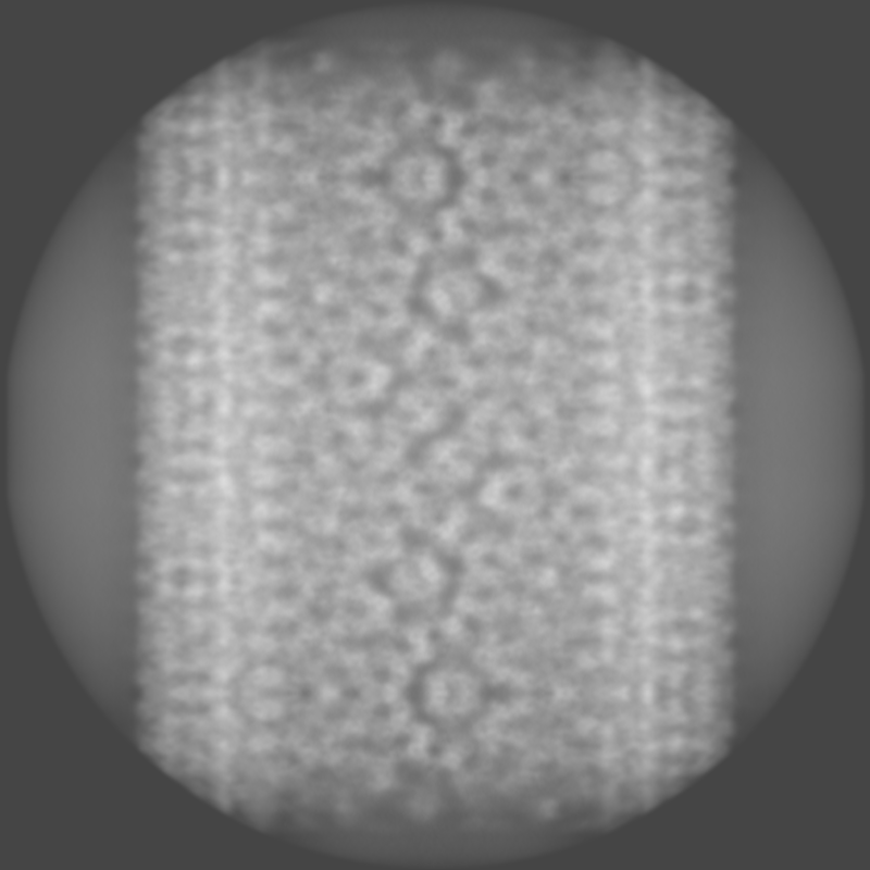
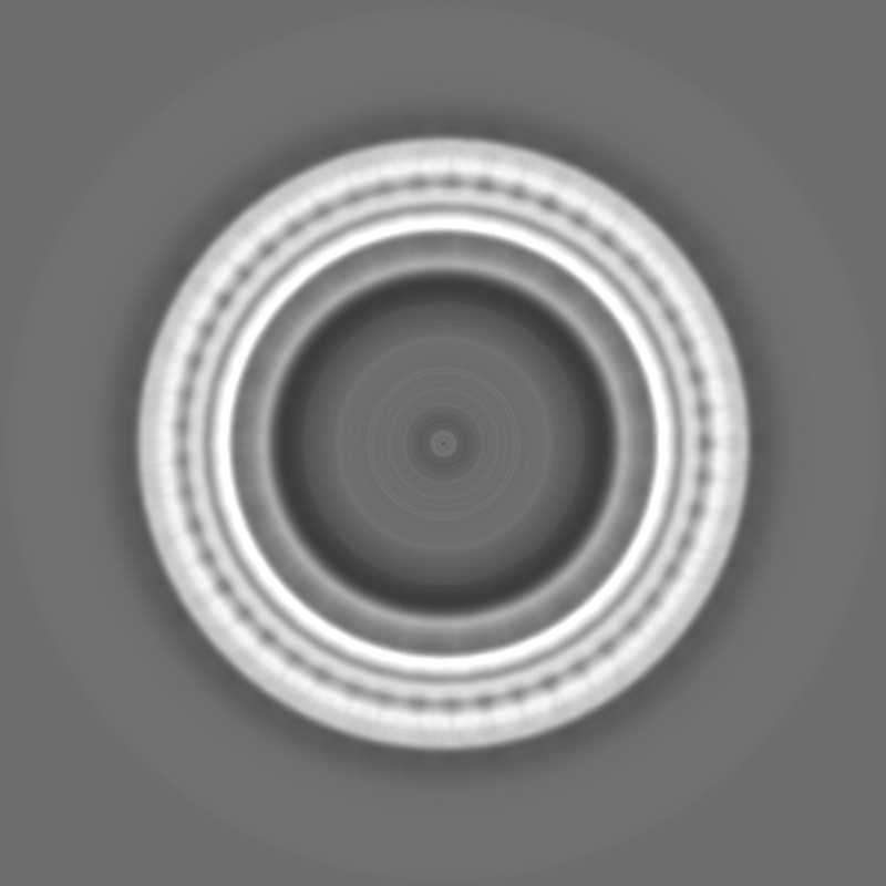
| 投影像・断面図 |
| ||||||||||||
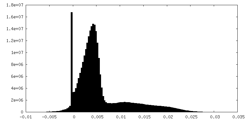
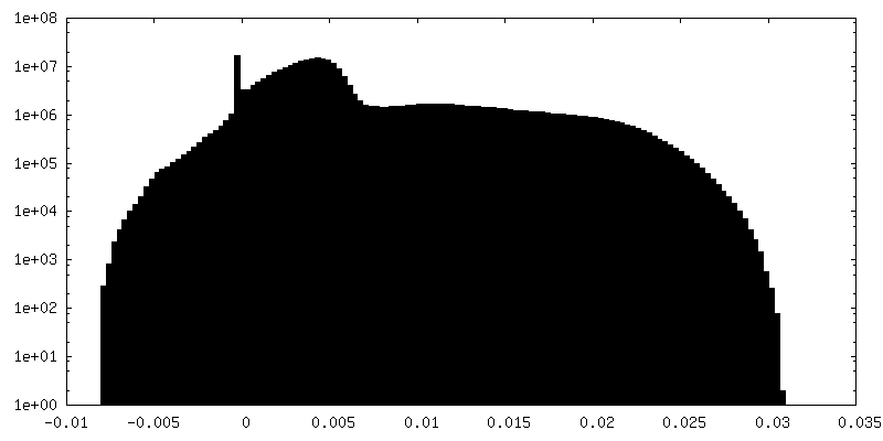
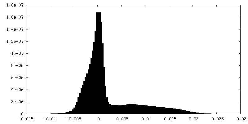
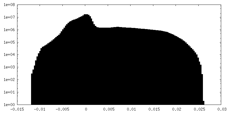
| 密度ヒストグラム |
 Z
Z Y
Y X
X


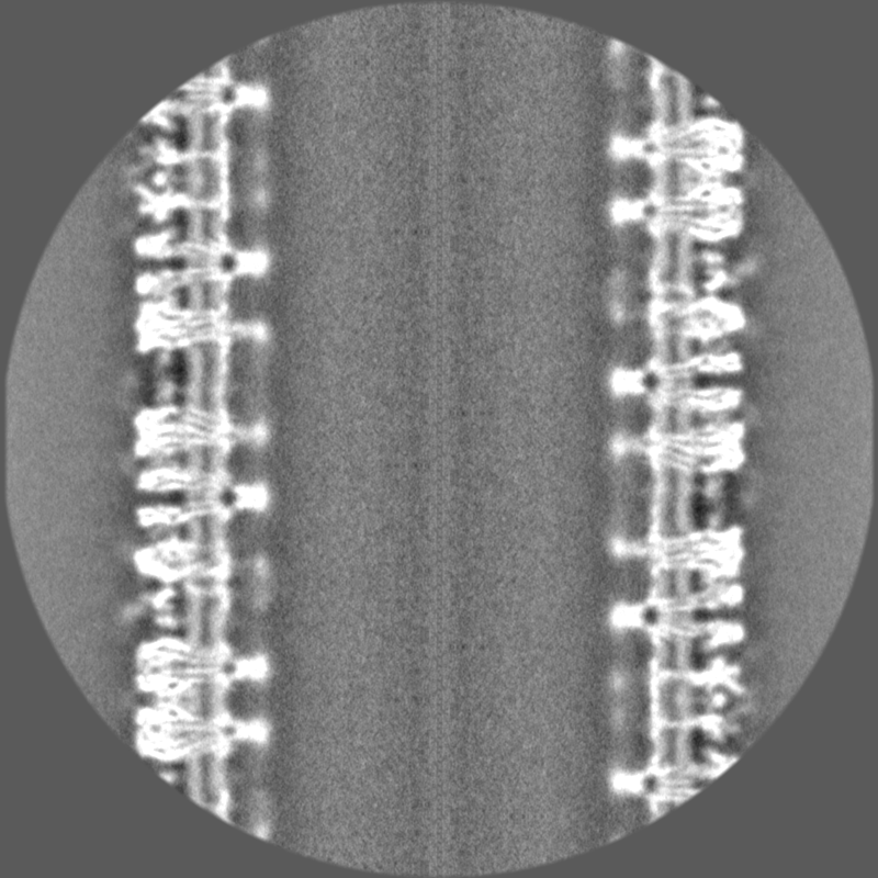
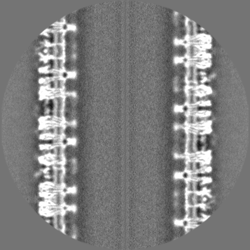
-ハーフマップ: Second half-map
| ファイル | emd_14946_half_map_2.map | ||||||||||||
|---|---|---|---|---|---|---|---|---|---|---|---|---|---|
| 注釈 | Second half-map | ||||||||||||
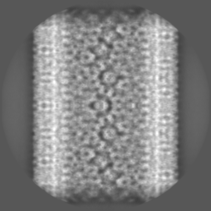
| 投影像・断面図 |
| ||||||||||||
| 密度ヒストグラム |
- 試料の構成要素
-全体 : Cholinergic cell membrane
| 全体 | 名称: Cholinergic cell membrane |
|---|---|
| 要素 |
|
-超分子 #1: Cholinergic cell membrane
| 超分子 | 名称: Cholinergic cell membrane / タイプ: organelle_or_cellular_component / ID: 1 / 親要素: 0 / 含まれる分子: #1 詳細: Tubular vesicles obtained from acetycholine receptor-rich cell membranes, isolated from Torpedo electric organ |
|---|---|
| 由来(天然) | 生物種: |
-実験情報
-構造解析
| 手法 | クライオ電子顕微鏡法 |
|---|---|
 解析 解析 | らせん対称体再構成法 |
| 試料の集合状態 | helical array |
-試料調製
| 緩衝液 | pH: 7 / 構成要素 - 濃度: 100.0 mM / 構成要素 - 式: C2H6AsNaO2 / 構成要素 - 名称: sodium cacodylate |
|---|---|
| グリッド | モデル: Quantifoil R1.2/1.3 / 材質: COPPER / メッシュ: 300 / 支持フィルム - 材質: CARBON / 支持フィルム - トポロジー: HOLEY ARRAY / 支持フィルム - Film thickness: 20 / 前処理 - タイプ: GLOW DISCHARGE / 前処理 - 時間: 60 sec. / 前処理 - 雰囲気: OTHER |
| 凍結 | 凍結剤: ETHANE / チャンバー内湿度: 95 % / チャンバー内温度: 283 K / 装置: HOMEMADE PLUNGER 詳細: blot until filter paper loses water-contact to grid surface (~5 secs).. |
| 詳細 | The tubular vesicles are comprised of nicotinic acetylcholine receptors in a cholesterol-rich phospholipid bilayer. |
- 電子顕微鏡法
| 顕微鏡 | FEI TITAN KRIOS |
|---|---|
| 温度 | 最低: 90.0 K / 最高: 90.0 K |
| 撮影 | フィルム・検出器のモデル: FEI FALCON III (4k x 4k) 検出モード: INTEGRATING / デジタル化 - サイズ - 横: 4096 pixel / デジタル化 - サイズ - 縦: 4096 pixel / 撮影したグリッド数: 160 / 実像数: 1993 / 平均露光時間: 2.0 sec. / 平均電子線量: 40.0 e/Å2 詳細: Images were collected in movie mode at 40 frames per second |
| 電子線 | 加速電圧: 300 kV / 電子線源:  FIELD EMISSION GUN FIELD EMISSION GUN |
| 電子光学系 | C2レンズ絞り径: 50.0 µm 最大 デフォーカス(補正後): 2.8000000000000003 µm 最小 デフォーカス(補正後): 1.4000000000000001 µm 照射モード: FLOOD BEAM / 撮影モード: BRIGHT FIELD / Cs: 2.7 mm 最大 デフォーカス(公称値): 2.8000000000000003 µm 最小 デフォーカス(公称値): 1.4000000000000001 µm 倍率(公称値): 59000 |
| 試料ステージ | 試料ホルダーモデル: FEI TITAN KRIOS AUTOGRID HOLDER ホルダー冷却材: NITROGEN |
| 実験機器 |  モデル: Titan Krios / 画像提供: FEI Company |
