 ムービー
ムービー コントローラー


+ データを開く

- 基本情報
基本情報
| 登録情報 |  | |||||||||
|---|---|---|---|---|---|---|---|---|---|---|
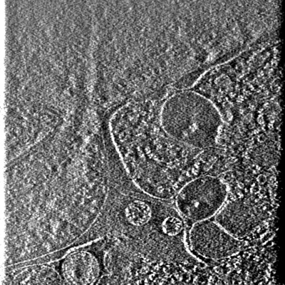
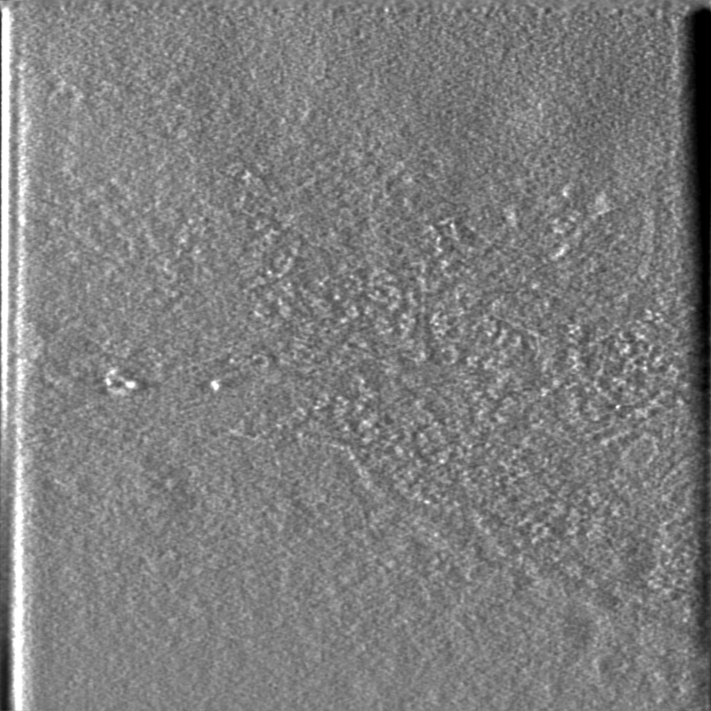
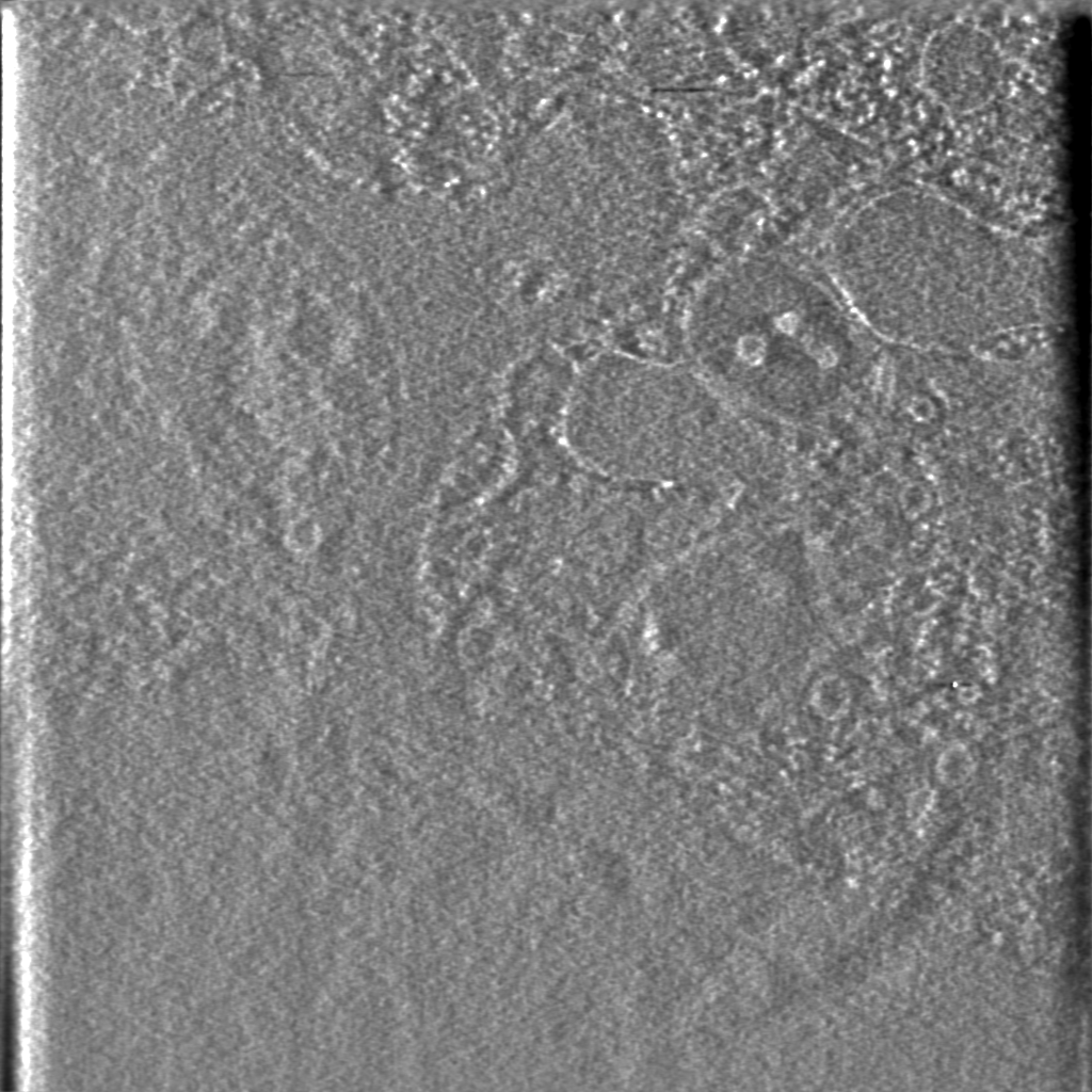
| タイトル | Cryo-STEM tomography of fibroblast nuclear periphery | |||||||||
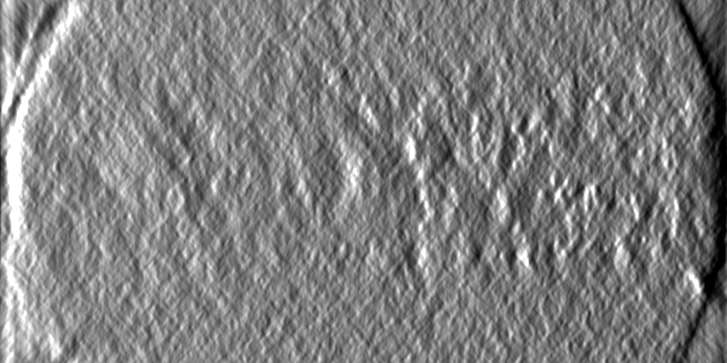
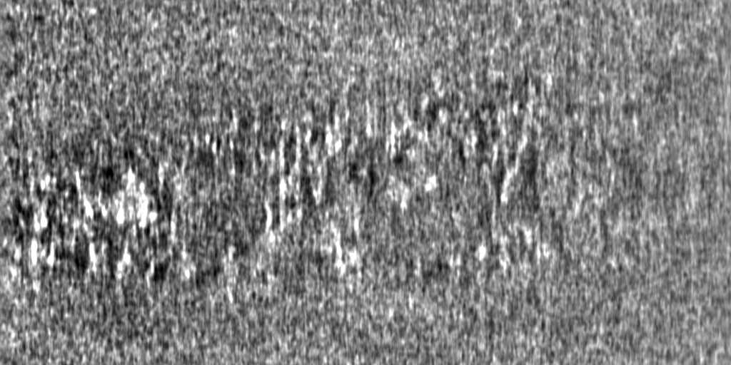
 マップデータ マップデータ | Cryo-STEM tomography (CSTET) of an intact fibroblast showing nuclear periphery, processed by 3D deconvolution. | |||||||||
 試料 試料 |
| |||||||||
 キーワード キーワード | nucleus / chromatin / nuclear envelope / mitochondria / STEM / CSTET / DNA | |||||||||
| 生物種 |  Homo sapiens (ヒト) Homo sapiens (ヒト) | |||||||||
| 手法 | 電子線トモグラフィー法 / クライオ電子顕微鏡法 | |||||||||
 データ登録者 データ登録者 | Wolf SG / Fass D / Elbaum M | |||||||||
| 資金援助 |  イスラエル, 1件 イスラエル, 1件
| |||||||||
 引用 引用 | ジャーナル: Proc Natl Acad Sci U S A / 年: 2020 タイトル: Three-dimensional deconvolution processing for STEM cryotomography. 著者: Barnali Waugh / Sharon G Wolf / Deborah Fass / Eric Branlund / Zvi Kam / John W Sedat / Michael Elbaum /  要旨: The complex environment of biological cells and tissues has motivated development of three-dimensional (3D) imaging in both light and electron microscopies. To this end, one of the primary tools in ...The complex environment of biological cells and tissues has motivated development of three-dimensional (3D) imaging in both light and electron microscopies. To this end, one of the primary tools in fluorescence microscopy is that of computational deconvolution. Wide-field fluorescence images are often corrupted by haze due to out-of-focus light, i.e., to cross-talk between different object planes as represented in the 3D image. Using prior understanding of the image formation mechanism, it is possible to suppress the cross-talk and reassign the unfocused light to its proper source post facto. Electron tomography based on tilted projections also exhibits a cross-talk between distant planes due to the discrete angular sampling and limited tilt range. By use of a suitably synthesized 3D point spread function, we show here that deconvolution leads to similar improvements in volume data reconstructed from cryoscanning transmission electron tomography (CSTET), namely a dramatic in-plane noise reduction and improved representation of features in the axial dimension. Contrast enhancement is demonstrated first with colloidal gold particles and then in representative cryotomograms of intact cells. Deconvolution of CSTET data collected from the periphery of an intact nucleus revealed partially condensed, extended structures in interphase chromatin. #1: ジャーナル: To Be Publishedタイトル: A Proposed Unified Interphase Nucleus Chromosome Structure: Preliminary Preponderance of Evidence 著者: Sedat J / McDonald A / Cang H / Lucas J / Arigovindan M / Kam Z / Murre C / Elbaum M | |||||||||
| 履歴 |
|
- 構造の表示
構造の表示
| 添付画像 |
|---|
- ダウンロードとリンク
-EMDBアーカイブ
| マップデータ | emd_14924.map.gz | 1.7 GB |  EMDBマップデータ形式 EMDBマップデータ形式 | |
|---|---|---|---|---|
| ヘッダ (付随情報) | emd-14924-v30.xmlemd-14924.xml | 11.8 KB 11.8 KB | 表示 表示 | EMDBヘッダ |
| 画像 |  emd_14924.png emd_14924.png | 234.4 KB | ||
| Filedesc metadata | emd-14924.cif.gz | 4.5 KB | ||
| アーカイブディレクトリ |  http://ftp.pdbj.org/pub/emdb/structures/EMD-14924ftp://ftp.pdbj.org/pub/emdb/structures/EMD-14924 http://ftp.pdbj.org/pub/emdb/structures/EMD-14924ftp://ftp.pdbj.org/pub/emdb/structures/EMD-14924 | HTTPS FTP |
-検証レポート
| 文書・要旨 | emd_14924_validation.pdf.gz | 325.8 KB | 表示 | EMDB検証レポート |
|---|---|---|---|---|
| 文書・詳細版 | emd_14924_full_validation.pdf.gz | 325.4 KB | 表示 | |
| XML形式データ | emd_14924_validation.xml.gz | 4.5 KB | 表示 | |
| CIF形式データ | emd_14924_validation.cif.gz | 5 KB | 表示 | |
| アーカイブディレクトリ | https://ftp.pdbj.org/pub/emdb/validation_reports/EMD-14924ftp://ftp.pdbj.org/pub/emdb/validation_reports/EMD-14924 | HTTPS FTP |
-リンク
| EMDBのページ | EMDB (EBI/PDBe) / EMDataResource |
|---|
-マップ
| ファイル | ダウンロード / ファイル: emd_14924.map.gz / 形式: CCP4 / 大きさ: 2 GB / タイプ: IMAGE STORED AS FLOATING POINT NUMBER (4 BYTES) | ||||||||||||||||||||||||||||||||
|---|---|---|---|---|---|---|---|---|---|---|---|---|---|---|---|---|---|---|---|---|---|---|---|---|---|---|---|---|---|---|---|---|---|
| 注釈 | Cryo-STEM tomography (CSTET) of an intact fibroblast showing nuclear periphery, processed by 3D deconvolution. | ||||||||||||||||||||||||||||||||
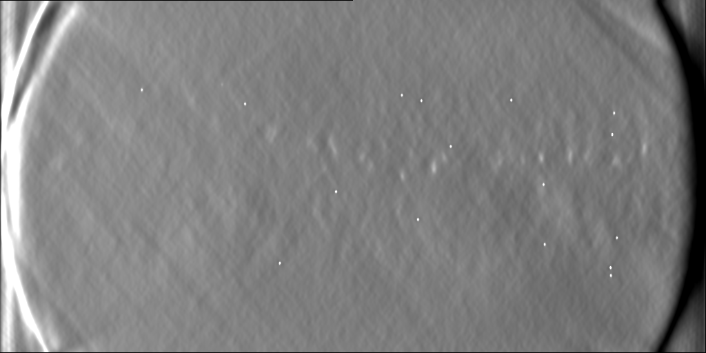
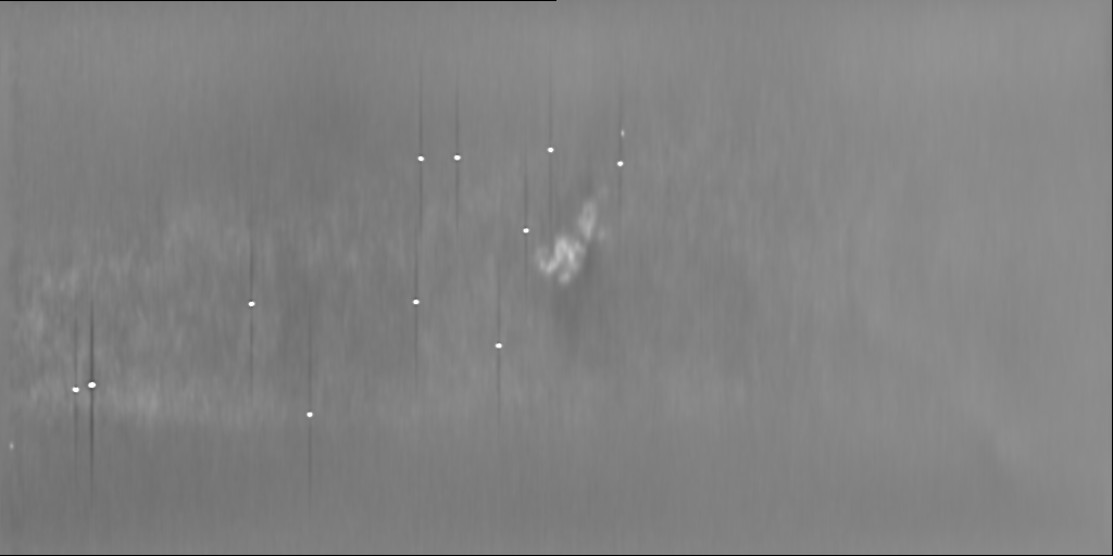
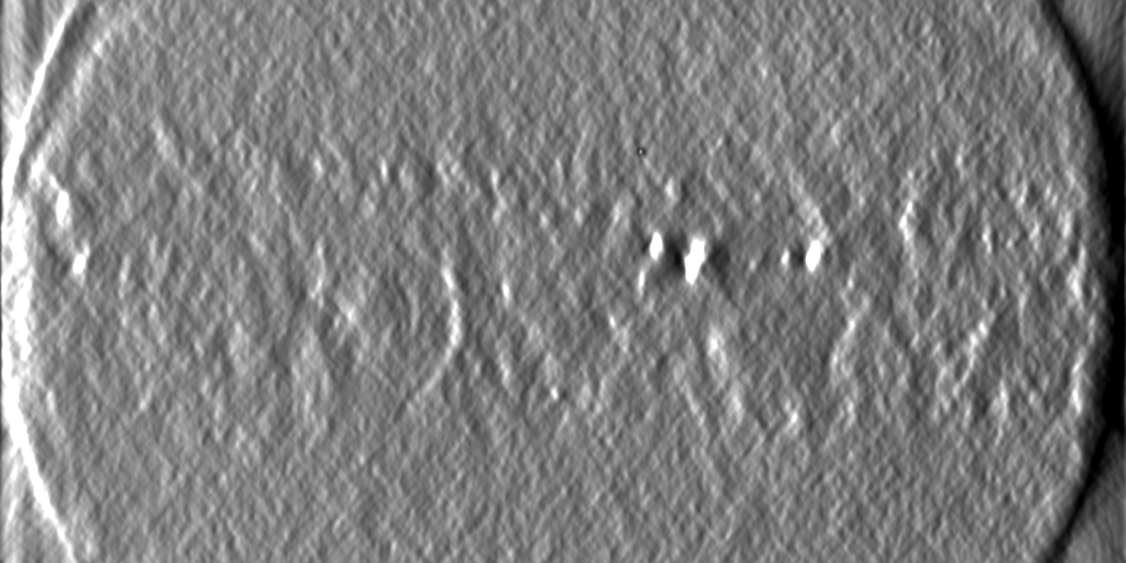
| 投影像・断面図 | 画像のコントロール
画像は Spider により作成 これらの図は立方格子座標系で作成されたものです | ||||||||||||||||||||||||||||||||
| ボクセルのサイズ | X=Y=Z: 33.51 Å | ||||||||||||||||||||||||||||||||
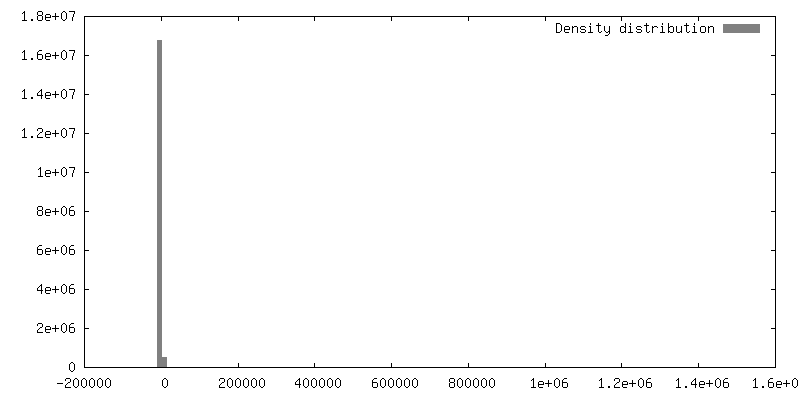
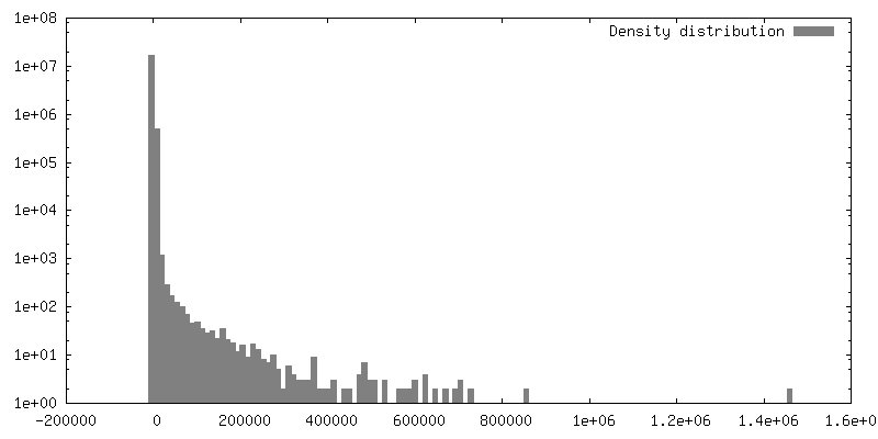
| 密度 |
| ||||||||||||||||||||||||||||||||
| 対称性 | 空間群: 1 | ||||||||||||||||||||||||||||||||
| 詳細 | EMDB XML:
|
 Z (Sec.)
 Y (Row.)
 X (Col.)


-添付データ
- 試料の構成要素
-全体 : WI-38 fibroblast cell
| 全体 | 名称: WI-38 fibroblast cell |
|---|---|
| 要素 |
|
-超分子 #1: WI-38 fibroblast cell
| 超分子 | 名称: WI-38 fibroblast cell / タイプ: cell / ID: 1 / 親要素: 0 / 詳細: cells cultured directly on gold EM grid |
|---|---|
| 由来(天然) | 生物種: Homo sapiens (ヒト) / 器官: lung / 組織: cell culture |
-実験情報
-構造解析
| 手法 | クライオ電子顕微鏡法 |
|---|---|
 解析 解析 | 電子線トモグラフィー法 |
| 試料の集合状態 | cell |
-試料調製
| 緩衝液 | pH: 7.4 詳細: Minimal Essential Medium (Gibco) supplemented with 15% fetal calf serum, L-glutamine, and penicillin/streptomycin. |
|---|---|
| グリッド | モデル: Quantifoil R3.5/1 / 材質: GOLD / メッシュ: 200 / 前処理 - タイプ: GLOW DISCHARGE / 前処理 - 時間: 60 sec. |
| 凍結 | 凍結剤: ETHANE / 装置: LEICA EM GP |
| 詳細 | WI-38 embryonic lung fibroblast cells (obtained from Coriell Institute) were cultured directly on gold EM grids and imaged intact by cryo-STEM tomography (CSTET). |
| 切片作成 | その他: NO SECTIONING |
| 位置合わせマーカー | Manufacturer: obtained from L Duchesne (https://doi.org/10.1021/la802876u ) 直径: 10 nm |
- 電子顕微鏡法
| 顕微鏡 | FEI TECNAI F20 |
|---|---|
| 特殊光学系 | 詳細: STEM data collection in BF mode. |
| 詳細 | NanoProbe STEM acquisition. |
| 撮影 | フィルム・検出器のモデル: OTHER / デジタル化 - サイズ - 横: 2048 pixel / デジタル化 - サイズ - 縦: 2048 pixel / 撮影したグリッド数: 1 / 実像数: 61 / 平均露光時間: 12.6 sec. / 平均電子線量: 3.8 e/Å2 / 詳細: Cryo-STEM tilt series |
| 電子線 | 加速電圧: 200 kV / 電子線源:  FIELD EMISSION GUN FIELD EMISSION GUN |
| 電子光学系 | C2レンズ絞り径: 20.0 µm / 照射モード: SPOT SCAN / 撮影モード: BRIGHT FIELD / Cs: 2.0 mm / 最大 デフォーカス(公称値): 0.0 µm / 最小 デフォーカス(公称値): 0.0 µm |
| 試料ステージ | 試料ホルダーモデル: GATAN 626 SINGLE TILT LIQUID NITROGEN CRYO TRANSFER HOLDER ホルダー冷却材: NITROGEN |
| 実験機器 |  モデル: Tecnai F20 / 画像提供: FEI Company |
-画像解析
| 詳細 | Gatan STEM BF/ADF detector |
|---|---|
| 最終 再構成 | アルゴリズム: BACK PROJECTION / ソフトウェア: (名称: TOMO3D, PRIISM/IVE) 詳細: The original alignment was done using etomo (IMOD), and then optimized using tomoalign as a "thin" specimen. A new aligned stack (.ali) was produced using tomowarpalign (tomoalign) and ...詳細: The original alignment was done using etomo (IMOD), and then optimized using tomoalign as a "thin" specimen. A new aligned stack (.ali) was produced using tomowarpalign (tomoalign) and reconstructed by SIRT using tomo3d. The reconstruction was then binned (by 2) and processed by 3D deconvolution as described in Waugh et al (doi:10.1073/pnas.2000700117) with smoothing parameter 0.1. 使用した粒子像数: 56 |
