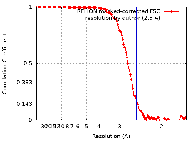
Journal: Proc Natl Acad Sci U S A / Year: 2022 Title: PTX3 structure determination using a hybrid cryoelectron microscopy and AlphaFold approach offers insights into ligand binding and complement activation. Authors: Dylan P Noone / Douwe J Dijkstra / Teun T van der Klugt / Peter A van Veelen / Arnoud H de Ru / Paul J Hensbergen / Leendert A Trouw / Thomas H Sharp / Abstract: Pattern recognition molecules (PRMs) form an important part of innate immunity, where they facilitate the response to infections and damage by triggering processes such as inflammation. The pentraxin ...Pattern recognition molecules (PRMs) form an important part of innate immunity, where they facilitate the response to infections and damage by triggering processes such as inflammation. The pentraxin family of soluble PRMs comprises long and short pentraxins, with the former containing unique N-terminal regions unrelated to other proteins or each other. No complete high-resolution structural information exists about long pentraxins, unlike the short pentraxins, where there is an abundance of both X-ray and cryoelectron microscopy (cryo-EM)-derived structures. This study presents a high-resolution structure of the prototypical long pentraxin, PTX3. Cryo-EM yielded a 2.5-Å map of the C-terminal pentraxin domains that revealed a radically different quaternary structure compared to other pentraxins, comprising a glycosylated D4 symmetrical octameric complex stabilized by an extensive disulfide network. The cryo-EM map indicated α-helices that extended N terminal of the pentraxin domains that were not fully resolved. AlphaFold was used to predict the remaining N-terminal structure of the octameric PTX3 complex, revealing two long tetrameric coiled coils with two hinge regions, which was validated using classification of cryo-EM two-dimensional averages. The resulting hybrid cryo-EM/AlphaFold structure allowed mapping of ligand binding sites, such as C1q and fibroblast growth factor-2, as well as rationalization of previous biochemical data. Given the relevance of PTX3 in conditions ranging from COVID-19 prognosis, cancer progression, and female infertility, this structure could be used to inform the understanding and rational design of therapies for these disorders and processes.
Protein or peptide: Pentraxin-related protein PTX3
Ligand: 2-acetamido-2-deoxy-beta-D-glucopyranose
Ligand: water
-
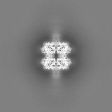
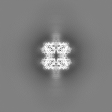
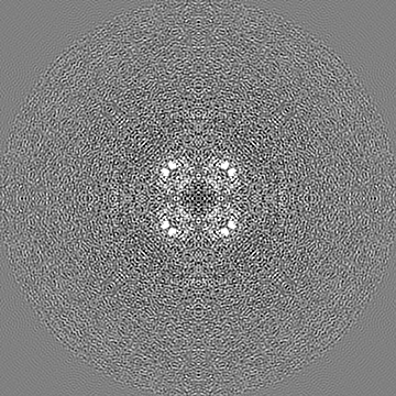
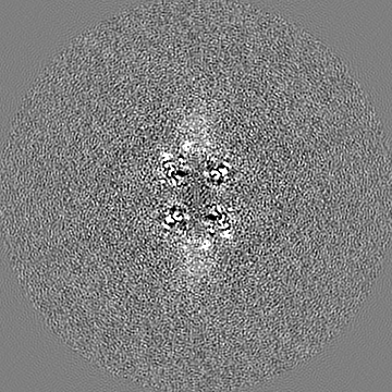
Supramolecule #1: Long pentraxin 3 pentraxin domain
Supramolecule
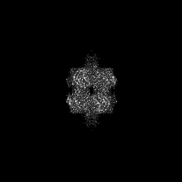
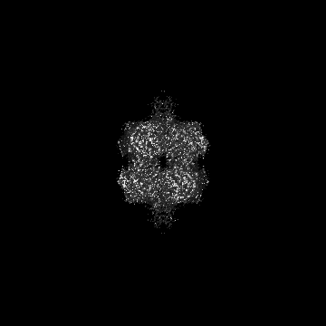
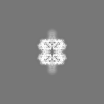
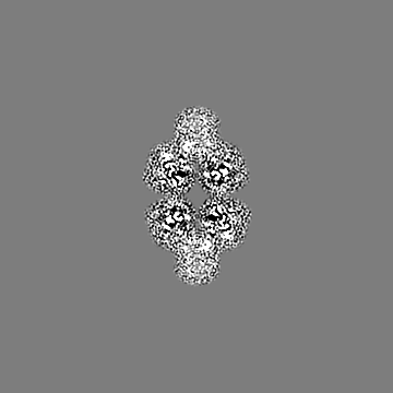
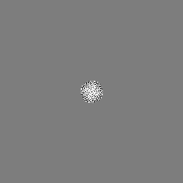
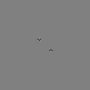
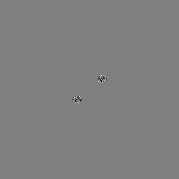
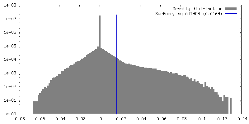
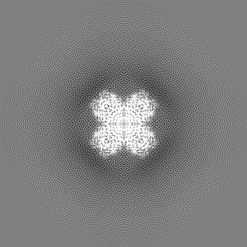
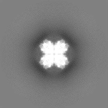
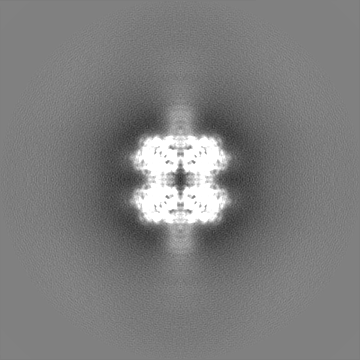
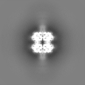
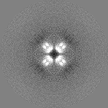
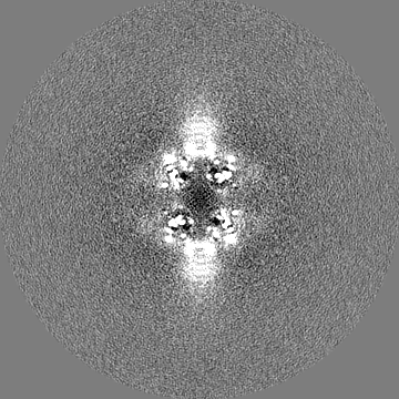
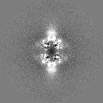
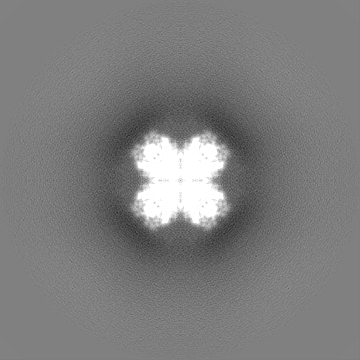
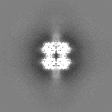
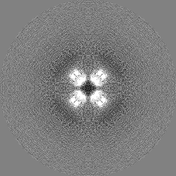
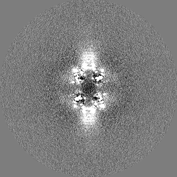
Name: Long pentraxin 3 pentraxin domain / type: complex / ID: 1 / Parent: 0 / Macromolecule list: #1 Details: CryoEM derived map of the pentraxin domains of PTX3. At higher isosurface threshold values the start of the coiled coil N-terminal domain can be seen.
Source (natural)
Organism: Homo sapiens (human)
Molecular weight
Theoretical: 336 KDa
-
Macromolecule #1: Pentraxin-related protein PTX3
Macromolecule
Name: Pentraxin-related protein PTX3 / type: protein_or_peptide / ID: 1 Details: Pentraxin domain of the long pentraxin 3. No N-terminal domain is included in this coordinate file. Number of copies: 8 / Enantiomer: LEVO
Model: Quantifoil R1.2/1.3 / Material: COPPER / Mesh: 400 / Support film - Material: GRAPHENE OXIDE / Support film - topology: HOLEY
Vitrification
Cryogen name: ETHANE / Chamber humidity: 65 % / Chamber temperature: 277.15 K / Instrument: LEICA EM GP
Details
PTX3 at concentrations between 1-2 uM in 20 mM Tris-HCl, 500 mM NaCl, pH 8.0 was concentrated in a 50 kDa molecular weight cut-off spin filter to concentrations between 60-70 uM.
-
Electron microscopy
Microscope
FEI TITAN KRIOS
Temperature
Min: 90.15 K / Max: 103.15 K
Specialist optics
Energy filter - Name: GIF Bioquantum / Energy filter - Slit width: 20 eV
Image recording
Film or detector model: GATAN K3 BIOQUANTUM (6k x 4k) / Number grids imaged: 1 / Number real images: 4612 / Average electron dose: 50.0 e/Å2
Electron beam
Acceleration voltage: 300 kV / Electron source: FIELD EMISSION GUN
In the structure databanks used in Yorodumi, some data are registered as the other names, "COVID-19 virus" and "2019-nCoV". Here are the details of the virus and the list of structure data.
Jan 31, 2019. EMDB accession codes are about to change! (news from PDBe EMDB page)
EMDB accession codes are about to change! (news from PDBe EMDB page)
The allocation of 4 digits for EMDB accession codes will soon come to an end. Whilst these codes will remain in use, new EMDB accession codes will include an additional digit and will expand incrementally as the available range of codes is exhausted. The current 4-digit format prefixed with “EMD-” (i.e. EMD-XXXX) will advance to a 5-digit format (i.e. EMD-XXXXX), and so on. It is currently estimated that the 4-digit codes will be depleted around Spring 2019, at which point the 5-digit format will come into force.
The EM Navigator/Yorodumi systems omit the EMD- prefix.
Related info.:Q: What is EMD? / ID/Accession-code notation in Yorodumi/EM Navigator
Yorodumi is a browser for structure data from EMDB, PDB, SASBDB, etc.
This page is also the successor to EM Navigator detail page, and also detail information page/front-end page for Omokage search.
The word "yorodu" (or yorozu) is an old Japanese word meaning "ten thousand". "mi" (miru) is to see.
Related info.:EMDB / PDB / SASBDB / Comparison of 3 databanks / Yorodumi Search / Aug 31, 2016. New EM Navigator & Yorodumi / Yorodumi Papers / Jmol/JSmol / Function and homology information / Changes in new EM Navigator and Yorodumi
 Movie
Movie Controller


 Open data
 Basic information
 Map data
 Sample
 Keywords
 Function and homology information
 Homo sapiens (human)
 Authors
 Citation
 Structure visualization
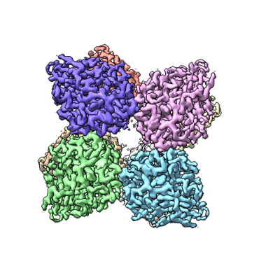
 Downloads & links
 emd_14774.png
 http://ftp.pdbj.org/pub/emdb/structures/EMD-14774
 Z (Sec.)
 Y (Row.)
 X (Col.)


 Sample components

 Processing
 Electron microscopy
 FIELD EMISSION GUN