 Movie
Movie Controller

[English] 日本語
 Yorodumi
- EMDB-14686: Subtomogram average of the chikungunya virus neck complex, unsymm... -
+ Open data

- Basic information
| Entry |  | |||||||||
|---|---|---|---|---|---|---|---|---|---|---|
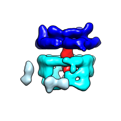
| Title | Subtomogram average of the chikungunya virus neck complex, unsymmetrized | |||||||||
 Map data Map data | Subtomogram average of chikungunya virus neck complex, without symmetrization. | |||||||||
 Sample Sample |
| |||||||||
| Biological species |   Chikungunya virus Chikungunya virus | |||||||||
| Method | subtomogram averaging / cryo EM / Resolution: 34.0 Å | |||||||||
 Authors Authors | Laurent T / Carlson LA | |||||||||
| Funding support |  Sweden, 1 items Sweden, 1 items
| |||||||||
 Citation Citation | Journal: Elife / Year: 2022 Title: Architecture of the chikungunya virus replication organelle. Authors: Timothée Laurent / Pravin Kumar / Susanne Liese / Farnaz Zare / Mattias Jonasson / Andreas Carlson / Lars-Anders Carlson /   Abstract: are mosquito-borne viruses that cause serious disease in humans and other mammals. Along with its mosquito vector, the s chikungunya virus (CHIKV) has spread explosively in the last 20 years, and ... are mosquito-borne viruses that cause serious disease in humans and other mammals. Along with its mosquito vector, the s chikungunya virus (CHIKV) has spread explosively in the last 20 years, and there is no approved treatment for chikungunya fever. On the plasma membrane of the infected cell, CHIKV generates dedicated organelles for viral RNA replication, so-called spherules. Whereas structures exist for several viral proteins that make up the spherule, the architecture of the full organelle is unknown. Here, we use cryo-electron tomography to image CHIKV spherules in their cellular context. This reveals that the viral protein nsP1 serves as a base for the assembly of a larger protein complex at the neck of the membrane bud. Biochemical assays show that the viral helicase-protease nsP2, while having no membrane affinity on its own, is recruited to membranes by nsP1. The tomograms further reveal that full-sized spherules contain a single copy of the viral genome in double-stranded form. Finally, we present a mathematical model that explains the membrane remodeling of the spherule in terms of the pressure exerted on the membrane by the polymerizing RNA, which provides a good agreement with the experimental data. The energy released by RNA polymerization is found to be sufficient to remodel the membrane to the characteristic spherule shape. | |||||||||
| History |
|
- Structure visualization
| Supplemental images |
|---|
- Downloads & links
-EMDB archive
| Map data | emd_14686.map.gz | 1.9 MB |  EMDB map data format EMDB map data format | |
|---|---|---|---|---|
| Header (meta data) | emd-14686-v30.xmlemd-14686.xml | 14.8 KB 14.8 KB | Display Display | EMDB header |
| Images |  emd_14686.png emd_14686.png | 58.1 KB | ||
| Masks | emd_14686_msk_1.map | 52.7 MB | Mask map | |
| Others | emd_14686_half_map_1.map.gzemd_14686_half_map_2.map.gz | 48.9 MB 48.9 MB | ||
| Archive directory |  http://ftp.pdbj.org/pub/emdb/structures/EMD-14686ftp://ftp.pdbj.org/pub/emdb/structures/EMD-14686 http://ftp.pdbj.org/pub/emdb/structures/EMD-14686ftp://ftp.pdbj.org/pub/emdb/structures/EMD-14686 | HTTPS FTP |
-Validation report
| Summary document | emd_14686_validation.pdf.gz | 756.7 KB | Display | EMDB validaton report |
|---|---|---|---|---|
| Full document | emd_14686_full_validation.pdf.gz | 756.3 KB | Display | |
| Data in XML | emd_14686_validation.xml.gz | 11.9 KB | Display | |
| Data in CIF | emd_14686_validation.cif.gz | 14 KB | Display | |
| Arichive directory | https://ftp.pdbj.org/pub/emdb/validation_reports/EMD-14686ftp://ftp.pdbj.org/pub/emdb/validation_reports/EMD-14686 | HTTPS FTP |
-Related structure data


-Links
| EMDB pages | EMDB (EBI/PDBe) / EMDataResource |
|---|
-Map
| File | Download / File: emd_14686.map.gz / Format: CCP4 / Size: 52.7 MB / Type: IMAGE STORED AS FLOATING POINT NUMBER (4 BYTES) | ||||||||||||||||||||||||||||||||||||
|---|---|---|---|---|---|---|---|---|---|---|---|---|---|---|---|---|---|---|---|---|---|---|---|---|---|---|---|---|---|---|---|---|---|---|---|---|---|
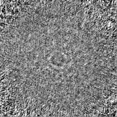
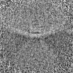
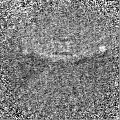
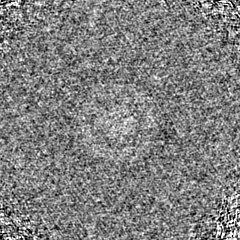
| Annotation | Subtomogram average of chikungunya virus neck complex, without symmetrization. | ||||||||||||||||||||||||||||||||||||
| Projections & slices | Image control
Images are generated by Spider. | ||||||||||||||||||||||||||||||||||||
| Voxel size | X=Y=Z: 4.29 Å | ||||||||||||||||||||||||||||||||||||
| Density |
| ||||||||||||||||||||||||||||||||||||
| Symmetry | Space group: 1 | ||||||||||||||||||||||||||||||||||||
| Details | EMDB XML:
|
 Z (Sec.)
 Y (Row.)
 X (Col.)


-Supplemental data
-Mask #1
| File | emd_14686_msk_1.map | ||||||||||||
|---|---|---|---|---|---|---|---|---|---|---|---|---|---|
| Projections & Slices |
| ||||||||||||
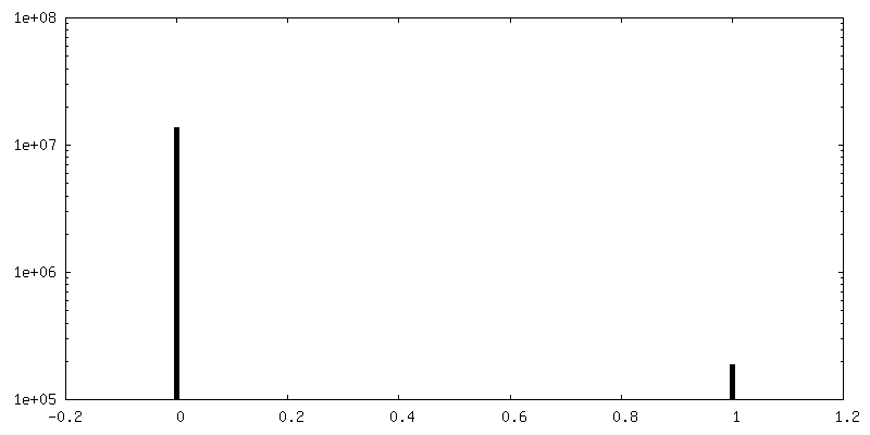
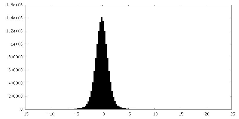
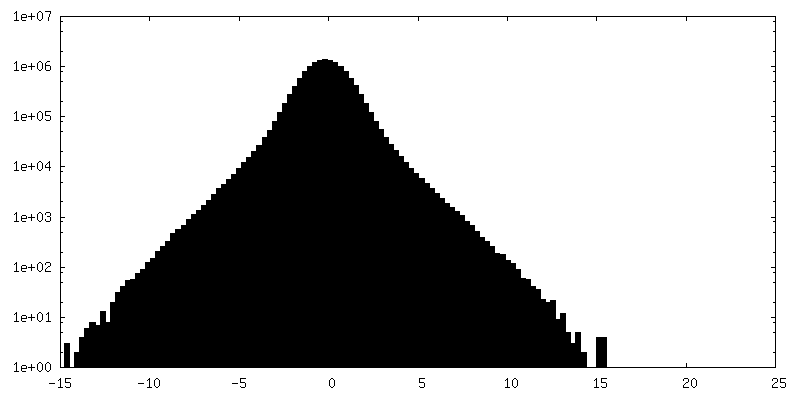
| Density Histograms |


-Half map: Half map 1
| File | emd_14686_half_map_1.map | ||||||||||||
|---|---|---|---|---|---|---|---|---|---|---|---|---|---|
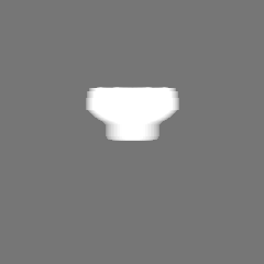
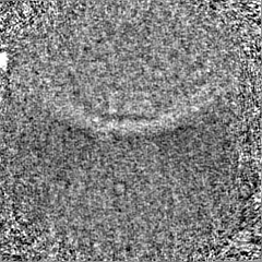
| Annotation | Half map 1 | ||||||||||||
| Projections & Slices |
| ||||||||||||
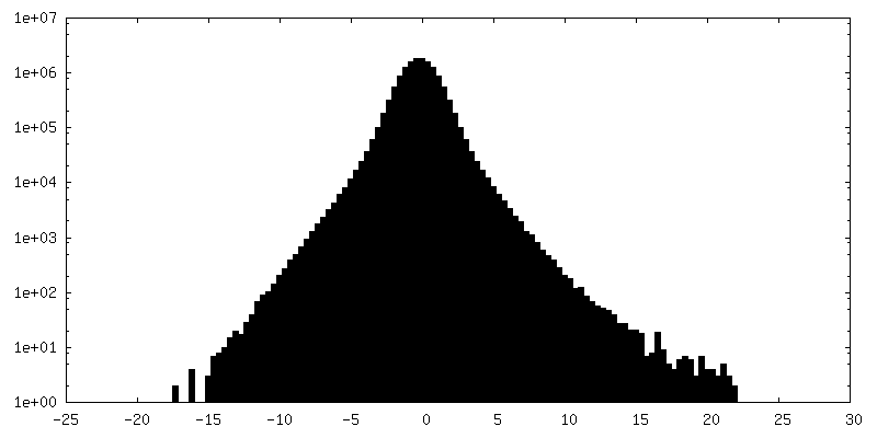
| Density Histograms |


-Half map: half map 2
| File | emd_14686_half_map_2.map | ||||||||||||
|---|---|---|---|---|---|---|---|---|---|---|---|---|---|
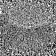
| Annotation | half map 2 | ||||||||||||
| Projections & Slices |
| ||||||||||||
| Density Histograms |


- Sample components
-Entire : Chikungunya virus neck complex
| Entire | Name: Chikungunya virus neck complex |
|---|---|
| Components |
|
-Supramolecule #1: Chikungunya virus neck complex
| Supramolecule | Name: Chikungunya virus neck complex / type: organelle_or_cellular_component / ID: 1 / Parent: 0 Details: Protein complex at the membrane neck of chikungunya virus spherules. |
|---|---|
| Source (natural) | Organism: Chikungunya virus |
-Experimental details
-Structure determination
| Method | cryo EM |
|---|---|
 Processing Processing | subtomogram averaging |
| Aggregation state | cell |
-Sample preparation
| Buffer | pH: 7.4 |
|---|---|
| Grid | Model: Quantifoil / Material: GOLD / Mesh: 200 / Support film - Material: CARBON / Support film - topology: HOLEY ARRAY / Support film - Film thickness: 10.0 nm / Pretreatment - Type: GLOW DISCHARGE / Pretreatment - Atmosphere: AIR / Pretreatment - Pressure: 0.0037 kPa |
| Vitrification | Cryogen name: ETHANE-PROPANE / Chamber humidity: 70 % / Chamber temperature: 295 K / Instrument: FEI VITROBOT MARK IV / Details: 5 seconds blot. |
- Electron microscopy
| Microscope | FEI TITAN KRIOS |
|---|---|
| Specialist optics | Energy filter - Name: GIF Bioquantum / Energy filter - Slit width: 20 eV |
| Image recording | Film or detector model: GATAN K2 SUMMIT (4k x 4k) / Detector mode: SUPER-RESOLUTION / Average electron dose: 1.5 e/Å2 |
| Electron beam | Acceleration voltage: 300 kV / Electron source:  FIELD EMISSION GUN FIELD EMISSION GUN |
| Electron optics | C2 aperture diameter: 70.0 µm / Illumination mode: FLOOD BEAM / Imaging mode: BRIGHT FIELD / Cs: 2.7 mm / Nominal defocus max: 5.0 µm / Nominal defocus min: 3.0 µm / Nominal magnification: 33000 |
| Sample stage | Specimen holder model: FEI TITAN KRIOS AUTOGRID HOLDER / Cooling holder cryogen: NITROGEN |
| Experimental equipment |  Model: Titan Krios / Image courtesy: FEI Company |
-Image processing
| Final reconstruction | Applied symmetry - Point group: C1 (asymmetric) / Resolution.type: BY AUTHOR / Resolution: 34.0 Å / Resolution method: FSC 0.143 CUT-OFF / Software - Name: Dynamo Details: 34A resolution was calculated using the uploaded mask Number subtomograms used: 64 |
|---|---|
| Extraction | Number tomograms: 9 / Number images used: 76 / Method: manual |
| CTF correction | Software: (Name: CTFFIND, CTFPHASEFLIP) |
| Final angle assignment | Type: ANGULAR RECONSTITUTION / Software - Name: Dynamo |
