 ムービー
ムービー コントローラー


+ データを開く

- 基本情報
基本情報
| 登録情報 | データベース: EMDB / ID: EMD-1454 | |||||||||
|---|---|---|---|---|---|---|---|---|---|---|
| タイトル | Human T-lymphotropic virus-1 visualized at the virological synapse by electron tomography. | |||||||||
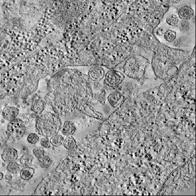
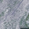
 マップデータ マップデータ | The tomogram shows a virological synapse between a chronically HTVL-1 infected MS9 cell (cell line)(top) and a Jurkat cell (bottom). Numerous HTLV-1 particles can be seen within the synaptic cleft. | |||||||||
 試料 試料 |
| |||||||||
| 生物種 | cell line (unknown) | |||||||||
| 手法 | 電子線トモグラフィー法 / ネガティブ染色法 / 解像度: 50.0 Å | |||||||||
 データ登録者 データ登録者 | Majorovits E / Nejmeddine M / Tanaka Y / Taylor GP / Fuller SD / Bangham CRM | |||||||||
 引用 引用 | ジャーナル: PLoS One / 年: 2008 タイトル: Human T-lymphotropic virus-1 visualized at the virological synapse by electron tomography. 著者: Endre Majorovits / Mohamed Nejmeddine / Yuetsu Tanaka / Graham P Taylor / Stephen D Fuller / Charles R M Bangham /  要旨: Human T-lymphotropic virus 1 (HTLV-1) is transmitted directly between cells via an organized cell-cell contact called a virological synapse (VS). The VS has been studied by light microscopy, but the ...Human T-lymphotropic virus 1 (HTLV-1) is transmitted directly between cells via an organized cell-cell contact called a virological synapse (VS). The VS has been studied by light microscopy, but the ultrastructure of the VS and the nature of the transmitted viral particle have remained unknown. Cell-free enveloped virions of HTLV-1 are undetectable in the serum of individuals infected with the human T-lymphotropic virus 1 (HTLV-1) and during in vitro culture of naturally infected lymphocytes. However, the viral envelope protein is required for infectivity of HTLV-1, suggesting that complete, enveloped HTLV-1 virions are transferred across the synapse. Here, we use electron tomography combined with immunostaining of viral protein to demonstrate the presence of enveloped HTLV-1 particles within the VS formed between naturally infected lymphocytes. We show in 3D that HTLV-1 particles can be detected in multiple synaptic clefts at different locations simultaneously within the same VS. The synaptic clefts are surrounded by the tightly apposed plasma membranes of the two cells. HTLV-1 virions can contact the recipient cell membrane before detaching from the infected cell. The results show that the HTLV-1 virological synapse that forms spontaneously between lymphocytes of HTLV-1 infected individuals allows direct cell-cell transmission of the virus by triggered, directional release of enveloped HTLV-1 particles into confined intercellular spaces. | |||||||||
| 履歴 |
|
- 構造の表示
構造の表示
| ムービー |
 ムービービューア ムービービューア |
|---|---|
| 構造ビューア | EMマップ: SurfViewMolmilJmol/JSmol |
| 添付画像 |
- ダウンロードとリンク
-EMDBアーカイブ
| マップデータ | emd_1454.map.gz | 106 MB | EMDBマップデータ形式 | |
|---|---|---|---|---|
| ヘッダ (付随情報) | emd-1454-v30.xmlemd-1454.xml | 8.6 KB 8.6 KB | 表示 表示 | EMDBヘッダ |
| 画像 |  1454.gif 1454.gif | 193.6 KB | ||
| アーカイブディレクトリ |  http://ftp.pdbj.org/pub/emdb/structures/EMD-1454ftp://ftp.pdbj.org/pub/emdb/structures/EMD-1454 http://ftp.pdbj.org/pub/emdb/structures/EMD-1454ftp://ftp.pdbj.org/pub/emdb/structures/EMD-1454 | HTTPS FTP |
-検証レポート
| 文書・要旨 | emd_1454_validation.pdf.gz | 168.4 KB | 表示 | EMDB検証レポート |
|---|---|---|---|---|
| 文書・詳細版 | emd_1454_full_validation.pdf.gz | 167.6 KB | 表示 | |
| XML形式データ | emd_1454_validation.xml.gz | 4.5 KB | 表示 | |
| アーカイブディレクトリ | https://ftp.pdbj.org/pub/emdb/validation_reports/EMD-1454ftp://ftp.pdbj.org/pub/emdb/validation_reports/EMD-1454 | HTTPS FTP |
-関連構造データ
-リンク
| EMDBのページ | EMDB (EBI/PDBe) / EMDataResource |
|---|
-マップ
| ファイル | ダウンロード / ファイル: emd_1454.map.gz / 形式: CCP4 / 大きさ: 166.6 MB / タイプ: IMAGE STORED AS FLOATING POINT NUMBER (4 BYTES) | ||||||||||||||||||||||||||||||||||||||||||||||||||||||||||||||||||||
|---|---|---|---|---|---|---|---|---|---|---|---|---|---|---|---|---|---|---|---|---|---|---|---|---|---|---|---|---|---|---|---|---|---|---|---|---|---|---|---|---|---|---|---|---|---|---|---|---|---|---|---|---|---|---|---|---|---|---|---|---|---|---|---|---|---|---|---|---|---|
| 注釈 | The tomogram shows a virological synapse between a chronically HTVL-1 infected MS9 cell (cell line)(top) and a Jurkat cell (bottom). Numerous HTLV-1 particles can be seen within the synaptic cleft. | ||||||||||||||||||||||||||||||||||||||||||||||||||||||||||||||||||||
| ボクセルのサイズ | X=Y=Z: 31.4 Å | ||||||||||||||||||||||||||||||||||||||||||||||||||||||||||||||||||||
| 密度 |
| ||||||||||||||||||||||||||||||||||||||||||||||||||||||||||||||||||||
| 対称性 | 空間群: 1 | ||||||||||||||||||||||||||||||||||||||||||||||||||||||||||||||||||||
| 詳細 | EMDB XML:
CCP4マップ ヘッダ情報:
| ||||||||||||||||||||||||||||||||||||||||||||||||||||||||||||||||||||
-添付データ
- 試料の構成要素
-全体 : HTLV-1 virological snapse formed between a chronically HTLV-1 inf...
| 全体 | 名称: HTLV-1 virological snapse formed between a chronically HTLV-1 infected MS9 cell and a target cell |
|---|---|
| 要素 |
|
-超分子 #1000: HTLV-1 virological snapse formed between a chronically HTLV-1 inf...
| 超分子 | 名称: HTLV-1 virological snapse formed between a chronically HTLV-1 infected MS9 cell and a target cell タイプ: sample / ID: 1000 / Number unique components: 1 |
|---|
-超分子 #1: virological synapse
| 超分子 | 名称: virological synapse / タイプ: organelle_or_cellular_component / ID: 1 / コピー数: 1 / 組換発現: No / データベース: NCBI |
|---|---|
| 由来(天然) | 生物種: cell line (unknown) / 株: MS9 / 別称: cell line / 細胞: MS9 / Organelle: plasma membrane / 細胞中の位置: plasma membrane |
-実験情報
-構造解析
| 手法 | ネガティブ染色法 |
|---|---|
 解析 解析 | 電子線トモグラフィー法 |
-試料調製
| 緩衝液 | pH: 7.2 / 詳細: sodium cacodylate buffer 0.1M |
|---|---|
| 染色 | タイプ: NEGATIVE / 詳細: 1% OsO4 staining 2% MgUranylAc |
| グリッド | 詳細: Formvar coated Cu/Pd 150 parallel bar grid |
| 凍結 | 凍結剤: NONE |
- 電子顕微鏡法
| 顕微鏡 | FEI TECNAI F30 |
|---|---|
| 温度 | 平均: 290 K |
| 日付 | 2006年11月3日 |
| 撮影 | カテゴリ: CCD フィルム・検出器のモデル: GENERIC GATAN (2k x 2k) |
| 電子線 | 加速電圧: 300 kV / 電子線源:  FIELD EMISSION GUN FIELD EMISSION GUN |
| 電子光学系 | 倍率(補正後): 19900 / 照射モード: FLOOD BEAM / 撮影モード: BRIGHT FIELD / Cs: 2 mm / 最大 デフォーカス(公称値): 0.2 µm / 最小 デフォーカス(公称値): 0.2 µm / 倍率(公称値): 20000 |
| 試料ステージ | 試料ホルダー: single-tilt holder / 試料ホルダーモデル: OTHER / Tilt series - Axis1 - Min angle: 60 ° / Tilt series - Axis1 - Max angle: 60 ° / Tilt series - Axis1 - Angle increment: 1 ° |
| 実験機器 |  モデル: Tecnai F30 / 画像提供: FEI Company |
-画像解析
| 最終 再構成 | アルゴリズム: OTHER / 解像度のタイプ: BY AUTHOR / 解像度: 50.0 Å / ソフトウェア - 名称: IMOD / 使用した粒子像数: 120 |
|---|
