Biotechnology and Biological Sciences Research Council (BBSRC)
BB/S003339/1
United Kingdom
Citation
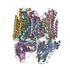
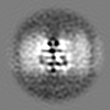
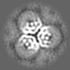
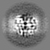
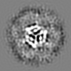
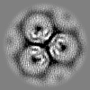
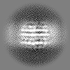
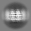
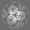
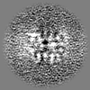
Journal: Nat Commun / Year: 2022 Title: Structure and activity of particulate methane monooxygenase arrays in methanotrophs. Authors: Yanan Zhu / Christopher W Koo / C Keith Cassidy / Matthew C Spink / Tao Ni / Laura C Zanetti-Domingues / Benji Bateman / Marisa L Martin-Fernandez / Juan Shen / Yuewen Sheng / Yun Song / ...Authors: Yanan Zhu / Christopher W Koo / C Keith Cassidy / Matthew C Spink / Tao Ni / Laura C Zanetti-Domingues / Benji Bateman / Marisa L Martin-Fernandez / Juan Shen / Yuewen Sheng / Yun Song / Zhengyi Yang / Amy C Rosenzweig / Peijun Zhang / Abstract: Methane-oxidizing bacteria play a central role in greenhouse gas mitigation and have potential applications in biomanufacturing. Their primary metabolic enzyme, particulate methane monooxygenase ...Methane-oxidizing bacteria play a central role in greenhouse gas mitigation and have potential applications in biomanufacturing. Their primary metabolic enzyme, particulate methane monooxygenase (pMMO), is housed in copper-induced intracytoplasmic membranes (ICMs), of which the function and biogenesis are not known. We show by serial cryo-focused ion beam (cryoFIB) milling/scanning electron microscope (SEM) volume imaging and lamellae-based cellular cryo-electron tomography (cryoET) that these ICMs are derived from the inner cell membrane. The pMMO trimer, resolved by cryoET and subtomogram averaging to 4.8 Å in the ICM, forms higher-order hexagonal arrays in intact cells. Array formation correlates with increased enzymatic activity, highlighting the importance of studying the enzyme in its native environment. These findings also demonstrate the power of cryoET to structurally characterize native membrane enzymes in the cellular context.
In the structure databanks used in Yorodumi, some data are registered as the other names, "COVID-19 virus" and "2019-nCoV". Here are the details of the virus and the list of structure data.
Jan 31, 2019. EMDB accession codes are about to change! (news from PDBe EMDB page)
EMDB accession codes are about to change! (news from PDBe EMDB page)
The allocation of 4 digits for EMDB accession codes will soon come to an end. Whilst these codes will remain in use, new EMDB accession codes will include an additional digit and will expand incrementally as the available range of codes is exhausted. The current 4-digit format prefixed with “EMD-” (i.e. EMD-XXXX) will advance to a 5-digit format (i.e. EMD-XXXXX), and so on. It is currently estimated that the 4-digit codes will be depleted around Spring 2019, at which point the 5-digit format will come into force.
The EM Navigator/Yorodumi systems omit the EMD- prefix.
Related info.:Q: What is EMD? / ID/Accession-code notation in Yorodumi/EM Navigator
Yorodumi is a browser for structure data from EMDB, PDB, SASBDB, etc.
This page is also the successor to EM Navigator detail page, and also detail information page/front-end page for Omokage search.
The word "yorodu" (or yorozu) is an old Japanese word meaning "ten thousand". "mi" (miru) is to see.
Related info.:EMDB / PDB / SASBDB / Comparison of 3 databanks / Yorodumi Search / Aug 31, 2016. New EM Navigator & Yorodumi / Yorodumi Papers / Jmol/JSmol / Function and homology information / Changes in new EM Navigator and Yorodumi
 Movie
Movie Controller


 Open data
 Basic information
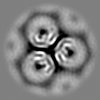
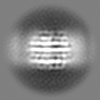
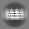
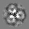
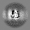
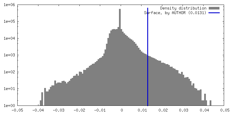
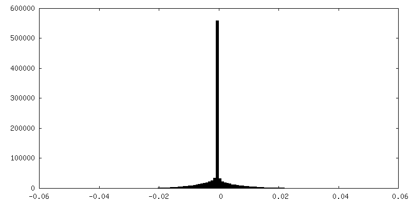
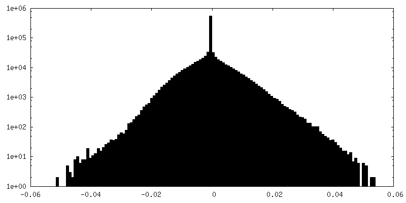
 Map data
 Sample
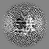
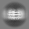
 Methylococcus capsulatus str. Bath (bacteria)
 Authors
 United Kingdom, 2 items
  Citation

 Structure visualization
 Downloads & links
 EMDB map data format
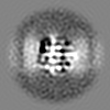
 emd_14530.png
 http://ftp.pdbj.org/pub/emdb/structures/EMD-14530
 Z (Sec.)
 Y (Row.)
 X (Col.)


 Sample components
 Processing
 Electron microscopy
 FIELD EMISSION GUN
