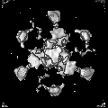
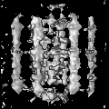
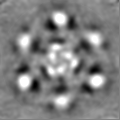
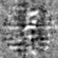
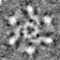
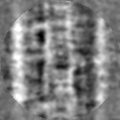
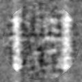
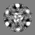
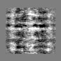
ジャーナル: J Muscle Res Cell Motil / 年: 2023 タイトル: Cryo-electron tomography of intact cardiac muscle reveals myosin binding protein-C linking myosin and actin filaments. 著者: Xinrui Huang / Iratxe Torre / Michele Chiappi / Zhan Yin / Anupama Vydyanath / Shuangyi Cao / Oliver Raschdorf / Morgan Beeby / Bonnie Quigley / Pieter P de Tombe / Jun Liu / Edward P Morris ...著者: Xinrui Huang / Iratxe Torre / Michele Chiappi / Zhan Yin / Anupama Vydyanath / Shuangyi Cao / Oliver Raschdorf / Morgan Beeby / Bonnie Quigley / Pieter P de Tombe / Jun Liu / Edward P Morris / Pradeep K Luther / 要旨: Myosin binding protein C (MyBP-C) is an accessory protein of the thick filament in vertebrate cardiac muscle arranged over 9 stripes of intervals of 430 Å in each half of the A-band in the region ...Myosin binding protein C (MyBP-C) is an accessory protein of the thick filament in vertebrate cardiac muscle arranged over 9 stripes of intervals of 430 Å in each half of the A-band in the region called the C-zone. Mutations in cardiac MyBP-C are a leading cause of hypertrophic cardiomyopathy the mechanism of which is unknown. It is a rod-shaped protein composed of 10 or 11 immunoglobulin- or fibronectin-like domains labelled C0 to C10 which binds to the thick filament via its C-terminal region. MyBP-C regulates contraction in a phosphorylation dependent fashion that may be through binding of its N-terminal domains with myosin or actin. Understanding the 3D organisation of MyBP-C in the sarcomere environment may provide new light on its function. We report here the fine structure of MyBP-C in relaxed rat cardiac muscle by cryo-electron tomography and subtomogram averaging of refrozen Tokuyasu cryosections. We find that on average MyBP-C connects via its distal end to actin across a disc perpendicular to the thick filament. The path of MyBP-C suggests that the central domains may interact with myosin heads. Surprisingly MyBP-C at Stripe 4 is different; it has weaker density than the other stripes which could result from a mainly axial or wavy path. Given that the same feature at Stripe 4 can also be found in several mammalian cardiac muscles and in some skeletal muscles, our finding may have broader implication and significance. In the D-zone, we show the first demonstration of myosin crowns arranged on a uniform 143 Å repeat.
 ムービー
ムービー コントローラー


 データを開く
 基本情報
基本情報
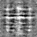
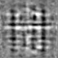
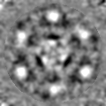
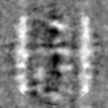
 マップデータ
 試料
試料 キーワード
キーワード
 データ登録者
 英国, 1件
  引用
引用


 構造の表示
構造の表示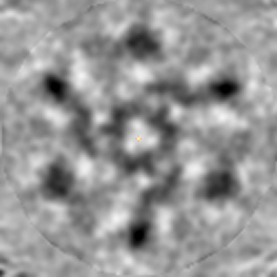
 ダウンロードとリンク
 EMDBマップデータ形式
 emd_14504.png
 http://ftp.pdbj.org/pub/emdb/structures/EMD-14504
 Z (Sec.)
 Y (Row.)
 X (Col.)


 試料の構成要素
 解析
解析 電子顕微鏡法
 FIELD EMISSION GUN

