Biotechnology and Biological Sciences Research Council (BBSRC)
BB/L014211/1
英国
Agence Nationale de la Recherche (ANR)
ANR-16-CE16-0011-03
フランス
Other government
EU-HEALTH-2013, DESIRE, Number 60253
Other government
COST Action CA16118
引用
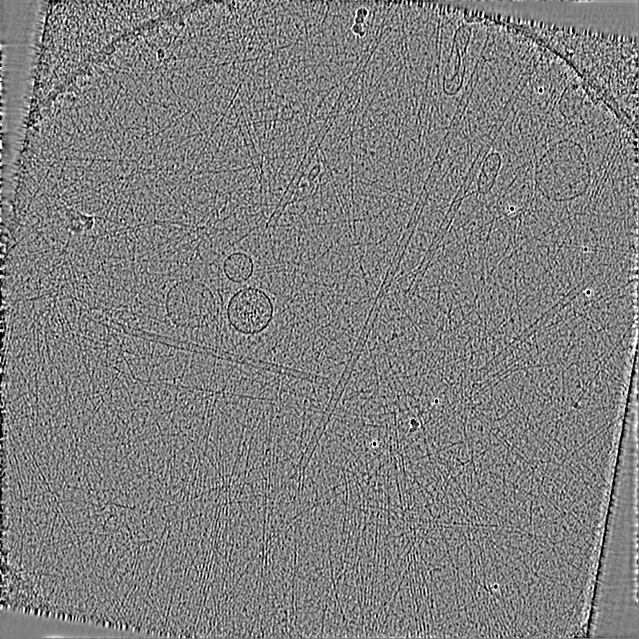
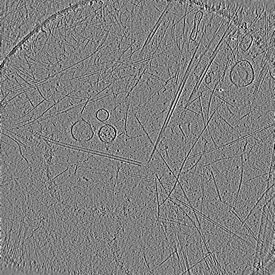
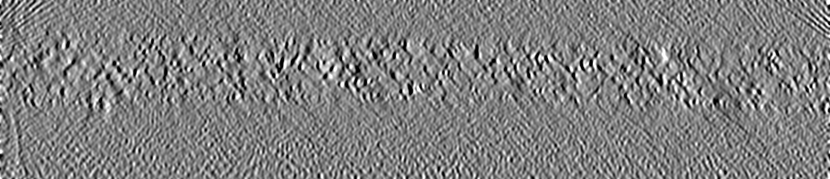
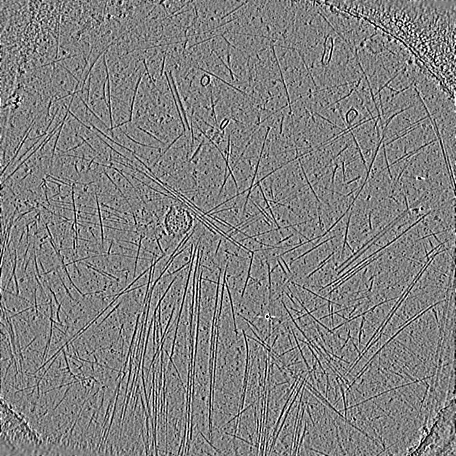
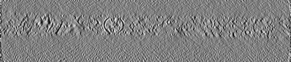
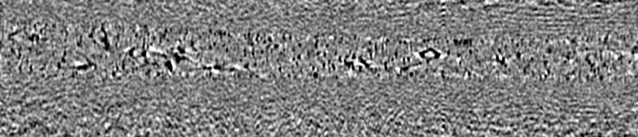
ジャーナル: J Cell Sci / 年: 2022 タイトル: Visualising the cytoskeletal machinery in neuronal growth cones using cryo-electron tomography. 著者: Joseph Atherton / Melissa Stouffer / Fiona Francis / Carolyn A Moores / 要旨: Neurons extend axons to form the complex circuitry of the mature brain. This depends on the coordinated response and continuous remodelling of the microtubule and F-actin networks in the axonal ...Neurons extend axons to form the complex circuitry of the mature brain. This depends on the coordinated response and continuous remodelling of the microtubule and F-actin networks in the axonal growth cone. Growth cone architecture remains poorly understood at nanoscales. We therefore investigated mouse hippocampal neuron growth cones using cryo-electron tomography to directly visualise their three-dimensional subcellular architecture with molecular detail. Our data showed that the hexagonal arrays of actin bundles that form filopodia penetrate and terminate deep within the growth cone interior. We directly observed the modulation of these and other growth cone actin bundles by alteration of individual F-actin helical structures. Microtubules with blunt, slightly flared or gently curved ends predominated in the growth cone, frequently contained lumenal particles and exhibited lattice defects. Investigation of the effect of absence of doublecortin, a neurodevelopmental cytoskeleton regulator, on growth cone cytoskeleton showed no major anomalies in overall growth cone organisation or in F-actin subpopulations. However, our data suggested that microtubules sustained more structural defects, highlighting the importance of microtubule integrity during growth cone migration.
 ムービー
ムービー コントローラー


 データを開く
 基本情報
基本情報
 マップデータ
 試料
試料 キーワード
キーワード
 データ登録者
 英国,
英国,  フランス, 6件
  引用
引用
 構造の表示
構造の表示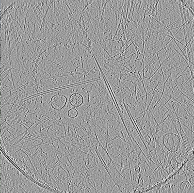
 ダウンロードとリンク
 EMDBマップデータ形式
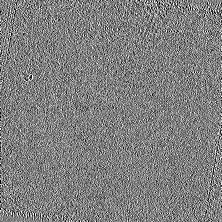
 emd_14396.png
 http://ftp.pdbj.org/pub/emdb/structures/EMD-14396


 Z (Sec.)
 Y (Row.)
 X (Col.)


 試料の構成要素
 解析
解析 電子顕微鏡法
 FIELD EMISSION GUN
