Biotechnology and Biological Sciences Research Council (BBSRC)
BB/L014211/1
United Kingdom
Agence Nationale de la Recherche (ANR)
ANR-16-CE16-0011-03
France
Other government
EU-HEALTH-2013, DESIRE, Number 60253
Other government
COST Action CA16118
Citation
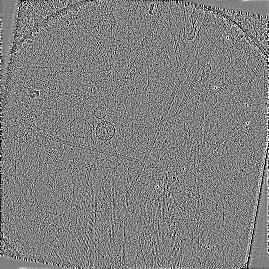
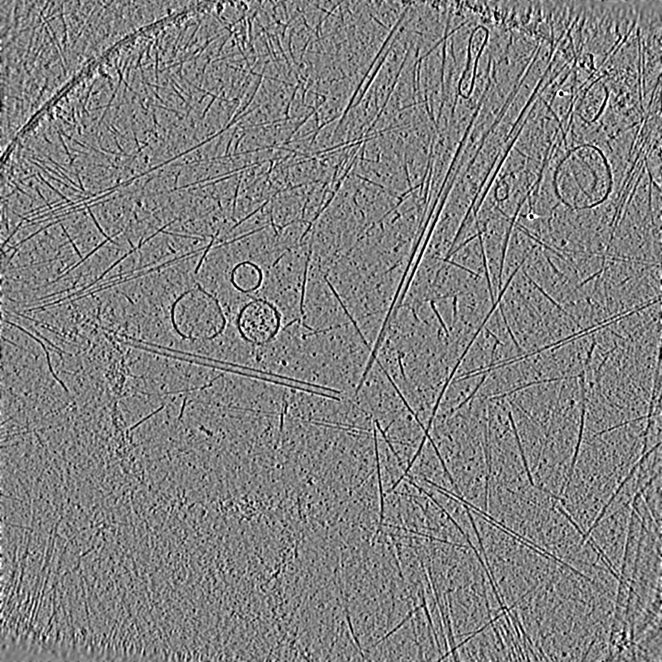
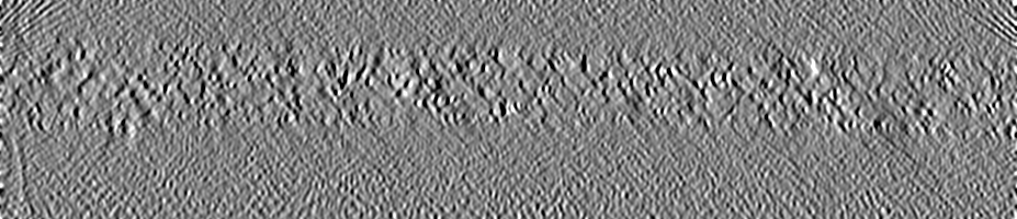
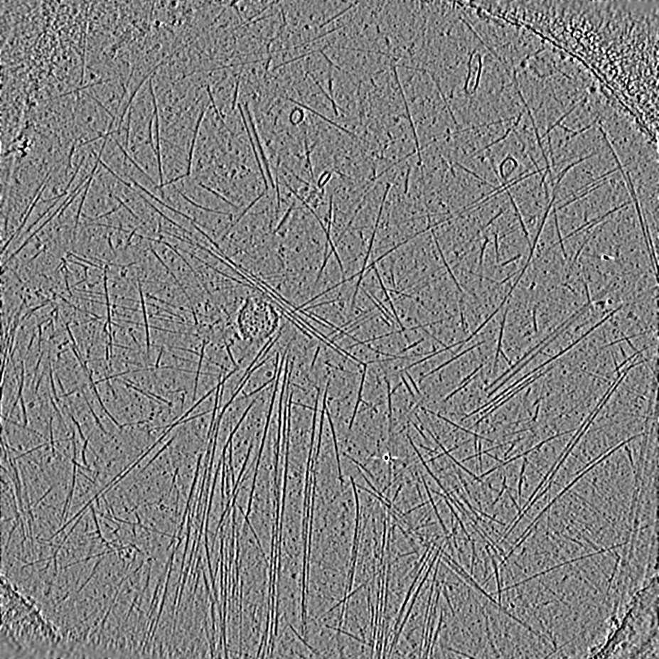
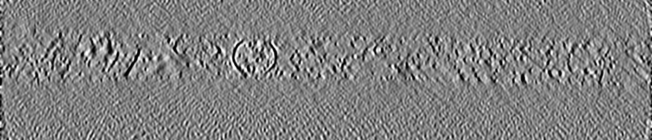
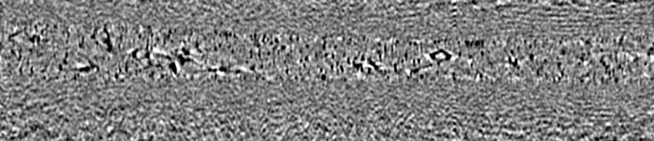
Journal: J Cell Sci / Year: 2022 Title: Visualising the cytoskeletal machinery in neuronal growth cones using cryo-electron tomography. Authors: Joseph Atherton / Melissa Stouffer / Fiona Francis / Carolyn A Moores / Abstract: Neurons extend axons to form the complex circuitry of the mature brain. This depends on the coordinated response and continuous remodelling of the microtubule and F-actin networks in the axonal ...Neurons extend axons to form the complex circuitry of the mature brain. This depends on the coordinated response and continuous remodelling of the microtubule and F-actin networks in the axonal growth cone. Growth cone architecture remains poorly understood at nanoscales. We therefore investigated mouse hippocampal neuron growth cones using cryo-electron tomography to directly visualise their three-dimensional subcellular architecture with molecular detail. Our data showed that the hexagonal arrays of actin bundles that form filopodia penetrate and terminate deep within the growth cone interior. We directly observed the modulation of these and other growth cone actin bundles by alteration of individual F-actin helical structures. Microtubules with blunt, slightly flared or gently curved ends predominated in the growth cone, frequently contained lumenal particles and exhibited lattice defects. Investigation of the effect of absence of doublecortin, a neurodevelopmental cytoskeleton regulator, on growth cone cytoskeleton showed no major anomalies in overall growth cone organisation or in F-actin subpopulations. However, our data suggested that microtubules sustained more structural defects, highlighting the importance of microtubule integrity during growth cone migration.
In the structure databanks used in Yorodumi, some data are registered as the other names, "COVID-19 virus" and "2019-nCoV". Here are the details of the virus and the list of structure data.
Jan 31, 2019. EMDB accession codes are about to change! (news from PDBe EMDB page)
EMDB accession codes are about to change! (news from PDBe EMDB page)
The allocation of 4 digits for EMDB accession codes will soon come to an end. Whilst these codes will remain in use, new EMDB accession codes will include an additional digit and will expand incrementally as the available range of codes is exhausted. The current 4-digit format prefixed with “EMD-” (i.e. EMD-XXXX) will advance to a 5-digit format (i.e. EMD-XXXXX), and so on. It is currently estimated that the 4-digit codes will be depleted around Spring 2019, at which point the 5-digit format will come into force.
The EM Navigator/Yorodumi systems omit the EMD- prefix.
Related info.:Q: What is EMD? / ID/Accession-code notation in Yorodumi/EM Navigator
Yorodumi is a browser for structure data from EMDB, PDB, SASBDB, etc.
This page is also the successor to EM Navigator detail page, and also detail information page/front-end page for Omokage search.
The word "yorodu" (or yorozu) is an old Japanese word meaning "ten thousand". "mi" (miru) is to see.
Related info.:EMDB / PDB / SASBDB / Comparison of 3 databanks / Yorodumi Search / Aug 31, 2016. New EM Navigator & Yorodumi / Yorodumi Papers / Jmol/JSmol / Function and homology information / Changes in new EM Navigator and Yorodumi
 Movie
Movie Controller

 Yorodumi
 Open data
 Basic information
 Map data
 Sample
 Keywords
 Authors
 United Kingdom,
  France, 6 items
  Citation
 Structure visualization
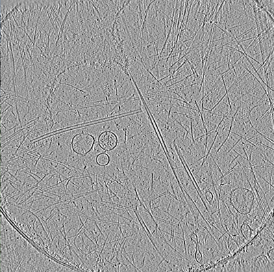
 Downloads & links
 EMDB map data format
 emd_14396.png
 http://ftp.pdbj.org/pub/emdb/structures/EMD-14396


 Z (Sec.)
 Y (Row.)
 X (Col.)


 Sample components
 Processing
 Electron microscopy
 FIELD EMISSION GUN
