 Movie
Movie Controller


+ Open data

- Basic information
| Entry |  | |||||||||
|---|---|---|---|---|---|---|---|---|---|---|
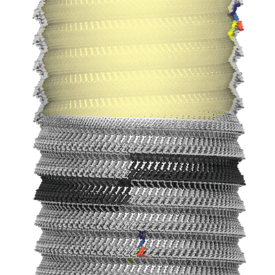
| Title | Cryo-EM structure of Bacillus megaterium gas vesicles | |||||||||
 Map data Map data | Main map, automatically B-factor sharpened with -30 A**2 in cryoSPARC / symmetrized with -3.874 degree rotation, 0.525 A rise | |||||||||
 Sample Sample |
| |||||||||
| Biological species |  Priestia megaterium NBRC 15308 = ATCC 14581 (bacteria) Priestia megaterium NBRC 15308 = ATCC 14581 (bacteria) | |||||||||
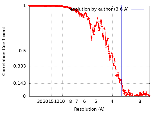
| Method | helical reconstruction / cryo EM / Resolution: 3.6 Å | |||||||||
 Authors Authors | Huber ST / Evers W / Jakobi AJ | |||||||||
| Funding support | 1 items
| |||||||||
 Citation Citation | Journal: Cell / Year: 2023 Title: Cryo-EM structure of gas vesicles for buoyancy-controlled motility. Authors: Stefan T Huber / Dion Terwiel / Wiel H Evers / David Maresca / Arjen J Jakobi /  Abstract: Gas vesicles are gas-filled nanocompartments that allow a diverse group of bacteria and archaea to control their buoyancy. The molecular basis of their properties and assembly remains unclear. Here, ...Gas vesicles are gas-filled nanocompartments that allow a diverse group of bacteria and archaea to control their buoyancy. The molecular basis of their properties and assembly remains unclear. Here, we report the 3.2 Å cryo-EM structure of the gas vesicle shell made from the structural protein GvpA that self-assembles into hollow helical cylinders closed off by cone-shaped tips. Two helical half shells connect through a characteristic arrangement of GvpA monomers, suggesting a mechanism of gas vesicle biogenesis. The fold of GvpA features a corrugated wall structure typical for force-bearing thin-walled cylinders. Small pores enable gas molecules to diffuse across the shell, while the exceptionally hydrophobic interior surface effectively repels water. Comparative structural analysis confirms the evolutionary conservation of gas vesicle assemblies and demonstrates molecular features of shell reinforcement by GvpC. Our findings will further research into gas vesicle biology and facilitate molecular engineering of gas vesicles for ultrasound imaging. | |||||||||
| History |
|
- Structure visualization
| Supplemental images |
|---|
- Downloads & links
-EMDB archive
| Map data | emd_14340.map.gz | 115.6 MB |  EMDB map data format EMDB map data format | |
|---|---|---|---|---|
| Header (meta data) | emd-14340-v30.xmlemd-14340.xml | 19.5 KB 19.5 KB | Display Display | EMDB header |
| FSC (resolution estimation) | emd_14340_fsc.xml | 17.7 KB | Display | FSC data file |
| Images |  emd_14340.png emd_14340.png | 245.5 KB | ||
| Masks | emd_14340_msk_1.map | 512 MB | Mask map | |
| Others | emd_14340_additional_1.map.gzemd_14340_half_map_1.map.gzemd_14340_half_map_2.map.gz | 115.5 MB 475.4 MB 475.5 MB | ||
| Archive directory |  http://ftp.pdbj.org/pub/emdb/structures/EMD-14340ftp://ftp.pdbj.org/pub/emdb/structures/EMD-14340 http://ftp.pdbj.org/pub/emdb/structures/EMD-14340ftp://ftp.pdbj.org/pub/emdb/structures/EMD-14340 | HTTPS FTP |
-Related structure data


-Links
| EMDB pages | EMDB (EBI/PDBe) / EMDataResource |
|---|
-Map
| File | Download / File: emd_14340.map.gz / Format: CCP4 / Size: 512 MB / Type: IMAGE STORED AS FLOATING POINT NUMBER (4 BYTES) | ||||||||||||||||||||||||||||||||||||
|---|---|---|---|---|---|---|---|---|---|---|---|---|---|---|---|---|---|---|---|---|---|---|---|---|---|---|---|---|---|---|---|---|---|---|---|---|---|
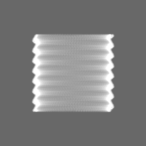
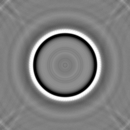
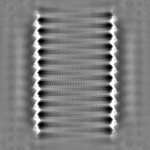
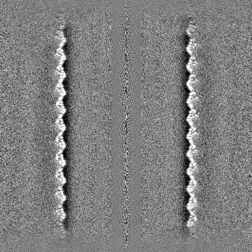
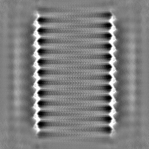
| Annotation | Main map, automatically B-factor sharpened with -30 A**2 in cryoSPARC / symmetrized with -3.874 degree rotation, 0.525 A rise | ||||||||||||||||||||||||||||||||||||
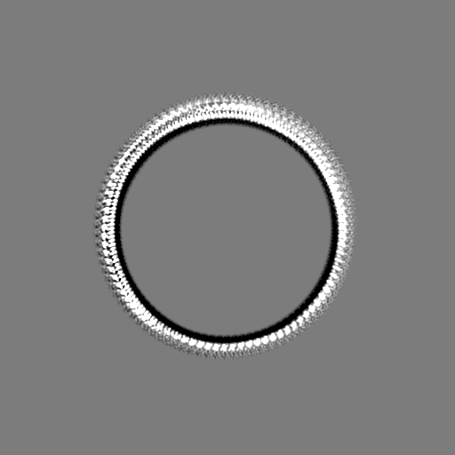
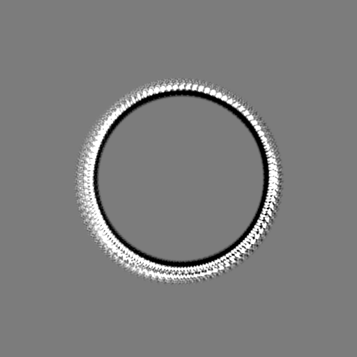
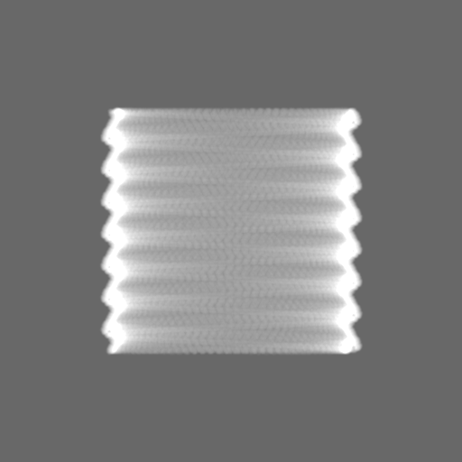
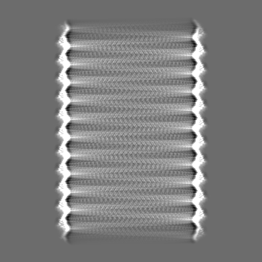
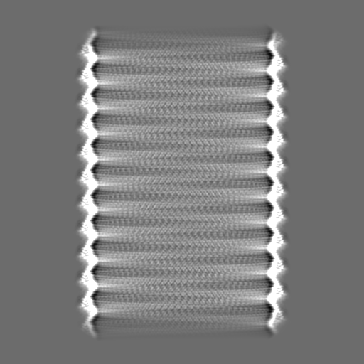
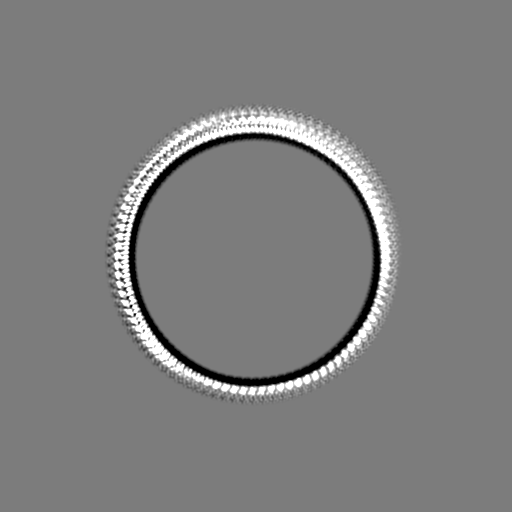
| Projections & slices | Image control
Images are generated by Spider. | ||||||||||||||||||||||||||||||||||||
| Voxel size | X=Y=Z: 1.37 Å | ||||||||||||||||||||||||||||||||||||
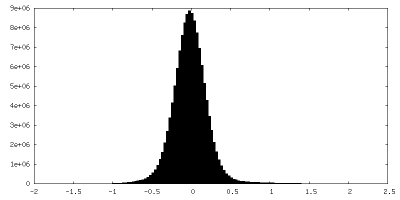
| Density |
| ||||||||||||||||||||||||||||||||||||
| Symmetry | Space group: 1 | ||||||||||||||||||||||||||||||||||||
| Details | EMDB XML:
|
 Z (Sec.)
 Y (Row.)
 X (Col.)


-Supplemental data
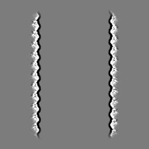
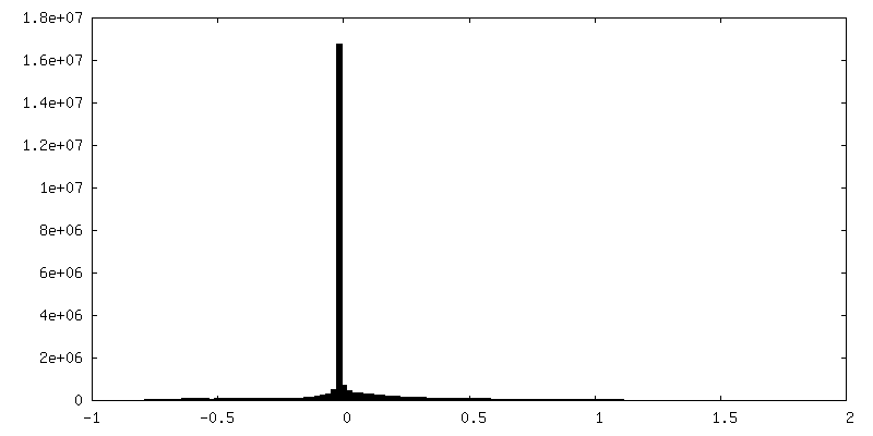
-Mask #1
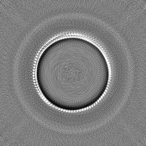
| File | emd_14340_msk_1.map | ||||||||||||
|---|---|---|---|---|---|---|---|---|---|---|---|---|---|
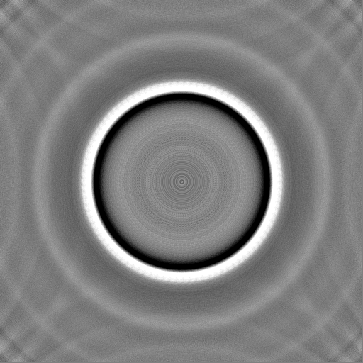
| Projections & Slices |
| ||||||||||||
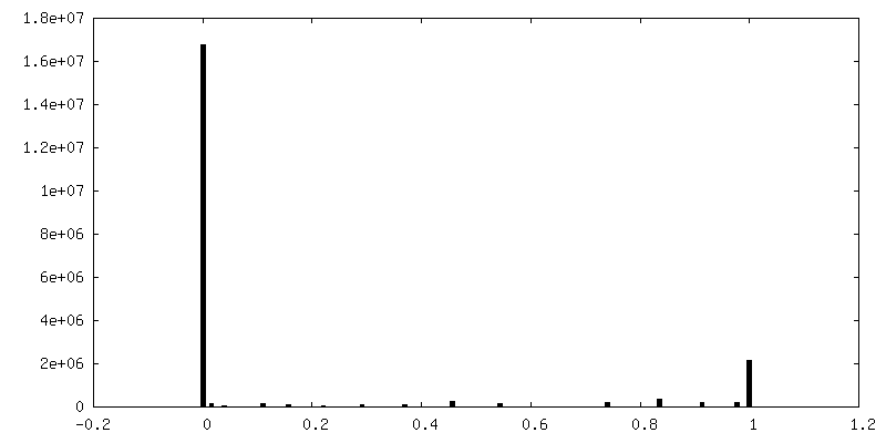
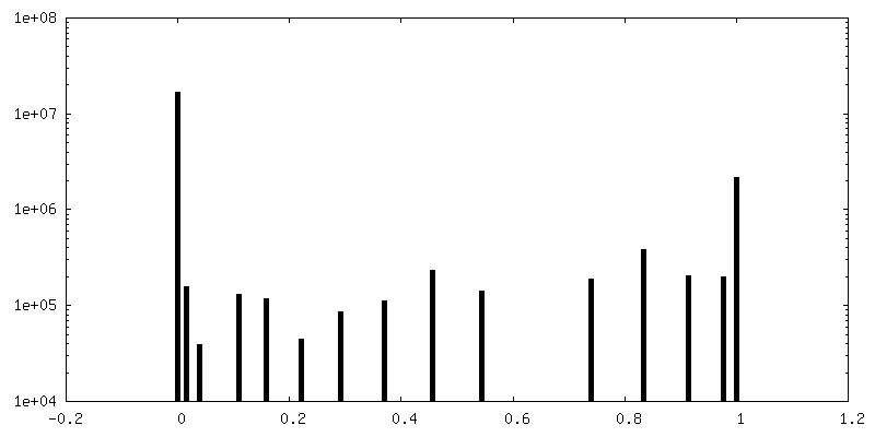
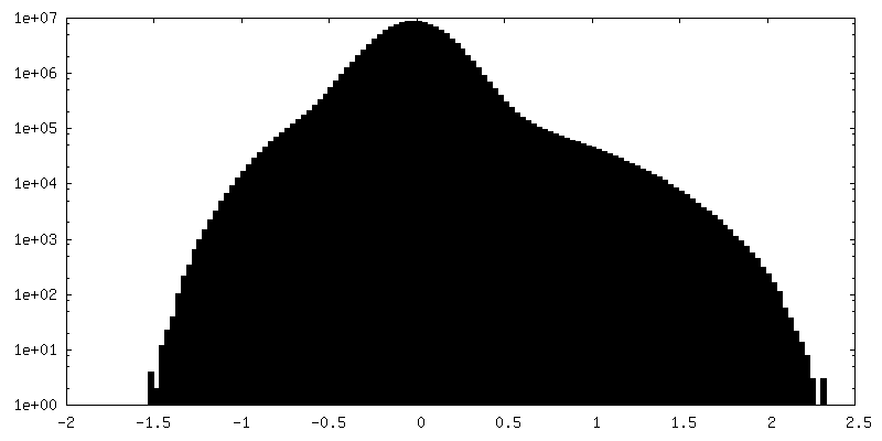
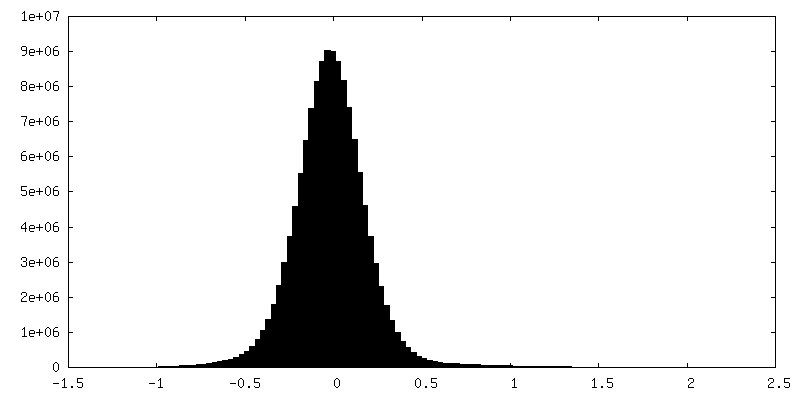
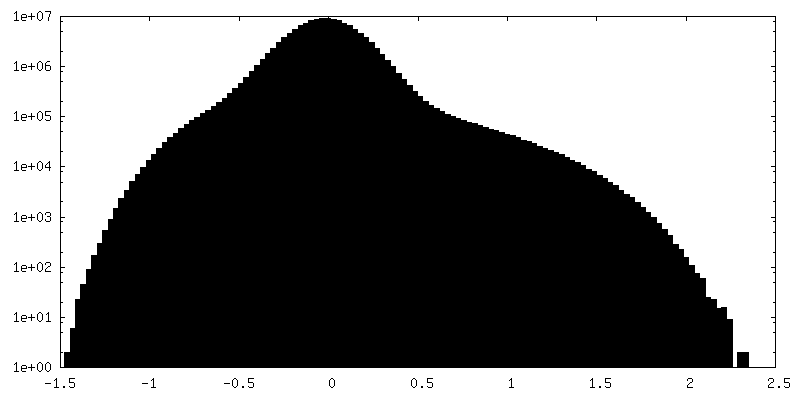
| Density Histograms |


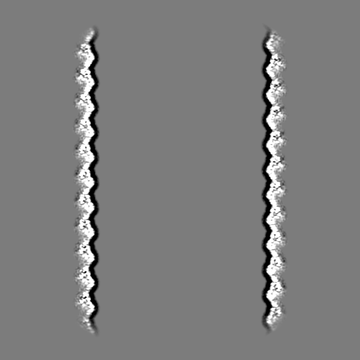
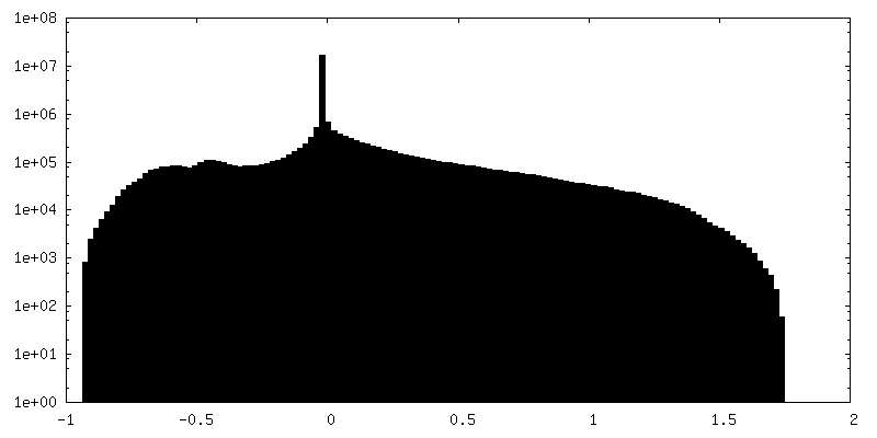
-Additional map: Unsharpened map / symmetrized with -3.874 degree rotation,...
| File | emd_14340_additional_1.map | ||||||||||||
|---|---|---|---|---|---|---|---|---|---|---|---|---|---|
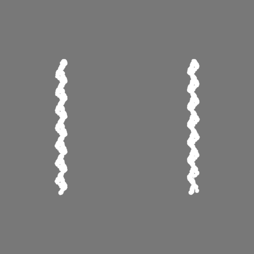
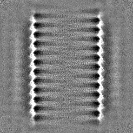
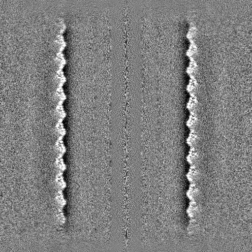
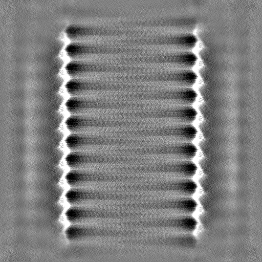
| Annotation | Unsharpened map / symmetrized with -3.874 degree rotation, 0.525 A rise | ||||||||||||
| Projections & Slices |
| ||||||||||||
| Density Histograms |

-Half map: Half Map A
| File | emd_14340_half_map_1.map | ||||||||||||
|---|---|---|---|---|---|---|---|---|---|---|---|---|---|
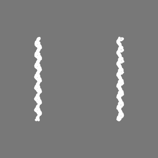
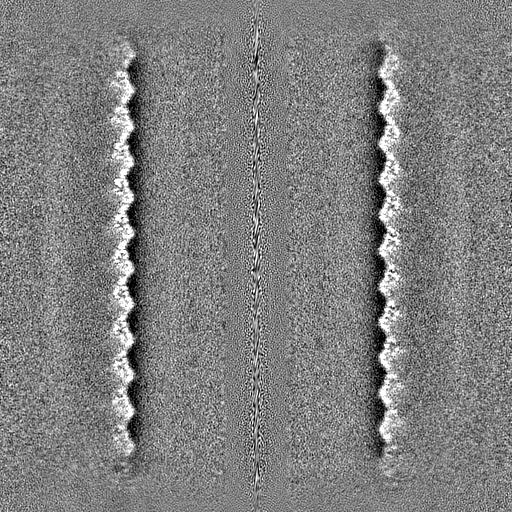
| Annotation | Half Map A | ||||||||||||
| Projections & Slices |
| ||||||||||||
| Density Histograms |

-Half map: Half Map B
| File | emd_14340_half_map_2.map | ||||||||||||
|---|---|---|---|---|---|---|---|---|---|---|---|---|---|
| Annotation | Half Map B | ||||||||||||
| Projections & Slices |
| ||||||||||||
| Density Histograms |

- Sample components
-Entire : Helical assembly of GvpB monomers forming the gas vesicle wall
| Entire | Name: Helical assembly of GvpB monomers forming the gas vesicle wall |
|---|---|
| Components |
|
-Supramolecule #1: Helical assembly of GvpB monomers forming the gas vesicle wall
| Supramolecule | Name: Helical assembly of GvpB monomers forming the gas vesicle wall type: complex / ID: 1 / Chimera: Yes / Parent: 0 / Macromolecule list: all |
|---|---|
| Source (natural) | Organism: Priestia megaterium NBRC 15308 = ATCC 14581 (bacteria) |
| Molecular weight | Theoretical: 9.99 MDa |
-Macromolecule #1: GvpA2/B from B.megaterium
| Macromolecule | Name: GvpA2/B from B.megaterium / type: protein_or_peptide / ID: 1 / Enantiomer: LEVO |
|---|---|
| Source (natural) | Organism: Priestia megaterium NBRC 15308 = ATCC 14581 (bacteria) |
| Recombinant expression | Organism: |
| Sequence | String: MSIQKSTNSS SLAEVIDRI L DKGIVIDA FA RVSVVGI EIL TIEARV VIAS VDTWL RYAEA VGLL RDDVEE NGL PERSNSS EG QPRFSI |
-Experimental details
-Structure determination
| Method | cryo EM |
|---|---|
 Processing Processing | helical reconstruction |
| Aggregation state | helical array |
-Sample preparation
| Concentration | 0.45 mg/mL | |||||||||
|---|---|---|---|---|---|---|---|---|---|---|
| Buffer | pH: 8 Component:
| |||||||||
| Vitrification | Cryogen name: ETHANE / Chamber humidity: 95 % / Chamber temperature: 293 K / Instrument: LEICA PLUNGER / Details: Blot times between 5 and 11 seconds.. | |||||||||
| Details | Concentration measured by OD(500)=3.12 against a sonicated blank. |
- Electron microscopy
| Microscope | FEI TITAN KRIOS |
|---|---|
| Image recording | Film or detector model: GATAN K3 BIOQUANTUM (6k x 4k) / Digitization - Dimensions - Width: 5760 pixel / Digitization - Dimensions - Height: 4092 pixel / Number grids imaged: 1 / Number real images: 4351 / Average exposure time: 2.4 sec. / Average electron dose: 30.0 e/Å2 Details: One shot per hole 1.37 A/pix 60 fractions over 30 e-/A2 |
| Electron beam | Acceleration voltage: 300 kV / Electron source:  FIELD EMISSION GUN FIELD EMISSION GUN |
| Electron optics | Illumination mode: FLOOD BEAM / Imaging mode: BRIGHT FIELD / Cs: 2.7 mm / Nominal defocus max: 1.25 µm / Nominal defocus min: 0.25 µm / Nominal magnification: 64000 |
| Sample stage | Specimen holder model: FEI TITAN KRIOS AUTOGRID HOLDER / Cooling holder cryogen: NITROGEN |
| Experimental equipment |  Model: Titan Krios / Image courtesy: FEI Company |
+Image processing
-Atomic model buiding 1
| Refinement | Space: REAL / Protocol: AB INITIO MODEL |
|---|
