+ データを開く
- 基本情報
基本情報
| 登録情報 | データベース: EMDB / ID: EMD-1420 | |||||||||
|---|---|---|---|---|---|---|---|---|---|---|
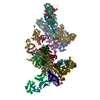
| タイトル | DNA poised for release in bacteriophage phi29. | |||||||||
 マップデータ マップデータ | 3D map of fiberless phi29 virion | |||||||||
 試料 試料 |
| |||||||||
| 生物種 |   Bacillus phage phi29 (ファージ) Bacillus phage phi29 (ファージ) | |||||||||
| 手法 | 単粒子再構成法 / クライオ電子顕微鏡法 / 解像度: 7.8 Å | |||||||||
 データ登録者 データ登録者 | Tang J / Olson N / Jardine P / Grimes S / Anderson D / Baker T | |||||||||
 引用 引用 |  ジャーナル: Structure / 年: 2008 ジャーナル: Structure / 年: 2008タイトル: DNA poised for release in bacteriophage phi29. 著者: Jinghua Tang / Norman Olson / Paul J Jardine / Shelley Grimes / Dwight L Anderson / Timothy S Baker /  要旨: We present here the first asymmetric, three-dimensional reconstruction of a tailed dsDNA virus, the mature bacteriophage phi29, at subnanometer resolution. This structure reveals the rich detail of ...We present here the first asymmetric, three-dimensional reconstruction of a tailed dsDNA virus, the mature bacteriophage phi29, at subnanometer resolution. This structure reveals the rich detail of the asymmetric interactions and conformational dynamics of the phi29 protein and DNA components, and provides novel insight into the mechanics of virus assembly. For example, the dodecameric head-tail connector protein undergoes significant rearrangement upon assembly into the virion. Specific interactions occur between the tightly packed dsDNA and the proteins of the head and tail. Of particular interest and novelty, an approximately 60A diameter toroid of dsDNA was observed in the connector-lower collar cavity. The extreme deformation that occurs over a small stretch of DNA is likely a consequence of the high pressure of the packaged genome. This toroid structure may help retain the DNA inside the capsid prior to its injection into the bacterial host. | |||||||||
| 履歴 |
|
- 構造の表示
構造の表示
| ムービー |
 ムービービューア ムービービューア |
|---|---|
| 構造ビューア | EMマップ:  SurfView SurfView Molmil Molmil Jmol/JSmol Jmol/JSmol |
| 添付画像 |
- ダウンロードとリンク
-EMDBアーカイブ
| マップデータ |  emd_1420.map.gz emd_1420.map.gz | 4.1 MB |  EMDBマップデータ形式 EMDBマップデータ形式 | |
|---|---|---|---|---|
| ヘッダ (付随情報) |  emd-1420-v30.xml emd-1420-v30.xml emd-1420.xml emd-1420.xml | 6.8 KB 6.8 KB | 表示 表示 |  EMDBヘッダ EMDBヘッダ |
| 画像 |  1420.gif 1420.gif | 28 KB | ||
| アーカイブディレクトリ |  http://ftp.pdbj.org/pub/emdb/structures/EMD-1420 http://ftp.pdbj.org/pub/emdb/structures/EMD-1420 ftp://ftp.pdbj.org/pub/emdb/structures/EMD-1420 ftp://ftp.pdbj.org/pub/emdb/structures/EMD-1420 | HTTPS FTP |
-検証レポート
| 文書・要旨 |  emd_1420_validation.pdf.gz emd_1420_validation.pdf.gz | 231.3 KB | 表示 |  EMDB検証レポート EMDB検証レポート |
|---|---|---|---|---|
| 文書・詳細版 |  emd_1420_full_validation.pdf.gz emd_1420_full_validation.pdf.gz | 230.4 KB | 表示 | |
| XML形式データ |  emd_1420_validation.xml.gz emd_1420_validation.xml.gz | 6.7 KB | 表示 | |
| アーカイブディレクトリ |  https://ftp.pdbj.org/pub/emdb/validation_reports/EMD-1420 https://ftp.pdbj.org/pub/emdb/validation_reports/EMD-1420 ftp://ftp.pdbj.org/pub/emdb/validation_reports/EMD-1420 ftp://ftp.pdbj.org/pub/emdb/validation_reports/EMD-1420 | HTTPS FTP |
-関連構造データ
- リンク
リンク
| EMDBのページ |  EMDB (EBI/PDBe) / EMDB (EBI/PDBe) /  EMDataResource EMDataResource |
|---|
- マップ
マップ
| ファイル |  ダウンロード / ファイル: emd_1420.map.gz / 形式: CCP4 / 大きさ: 101.6 MB / タイプ: IMAGE STORED AS FLOATING POINT NUMBER (4 BYTES) ダウンロード / ファイル: emd_1420.map.gz / 形式: CCP4 / 大きさ: 101.6 MB / タイプ: IMAGE STORED AS FLOATING POINT NUMBER (4 BYTES) | ||||||||||||||||||||||||||||||||||||||||||||||||||||||||||||||||||||
|---|---|---|---|---|---|---|---|---|---|---|---|---|---|---|---|---|---|---|---|---|---|---|---|---|---|---|---|---|---|---|---|---|---|---|---|---|---|---|---|---|---|---|---|---|---|---|---|---|---|---|---|---|---|---|---|---|---|---|---|---|---|---|---|---|---|---|---|---|---|
| 注釈 | 3D map of fiberless phi29 virion | ||||||||||||||||||||||||||||||||||||||||||||||||||||||||||||||||||||
| 投影像・断面図 | 画像のコントロール
画像は Spider により作成 | ||||||||||||||||||||||||||||||||||||||||||||||||||||||||||||||||||||
| ボクセルのサイズ | X=Y=Z: 3.68 Å | ||||||||||||||||||||||||||||||||||||||||||||||||||||||||||||||||||||
| 密度 |
| ||||||||||||||||||||||||||||||||||||||||||||||||||||||||||||||||||||
| 対称性 | 空間群: 1 | ||||||||||||||||||||||||||||||||||||||||||||||||||||||||||||||||||||
| 詳細 | EMDB XML:
CCP4マップ ヘッダ情報:
| ||||||||||||||||||||||||||||||||||||||||||||||||||||||||||||||||||||
-添付データ
- 試料の構成要素
-全体 : phi29 fiberless virion
| 全体 | 名称: phi29 fiberless virion |
|---|---|
| 要素 |
|
-超分子 #1000: phi29 fiberless virion
| 超分子 | 名称: phi29 fiberless virion / タイプ: sample / ID: 1000 / Number unique components: 1 |
|---|
-超分子 #1: Bacillus phage phi29
| 超分子 | 名称: Bacillus phage phi29 / タイプ: virus / ID: 1 / NCBI-ID: 10756 / 生物種: Bacillus phage phi29 / ウイルスタイプ: VIRION / ウイルス・単離状態: SPECIES / ウイルス・エンベロープ: No / ウイルス・中空状態: No |
|---|---|
| 宿主 | 生物種:  |
-実験情報
-構造解析
| 手法 | クライオ電子顕微鏡法 |
|---|---|
 解析 解析 | 単粒子再構成法 |
| 試料の集合状態 | particle |
- 試料調製
試料調製
| 凍結 | 凍結剤: ETHANE / 装置: REICHERT-JUNG PLUNGER |
|---|
- 電子顕微鏡法
| 顕微鏡 | FEI/PHILIPS CM200FEG/ST |
|---|---|
| 日付 | 2008年6月11日 |
| 撮影 | デジタル化 - スキャナー: ZEISS SCAI / デジタル化 - サンプリング間隔: 14 µm / 実像数: 78 / Od range: 1 / ビット/ピクセル: 8 |
| 電子線 | 加速電圧: 200 kV / 電子線源:  FIELD EMISSION GUN FIELD EMISSION GUN |
| 電子光学系 | 照射モード: FLOOD BEAM / 撮影モード: BRIGHT FIELD |
| 試料ステージ | 試料ホルダー: Eucentric / 試料ホルダーモデル: GATAN LIQUID NITROGEN |
- 画像解析
画像解析
| 最終 再構成 | 想定した対称性 - 点群: C1 (非対称) / 解像度のタイプ: BY AUTHOR / 解像度: 7.8 Å / 解像度の算出法: FSC 0.5 CUT-OFF / 使用した粒子像数: 12682 |
|---|
 ムービー
ムービー コントローラー


 Z (Sec.)
 Y (Row.)
 X (Col.)


