 Movie
Movie Controller


+ Open data

- Basic information
| Entry | Database: EMDB / ID: EMD-14179 | |||||||||
|---|---|---|---|---|---|---|---|---|---|---|
| Title | HeLa cell expressing NSP3, NSP4 and NSP6 of SARS-CoV-2 | |||||||||
 Map data Map data | EM tomography of HeLa cell expressing NSP3, NSP4 and NSP6 of SARS-CoV-2 | |||||||||
 Sample Sample |
| |||||||||
| Biological species |  Homo sapiens (human) Homo sapiens (human) | |||||||||
| Method | electron tomography / negative staining | |||||||||
 Authors Authors | Polishchuk R | |||||||||
| Funding support | 1 items
| |||||||||
 Citation Citation | Journal: Nature / Year: 2022 Title: The role of NSP6 in the biogenesis of the SARS-CoV-2 replication organelle. Authors: Simona Ricciardi / Andrea Maria Guarino / Laura Giaquinto / Elena V Polishchuk / Michele Santoro / Giuseppe Di Tullio / Cathal Wilson / Francesco Panariello / Vinicius C Soares / Suelen S G ...Authors: Simona Ricciardi / Andrea Maria Guarino / Laura Giaquinto / Elena V Polishchuk / Michele Santoro / Giuseppe Di Tullio / Cathal Wilson / Francesco Panariello / Vinicius C Soares / Suelen S G Dias / Julia C Santos / Thiago M L Souza / Giovanna Fusco / Maurizio Viscardi / Sergio Brandi / Patrícia T Bozza / Roman S Polishchuk / Rossella Venditti / Maria Antonietta De Matteis /   Abstract: SARS-CoV-2, like other coronaviruses, builds a membrane-bound replication organelle to enable RNA replication. The SARS-CoV-2 replication organelle is composed of double-membrane vesicles (DMVs) that ...SARS-CoV-2, like other coronaviruses, builds a membrane-bound replication organelle to enable RNA replication. The SARS-CoV-2 replication organelle is composed of double-membrane vesicles (DMVs) that are tethered to the endoplasmic reticulum (ER) by thin membrane connectors, but the viral proteins and the host factors involved remain unknown. Here we identify the viral non-structural proteins (NSPs) that generate the SARS-CoV-2 replication organelle. NSP3 and NSP4 generate the DMVs, whereas NSP6, through oligomerization and an amphipathic helix, zippers ER membranes and establishes the connectors. The NSP6(ΔSGF) mutant, which arose independently in the Alpha, Beta, Gamma, Eta, Iota and Lambda variants of SARS-CoV-2, behaves as a gain-of-function mutant with a higher ER-zippering activity. We identified three main roles for NSP6: first, to act as a filter in communication between the replication organelle and the ER, by allowing lipid flow but restricting the access of ER luminal proteins to the DMVs; second, to position and organize DMV clusters; and third, to mediate contact with lipid droplets (LDs) through the LD-tethering complex DFCP1-RAB18. NSP6 thus acts as an organizer of DMV clusters and can provide a selective means of refurbishing them with LD-derived lipids. Notably, both properly formed NSP6 connectors and LDs are required for the replication of SARS-CoV-2. Our findings provide insight into the biological activity of NSP6 of SARS-CoV-2 and of other coronaviruses, and have the potential to fuel the search for broad antiviral agents. | |||||||||
| History |
|
- Structure visualization
| Movie |
 Movie viewer Movie viewer |
|---|---|
| Supplemental images |
 UCSF Chimera
- Downloads & links
-EMDB archive
| Map data | emd_14179.map.gz | 930.5 MB | EMDB map data format | |
|---|---|---|---|---|
| Header (meta data) | emd-14179-v30.xmlemd-14179.xml | 9.1 KB 9.1 KB | Display Display | EMDB header |
| Images |  emd_14179.png emd_14179.png | 190.9 KB | ||
| Archive directory |  http://ftp.pdbj.org/pub/emdb/structures/EMD-14179ftp://ftp.pdbj.org/pub/emdb/structures/EMD-14179 http://ftp.pdbj.org/pub/emdb/structures/EMD-14179ftp://ftp.pdbj.org/pub/emdb/structures/EMD-14179 | HTTPS FTP |
-Related structure data
| EM raw data | EMPIAR-10935 (Title: Tilt series and tomograms of cells expressing different non structural proteins (NSPs) of SARS-CoV-2 Data size: 107.1 Data #1: reconstructed tomograms of HeLa cells co-expressing NSP3 and NSP4 of SARS-CoV-2 [reconstructed volumes] Data #2: tilt series of HeLa cells co-expressing NSP3 and NSP4 of SARS-CoV-2 [tilt series] Data #3: reconstructed tomograms of HeLa cells expressing NSP6 of SARS-CoV-2 [reconstructed volumes] Data #4: tilt series of HeLa cells expressing NSP6 of SARS-CoV-2 [tilt series] Data #5: reconstructed tomograms of HeLa cells co-expressing NSP3, NSP4 and NSP6 of SARS-CoV-2 [reconstructed volumes] Data #6: tilt series of HeLa cells co-expressing NSP3, NSP4 and NSP6 of SARS-CoV-2 [tilt series] Data #7: reconstructed tomograms of HeLa cells co-expressing NSP3, NSP4 and NSP6-deltaSGF of SARS-CoV-2 [reconstructed volumes] Data #8: tilt series of HeLa cells co-expressing NSP3, NSP4 and NSP6-deltaSGF of SARS-CoV-2 [tilt series] Data #9: reconstructed tomograms of Calu-3 cells infected with early lineage of SARS-CoV-2 [reconstructed volumes] Data #10: tilt series of Calu-3 cells infected with early lineage of SARS-CoV-2 [tilt series] Data #11: reconstructed tomograms of Calu-3 cells infected with gamma variant of SARS-CoV-2 [reconstructed volumes] Data #12: tilt series of Calu-3 cells infected with gamma variant of SARS-CoV-2 [tilt series]) |
|---|
-Links
| EMDB pages | EMDB (EBI/PDBe) / EMDataResource |
|---|
-Map
| File | Download / File: emd_14179.map.gz / Format: CCP4 / Size: 1.2 GB / Type: IMAGE STORED AS SIGNED INTEGER (2 BYTES) | ||||||||||||||||||||||||||||||||||||||||||||||||||||||||||||
|---|---|---|---|---|---|---|---|---|---|---|---|---|---|---|---|---|---|---|---|---|---|---|---|---|---|---|---|---|---|---|---|---|---|---|---|---|---|---|---|---|---|---|---|---|---|---|---|---|---|---|---|---|---|---|---|---|---|---|---|---|---|
| Annotation | EM tomography of HeLa cell expressing NSP3, NSP4 and NSP6 of SARS-CoV-2 | ||||||||||||||||||||||||||||||||||||||||||||||||||||||||||||
| Projections & slices | Image control
Images are generated by Spider. generated in cubic-lattice coordinate | ||||||||||||||||||||||||||||||||||||||||||||||||||||||||||||
| Voxel size | X=Y=Z: 6.908 Å | ||||||||||||||||||||||||||||||||||||||||||||||||||||||||||||
| Density |
| ||||||||||||||||||||||||||||||||||||||||||||||||||||||||||||
| Symmetry | Space group: 1 | ||||||||||||||||||||||||||||||||||||||||||||||||||||||||||||
| Details | EMDB XML:
CCP4 map header:
| ||||||||||||||||||||||||||||||||||||||||||||||||||||||||||||
 Z (Sec.)
 Y (Row.)
 X (Col.)


-Supplemental data
- Sample components
-Entire : Thick section of plastic embedded HeLa cells co-expressing SARS-C...
| Entire | Name: Thick section of plastic embedded HeLa cells co-expressing SARS-CoV-2 NSP3/4/6. |
|---|---|
| Components |
|
-Supramolecule #1: Thick section of plastic embedded HeLa cells co-expressing SARS-C...
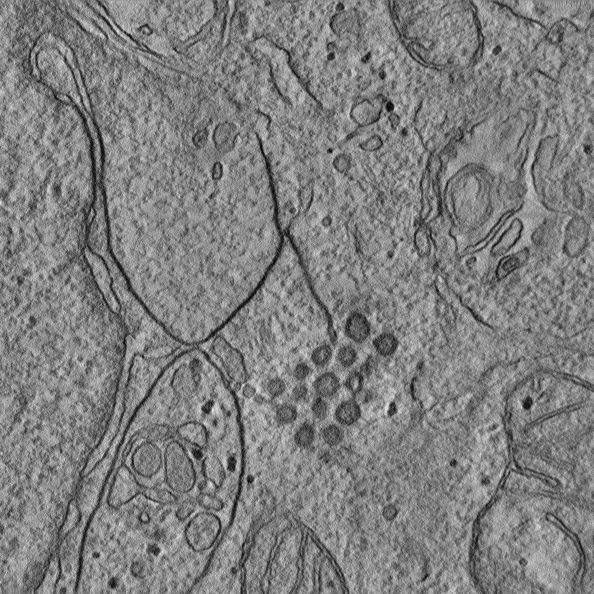
| Supramolecule | Name: Thick section of plastic embedded HeLa cells co-expressing SARS-CoV-2 NSP3/4/6. type: cell / ID: 1 / Parent: 0 Details: The tomogram shows double membrane vesicles connected to zippered ER domain. |
|---|---|
| Source (natural) | Organism: Homo sapiens (human) |
-Experimental details
-Structure determination
| Method | negative staining |
|---|---|
 Processing Processing | electron tomography |
| Aggregation state | cell |
-Sample preparation
| Buffer | pH: 7.2 |
|---|---|
| Staining | Type: POSITIVE / Material: Uranyl Acetate, OsO4 |
| Sugar embedding | Material: Epon |
| Details | Epon embedded pellet from cultured HeLa cells |
| Sectioning | Ultramicrotomy - Instrument: Leica EM UC7 / Ultramicrotomy - Temperature: 20 K / Ultramicrotomy - Final thickness: 250 nm |
| Fiducial marker | Manufacturer: Aurion / Diameter: 10 nm |
- Electron microscopy
| Microscope | FEI TECNAI 12 |
|---|---|
| Image recording | Film or detector model: OTHER / Average electron dose: 30.0 e/Å2 |
| Electron beam | Acceleration voltage: 120 kV / Electron source: LAB6 |
| Electron optics | Illumination mode: FLOOD BEAM / Imaging mode: BRIGHT FIELD / Nominal defocus max: 1.2 µm / Nominal defocus min: 1.0 µm |
| Sample stage | Specimen holder model: FISCHIONE 2550 |
-Image processing
| Details | IMOD |
|---|---|
| Final reconstruction | Number images used: 131 |
