ジャーナル: Proc Natl Acad Sci U S A / 年: 2022 タイトル: The structured organization of ' cell envelope. 著者: Domenica Farci / Patrycja Haniewicz / Dario Piano / 要旨: Surface layers (S-layers) are highly ordered coats of proteins localized on the cell surface of many bacterial species. In these structures, one or more proteins form elementary units that self- ...Surface layers (S-layers) are highly ordered coats of proteins localized on the cell surface of many bacterial species. In these structures, one or more proteins form elementary units that self-assemble into a crystalline monolayer tiling the entire cell surface. Here, the cell envelope of the radiation-resistant bacterium was studied by cryo-electron microscopy, finding the crystalline regularity of the S-layer extended into the layers below (outer membrane, periplasm, and inner membrane). The cell envelope appears to be highly packed and resulting from a three-dimensional crystalline distribution of protein complexes organized in close continuity yet allowing a certain degree of free space. The presented results suggest how S-layers, at least in some species, are mesoscale assemblies behaving as structural and functional scaffolds essential for the entire cell envelope.
 ムービー
ムービー コントローラー


 データを開く
 基本情報
基本情報
 マップデータ
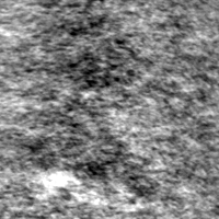
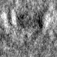
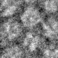
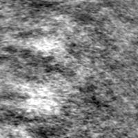
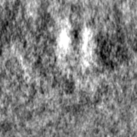
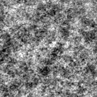
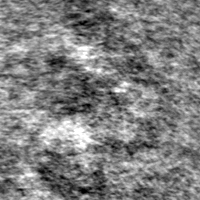
 試料
試料 Deinococcus radiodurans R1 (放射線耐性)
 データ登録者
 ポーランド, 2件
  引用
引用

 構造の表示
構造の表示
 ダウンロードとリンク
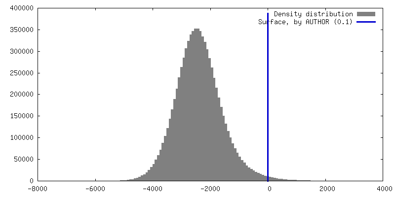
 EMDBマップデータ形式
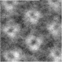
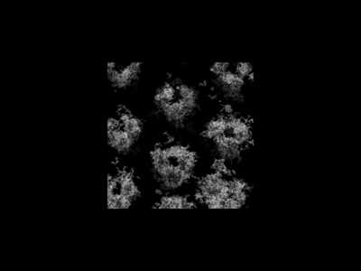 emd_14095.png
 http://ftp.pdbj.org/pub/emdb/structures/EMD-14095

 Z (Sec.)
 Y (Row.)
 X (Col.)


 試料の構成要素
 解析
解析 電子顕微鏡法
 FIELD EMISSION GUN
